Case Report Asbestos-Related Diseases in Thailand and Review Literature Ponglada Subhannachart MD*, Narongpon Dumavibhat MD**,***, Somkiat Siriruttanapruk MD, PhD**** * Department of Radiology, Central Chest Institute of Thailand, Nonthaburi, Thailand ** Department of Preventive and Social Medicine, Faculty of Medicine, Siriraj Hospital, Mahidol University, Bangkok, Thailand *** Department of Environmental Medicine, Kochi Medical School, Kochi University, Kochi, Japan **** Bureau of Occupational and Environmental Diseases, Ministry of Public Health, Nonthaburi, Thailand Asbestos is a harmful substance that can cause both malignancy and non-malignancy in humans. Although it has been used in Thailand for several years, few cases of asbestos-related diseases were reported. Concerning about high consumption and long exposure of asbestos in the country, the incurable but preventable diseases caused by asbestos will be the health problem in the near future. The authors presented 2 cases with asbestos-related diseases, one diagnosed as malignant mesothelioma and the other as asbestosis. Keywords: Asbestos in Thailand, Asbestosis, Asbestos-related diseases, Mesothelioma
J Med Assoc Thai 2012; 95 (Suppl. 8): S71-S76 Full text. e-Journal: http://jmat.mat.co.th Asbestos, a group of mineral fibers, has been used in many products for centuries. It can cause malignant and non-malignant diseases of the lungs and pleura e.g. pleuritis, pleural plaque, asbestosis, lung cancer and malignant mesothelioma (MM). Developed countries, which use asbestos before the others, have already got experiences with asbestos-related diseases (ARD). Although most of them stop using asbestos, the incidence of ARD still increases. The reason is the long latency period of ARD which usually ranges from 20 to 40 years. Even though asbestos has been used in Thailand for more than 70 years, only a few cases of ARD were recognized(1,2). The aim of the present study was to present 2 cases of ARD who were diagnosed recently. This may raise concern about the health problem from asbestos in Thailand.
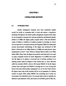
at a fiber cement factory in production line since 1985 and continued working until the presentation of symptoms. Decreased breath sounds with dullness to percussion were detected at the lower part of the right hemithorax. Chest radiograph revealed haziness of the lateral part of the right chest, suggestive of loculated right pleural effusion or pleural mass (Fig. 1). Computed Tomography of the chest revealed loculated right pleural effusion with circumferential pleural thickening at both chest wall and mediastinal sites associated with partial right lung volume loss. Multiple nodules at
Case Report Case 1 A 51-year-old Thai male presented in May 2008 with a 1-month history of progressive dyspnea, non-productive cough and weight loss. He had worked Correspondence to: Dumavibhat N, Department of Preventive and Social Medicine, Faculty of Medicine Siriraj Hospital, Mahidol University, Bangkok 10700, Thailand. Phone: 0-2419-7284 E-mail:
[email protected]
J Med Assoc Thai Vol. 95 Suppl. 8 2012
Fig. 1
Frontal radiograph of the chest revealed haziness of lateral part of right chest, suggested of loculated right pleural effusion or pleural mass
S71
surface of the right lobe of the liver with liver mass, nodular thickening at right upper peritoneum, multiple paraaortic lymph nodes enlargement and right adrenal mass were detected (Fig. 2-4). He underwent routine chest radiography every year which was normal. Pleural biopsy was performed during the inpatient admission. Microscopic examination demonstrated moderately pleomorphic malignant spindle cells. The diagnosis of sarcomatoid mesothelioma was made after immunohistochemical staining which was positive for calretinin, vimentin, as well as thrombomodulin and negative for CK 5/6 and WT-1. The patient died of disease progression 4 months after diagnosis. Case 2 A 76-year-old Thai male, who had a 35 pack-
year history of smoking and underlying conditions including IHD, AF and old CVA for 5 years, went for a medical check-up in July 2011. He had worked as a mechanic at a fiber cement factory for 35 years and retired at the age of 60 years. His chest radiograph revealed minimal fine reticular opacities at both lung bases of s/s 1/0 according to ILO 2000 International Classification of Radiographs of Pneumoconioses, calcified pleural plaque at left chest wall and both diaphragms, mild cardiomegaly and hyperaeration of both lungs suggestive of pulmonary emphysema (Fig. 5). Computed tomography of the chest demonstrated intralobular and interlobular interstitial thickening at both lung bases with some honeycombing appearance, distortion of bronchovascular structure with parenchymal band and rounded atelectasis at right lung base adjacent to right diaphragmatic calcified pleural plaque. Multiple areas of calcified pleural plaque at chest wall pleura and diaphragmatic pleura were noted bilaterally (Fig. 6-9). Centrilobular emphysema, more pronounced at upper lungs, was also demonstrated. Spirometry showed severe airway obstruction with FEV1 = 1.08 L (53%), FVC = 2.70 L (97%) and FEV1/FVC = 40%. The patient refused to undergo bronchoscopy with transbronchial biopsy. Discussion Asbestos is a group of naturally occurring fibrous silicate minerals which is well-known for its
Fig. 2
Axial contrast-enhanced CT scan demonstrated loculated right pleural effusion, circumferential pleural thickening and multiple paraaortic lymph nodes enlargement
Fig. 4
Fig. 3
S72
Axial contrast-enhanced CT scan showed multiple nodules at surface of right lobe liver and right adrenal mass
Coronal reconstruction contrast-enhanced CT scan revealed loculated right pleural effusion, circumferential, lobulated pleural thickening encasing the right lung. Nodules at surface of right lobe liver with liver mass, nodular thickening at right upper peritoneum were detected
J Med Assoc Thai Vol. 95 Suppl. 8 2012
Fig. 5
Fig. 6
Fig. 7
Axial CT at lung bases (lung window) showed intralobular and interlobular interstitial thickening with some honeycombing appearance
Fig. 8
Axial CT (lung window) demonstrated rounded atelectasis at right lung base (arrow)
PA chest radiograph revealed minimal fine reticular opacities at both lung bases.Calcified pleural plaque at left chest wall and both diaphragms was noted (arrow)
Axial CT at lung bases (mediastinal window) revealed calcified pleural plaques at posterior left chest wall and both diaphragmatic pleura (arrow)
properties i.e. thermal and chemical resistance, flexibility, and high tensile strength. In addition, it is inexpensive compared with substitute with similar properties(3). There are 2 configurations of asbestos i.e. amphibole and serpentine. Amphibole minerals include 5 asbestos species i.e. amosite, crocidolite, tremolite, anthophyllite, and actinolite. The only type of serpentine groups is chrysotile, also known as white asbestos(4). Chrysotile, accounted for 95% of the asbestos used around the world, is the only type of asbestos in commercial use today(4). Asbestos has been commonly used in a wide
J Med Assoc Thai Vol. 95 Suppl. 8 2012
range of products e.g. roofing shingles, ceiling and floor tiles, paper products, insulation, cement products, textiles, as well as brake and clutch. It is well established that asbestos causes lung cancer and MM(5-7). There are approximately 125 million people in the world exposed to asbestos at the workplace. Furthermore, more than 107,000 workers exposed to asbestos die each year from asbestos-related lung cancer, MM and asbestosis(8). More important, ARD are also associated with exposure to asbestos in non-occupational or environmental setting. Currently, Thailand is the world top 5 in terms of importer and user of asbestos. Although it has been imported for more than 70 years, there were few reports of ARD in the country. Pleural mesothelioma was first diagnosed in Thailand in 1954(9). Since then, there were some sporadic cases but none of the patients had asbestos exposure history(10-13). In 2007, a case of MM
S73
Fig. 9
CT chest with sagittal reconstruction revealed rounded atelectasis at right lung base (arrow) adjacent to right diaphragmatic calcified pleural plaque
with history of asbestos exposure was firstly reported(1). In addition, in 2009, there was one study which reported 39 workers out of 907 (4.3%), who had abnormal chest imaging compatible with asbestosis and asbestosrelated pleural diseases in one fiber cement factory(2). In the present study, the authors reported 2 cases of ARD, i.e. MM and asbestosis. Both patients worked at the same factory but in different time periods. This factory is also the same one mentioned in the report in 2009 and it uses asbestos as a raw material in its products. Therefore, both patients had a definite history of asbestos exposure. Exposure to asbestos is the most common cause of MM, accounted for 80-90%(14). In some countries e.g. France, Belgium, the Netherlands, Japan, South Korea, patients with pathologically proved MM can get compensation even if they are not exposed to asbestos in an occupational setting. MM is a malignant tumor of mesothelium which lines pleural, peritoneal and pericardial cavities. The incidence of MM varies between countries. The highest incidence rate is approximately 30 cases per million reported or estimated for Australia, Belgium, and Great Britain(15). Unfortunately, there is no national data of MM in many countries especially the countries
S74
that use large amounts of asbestos e.g. Russia, China, India and Thailand. The predicted cumulative mortality of MM among these countries during 1994-2008 were approximately 21,300, 5,100, 2,200 and 545 cases, respectively(16). The latency period of the disease is unpredictable. It is usually more than 20 years. In some cases, the time between first exposure to asbestos and the onset of symptoms may be as long as 50-60 years(17). Due to the poor prognosis and poor response to treatment, most of the patients die within the first year after diagnosis. Chest radiograph of malignant pleural mesothelioma usually shows pleural effusion. Pleural thickening may also be seen and as the tumor progresses, a more lobulated outline is observed. The affected hemithorax becomes contracted and diffuse encasement of lung tissue. Tumor tissue sometimes extends into interlobar fissures. When the lesion progresses, chest wall, mediastinum and diaphragm may be involved as well as lymph nodes, the contralateral lung, the liver and adrenal glands(18-20). The most common finding in CT is pleural thickening which can be lobular or smooth. Other benign features such as pleural plaques or calcification can be seen in conjunction, but MM is not known to arise from plaques. CT can also be useful in demonstrating invasion of the tumor into the mediastinum, chest wall, diaphragm and pericardium, which aids in disease staging and treatment planning. In case 1, the lesions are very extensive with invasion of diaphragm, liver and metastasis to adrenal gland and contralateral media-stinal lymph nodes. Asbestosis, a fibrotic lung disease caused by inhalation of asbestos fibers, typically has a latency period of 15 years or more from initial exposure until diagnosis(21). Asbestos exposure may also be associated with fibrosis of the walls of the respiratory bronchioles and alveolar ducts, as very early asbestosis(21). The most common abnormalities seen on chest radiograph are interstitial lines (thickened intralobular and interlobular interstitial and centrilobular core structures), found approximately 84% of cases, followed by parenchymal bands (76%) and distortion of secondary pulmonary lobules (56%)(17). An early feature of asbestosis is a subpleural curvilinear opacity. High-resolution CT, a sensitive tool for detecting early asbestosis than plain chest radiograph, depicted changes in 80% of patients with clinical but not chest radiographic evidence of asbestosis and showed changes of asbestosis in one-third of patients with neither clinical nor chest radiographic evidence of asbestosis(18). Despite the increased sensitivity of
J Med Assoc Thai Vol. 95 Suppl. 8 2012
HRCT compared with radiography, it is important to recognize that the finding of a normal examination by computed tomography does not exclude the presence of asbestosis. The abnormal chest radiograph with interstitial lines association with pleural plaques increases the specificity of the diagnosis. The classic distribution of pleural plaques seen on chest radiographs is the posterolateral chest wall between the seventh and tenth ribs, lateral chest wall between the sixth and ninth ribs, the dome of the diaphragm, and the mediastinal pleura. Rounded atelectasis may appear with chronic pleural scarring. Typical CT features of rounded atelectasis are of a round or oval mass that abuts the pleura, a “comet tail” of bronchovascular structures going into the mass and thickening of the adjacent pleura(18). A recent study showed that the trend of asbestos-related lung diseases was associated with low ILO scores, long latencies, more severe degree in smokers and a normal or obstructive pattern of pulmonary function test(22). The additive effects of exposure to both cigarette smoke and asbestos fibers contributed to airway obstruction. The diagnosis of asbestosis is usually based on exposure history and radiological findings(21,23). Tissue pathology may be helpful when other diagnosis is suspicious(21). In case 2, chest imaging is typical for asbestosis and pleural plaque according to the ILO classification. Tobacco smoke in combination with asbestos cause centrilobular emphysema as well as airway obstruction in this patient. ARD vary in terms of clinical and radiologic presentation. The long latency period makes it more difficult to diagnose. Due to the sharp increase in asbestos use in Asia since 1970, a surge of ARD should be anticipated in the coming decades(24). Concerning the facts that there is no evidence for a threshold for the carcinogenic effect of asbestos and that increased cancer risks have been observed in populations exposed to a very low level, the World Health Organization (WHO) emphasizes that the most efficient way to eliminate ARD is to stop using all types of asbestos(25). To date, asbestos is banned in 52 countries but this number is quite small when compared with the number of countries which still use asbestos(4). Conclusion The report shows 2 cases of asbestos-related diseases; malignant mesothelioma and asbestosis. Both patients had a history with a long duration of asbestos exposure. Since the amount of asbestos imported and used in Thailand is quite high (approximately 150,000 tons a year), this will be one of the public
J Med Assoc Thai Vol. 95 Suppl. 8 2012
health problems in the near future. Therefore, preventative and control measures, such as, reducing and stopping the use of asbestos, substitution of safer materials and medical surveillance of the diseases, must be implemented urgently. Acknowledgment The authors wish to thank Dr. Kenji Morinaga, Yukinori Kusaka, Narufumi Suganuma, Chanon Mekjarasnapha, Sutarat Tungsagunwattana, Chomphunut Vijitsanguan, Rapeeporn Rojsaengroeng, Teerasak Kiatprathomchai and Chatkanok Dumavibhat for their precious discussion and comments. Potential conflicts of interest None. References 1. Wongvitvichot S, Jiamjarasrangsi W, Sriuranpong V. Occupational malignant mesothelioma in Thailand. J Health Sci 2009; 18: 155-62. 2. Subhannachart P, Vijitsanguan C, Tungsagunwattana S, Uttawet S. Abnormal chest imaging compatible with asbestosis and asbestos-related pleural diseases in fiber cement factory. Bull Dept Med Serv 2009; 34: 13-27. 3. Roggli VL, Coin P. Mineralogy of asbestos. In: Roggli VL, Oury TD, Sporn TA, editors. Pathology of asbestos-associated diseases. 2nd ed. New York: Springer; 2004: 1-16. 4. Collegium Ramazzini. Asbestos is still with us: Repeat call for a universal ban. Am J Ind Med 2011; 54: 168-73. 5. National Institute for Occupational Safety and Health. Revised recommended asbestos standard [Internet]. Atlanta: CDC; 1976 [cited 2011 Nov 11]. Available from: http://www.cdc.gov/niosh/docs/ 77-169/ 6. International Agency for Research on Cancer. IARC monographs on the evaluation of carcinogenic risks to humans- asbestos [Internet]. Vol 14. Lyon: IARC; 1977 [cited 2011 Nov 11]. Available from: http://monographs.iarc.fr/ENG/ Monographs/vol14/volume14.pdf 7. Agency for Toxic Substances and Disease Registry. Toxicological profile for asbestos [Internet]. Atlanta: ATSDR; 2001 [cited 2011 Nov 11]. Available from: http://www.atsdr.cdc.gov/ toxprofiles/TP.asp?id=30&tid=4 8. World Health Organization. Asbestos: elimination of asbestos-related diseases [Internet]. Geneva:
S75
9.
10.
11.
12.
13. 14.
15.
16.
17.
18.
WHO; 2010 [cited 2011 Nov 11]. Available from: http://www.who.int/mediacentre/factsheets/fs343/ en/index.html Bovornkitti S, Pacharee P. Pleural mesothelioma in Thailand. Internal Medicine Journal of Thailand 1981; 1: 39-45. Bovornkitti S, Prijyanonda B, Chatikavanij K, Suwanwilai C, Boonprasarn C. Pleural mesothelioma, fibrous type. Vajira Med J 1968; 12: 31-3. Bovornkitti S, Oonsombati P, Pacharee P, Limsila T. Pleural mesothelioma: report of one case. Siriraj Hosp Gaz 1969; 21: 1190-7. Bovornkitti S, Limsila T, Chaithirapan S, Stitnimankarn T. Primary pleural tumor: mesothelioma. Siriraj Hosp Gaz 1974; 26: 1360-72. Suttinont P, Bovornkitti S. Pleural mesothelioma in Thailand revisited. J Environ Med 1999; 1: 46-53. Sporn TA, Roggli VL. Mesothelioma. In: Roggli VL, Oury TD, Sporn TA, editors. Pathology of asbestos-associated diseases. 2nd ed. New York: Springer; 2004: 104-68. Bianchi C, Bianchi T. Malignant mesothelioma: global incidence and relationship with asbestos. Ind Health 2007; 45: 379-87. Park EK, Takahashi K, Hoshuyama T, Cheng TJ, Delgermaa V, Le GV, et al. Global magnitude of reported and unreported mesothelioma. Environ Health Perspect 2011; 119: 514-8. Craighead JE. Epidemiology of mesothelioma and historical background. In: Tannapfel A, editor. Malignant mesothelioma. Heidelberg: Springer; 2011: 13-25. Roach HD, Davies GJ, Attanoos R, Crane M,
19.
20.
21.
22.
23.
24.
25.
Adams H, Phillips S. Asbestos: when the dust settles an imaging review of asbestos-related disease. Radiographics 2002; 22: S167-84. Hassan R, Alexander R. Nonpleural mesotheliomas: mesothelioma of the peritoneum, tunica vaginalis, and pericardium. Hematol Oncol Clin North Am 2005; 19: 1067-87. Wang ZJ, Reddy GP, Gotway MB, Higgins CB, Jablons DM, Ramaswamy M, et al. Malignant pleural mesothelioma: evaluation with CT, MR imaging, and PET. Radiographics 2004; 24: 105-19. Roggli VL, Gibbs AR, Attanoos R, Churg A, Popper H, Cagle P, et al. Pathology of asbestosis- An update of the diagnostic criteria: Report of the asbestosis committee of the college of american pathologists and pulmonary pathology society. Arch Pathol Lab Med 2010; 134: 462-80. Ohar J, Sterling DA, Bleecker E, Donohue J. Changing patterns in asbestos-induced lung disease. Chest 2004; 125: 744-53. Kishimoto T, Kato K, Arakawa H, Ashizawa K, Inai K, Takeshima Y. Clinical, radiological, and pathological investigation of asbestosis. Int J Environ Res Public Health 2011; 8: 899-912. Le GV, Takahashi K, Park EK, Delgermaa V, Oak C, Qureshi AM, et al. Asbestos use and asbestosrelated diseases in Asia: past, present and future. Respirology 2011; 16: 767-75. World Health Organization. Elimination of asbestos-related diseases [Internet]. Geneva: WHO; 2006 [cited 2011 Nov 11]. Available from: http://www.who.int/occupational_health/ publications/asbestosrelateddisease/en/
โรคทีเ่ กีย่ วข้องกับสารแอสเบสทอส: รายงานผูป้ ว่ ย 2 ราย และทบทวนวรรณกรรม พงษ์ลดา สุพรรณชาติ, ณรงค์ภณ ทุมวิภาต, สมเกียรติ ศิรริ ตั นพฤกษ์ แอสเบสทอส เป็นสารซึ่งมีอันตรายต่อสุขภาพ สามารถก่อให้เกิดโรคในคนทั้งที่เป็นเนื้อร้ายและโรคอื่นๆ ที ่ ม ิ ใ ช่ เ นื ้ อ ร้ า ย ในประเทศไทยมี ก ารใช้ ส ารแอสเบสทอสมาหลายปี แ ต่ พ บว่ า ยั ง มี ก ารรายงานผู ้ ป ่ ว ยที ่ เ ป็ น โรคจากแอสเบสทอสน้อยมาก การใช้แอสเบสทอสปริมาณสูงเป็นระยะเวลายาวนานในประเทศไทยทําให้โรค ซึ่งมีสาเหตุจากแอสเบสทอสซึ่งเป็นโรคที่ป้องกันได้แต่ไม่สามารถรักษาให้หายได้อาจเป็นปัญหาในอนาคตอันใกล้นี้ ผูน้ พิ นธ์ได้รายงานผูป้ ว่ ยซึง่ มีประวัตสิ มั ผัสแอสเบสทอส 2 ราย ได้แก่ malignant mesothelioma และ asbestosis S76
J Med Assoc Thai Vol. 95 Suppl. 8 2012