CLINICAL OBSTETRICS AND GYNECOLOGY Volume 52, Number 4, 586–596 r 2009, Lippincott Williams & Wilkins
Appendicitis and Cholecystitis in Pregnancy NORIDELLE B. GILO, MD, DENNIS AMINI, MD, MS, and HELAIN J. LANDY, MD Division of Maternal-Fetal Medicine, Department of Obstetrics and Gynecology, Georgetown University Hospital, Washington, DC Abstract: Acute abdominal pain in pregnancy may be attributable to a broad range of nonobstetrical causes. The evaluation of an acute abdomen during pregnancy must include in the differential diagnosis appendicitis and cholecystitis, which are 2 of the most common reasons for nonobstetric surgical intervention in pregnancy. Both conditions may be associated with significant maternal and fetal morbidity and/or mortality. This study will provide a contemporary synopsis regarding the diagnosis and management of appendicitis and cholecystitis during pregnancy. Key words: appendicitis, cholecystitis, abdominal pain in pregnancy
Introduction Appendicitis and cholecystitis are the 2 most common conditions associated with an acute abdomen during pregnancy. Their accurate diagnosis is critical for properly managing pregnant women and their fetuses considering the potential for significant morbidity and even mortality. Abdominal surgical treatment, which Correspondence: Helain J. Landy, MD, Chair, Department of Obstetrics and Gynecology, Georgetown University Hospital, 3800 Reservoir Road, NW (3PHC), Washington, DC 20007-2113. E-mail: landyh @gunet.georgetown.edu CLINICAL OBSTETRICS AND GYNECOLOGY
586 | www.clinicalobgyn.com
/
occurs in approximately 0.2% of patients during pregnancy, is most often commonly performed in patients with these diagnoses.1 This article will describe the current information about the clinical and radiologic diagnosis and treatment pertaining to appendicitis and cholecystitis associated with pregnancy.
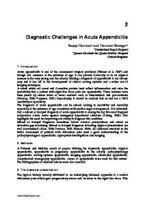
Appendicitis Appendicitis is a rare but emergent complication of pregnancy, which has been estimated to occur in approximately 1 of 1400 to 1 of 6600 deliveries.1–4 Compilation of reported data suggests a more frequent occurrence during the second trimester compared with other times during pregnancy (Fig. 1). Appendectomy accounts for roughly 25% of nonobstetrical operations making it the most common nonobstetrical surgery performed during pregnancy,1 which could be taken to reflect a relative inaccuracy in correctly diagnosing the condition during gestation. As pregnancy advances, the enlarging uterus gradually displaces the appendix caudally.6 Anatomic changes, VOLUME 52
/
NUMBER 4
/
DECEMBER 2009
Appendicitis and Cholecystitis in Pregnancy
587
Appendicitis cases compared by trimester 100 1st trimester 2nd trimester
80
3rd trimester
60 % 40
46
43 43
42
38
32 26
20
16
14
0 Total number of cases (N=1116)
Perforation (N=116)
Pathologically confirmed (N=663)
FIGURE 1. Comparison by trimester of total number of cases, pathologically confirmed cases, and appendiceal perforation among reports of appendicitis in pregnancy.1,2,4,5 Totals vary, as not all reports have included data for all categories.
compounded by efforts to limit radiologic exposure during pregnancy, pose difficulties with the radiologic confirmation of the diagnosis and must be remembered while evaluating the patient and studying radiologic images. Adding to potential diagnostic confusion, presenting complaints may be nonspecific (eg, malaise, nausea, vomiting, or vague abdominal discomfort), symptoms commonly attributed to a variety of other causes of abdominal pain during gestation. Overall, the prevalence of appendicitis is similar in gravid versus nongravid women. The reported risk of appendiceal rupture during pregnancy, ranging from 4% to 57%, is somewhat higher than that in the nonobstetric population (between 4% and 19%).1,3 Reported data of the cases during pregnancy indicate a less frequent occurrence of perforation during the first trimester (Fig. 1).2 Older reports describe appendiceal perforation during pregnancy as being associated with 20% to 35% fetal mortality rates, compared with a more favorable, though still signi-
ficant, fetal loss rate as high as 8% in more recent studies.1,2,5 Such data should be contrasted with much lower fetal mortality rates of 2% or lesser in cases of unruptured maternal appendicitis.1 Signs and symptoms of appendicitis in pregnancy may include fever, anorexia, nausea, vomiting, right lower abdominal pain, and leukocytosis. Symptoms, such as nausea and vomiting, may occur during normal pregnancy or in association with other conditions, thus, obscuring and delaying the proper diagnosis. Pain caused by peritoneal irritation is localized to the right lower quadrant as expected in early pregnancy. In fact, right lower quadrant pain is reported as the most common presenting symptom of appendicitis in pregnancy,3,5 although the pain may become less localized after the fifth month of gestation because of the displacement of the appendix superiorly and away from the right lower quadrant by the enlarging uterus. Some investigators have stressed that the pain does not necessarily ‘‘shift’’ to the right upper quadrant and that most www.clinicalobgyn.com
588
Gilo et al
patients have pain in the right lower quadrant in all trimesters.3,5 Back or flank pain may be the presenting complaint in patients with a retrocecal appendix, leading to a suspicion of pyelonephritis or nephrolithiasis. One study has reported that rebound and guarding on physical examination were the most reliable diagnostic finding for acute appendicitis among pregnant women. In another study of 67 cases of probable appendicitis in pregnancy, Mourad et al3 concluded that neither fever nor leukocytosis, a laboratory result that is not uncommon during gestation, are clear indicators of the diagnosis among pregnant women. If undiagnosed in a timely manner, the risk of appendiceal perforation and development of peritonitis may develop, in case the appendiceal diameter and inflammation increase, and with this increase higher risks of morbidity and mortality. Complications include preterm uterine contractions, preterm labor, preterm delivery, wound infection, sepsis, ileus, and pneumonia. Taking into account the combined results of several studies on appendicitis in pregnancy, it may be said that the risk of perforation occurs less frequently in the first trimester and equally during the second and third trimesters (Fig. 1). The enlarging uterus may limit the ability of the omentum and bowel to wall off the infection. The risk of preterm delivery after appendectomy during pregnancy has been reported by several researchers. In a study that involved nearly 800 women after 23 weeks’ gestation, Mazze and Kallen reported that 22% of patients delivered prematurely within 1 week of surgery. Encouragingly, these same researchers noted no additional increase in the preterm birth rate among the pregnancies that continue beyond the first postoperative week. After appendectomy during pregnancy, another group reported a 30% fetal loss rate in the first trimester and an 11% premature delivery rate in the second trimester, but www.clinicalobgyn.com
no such finding was reported in the third trimester.2 Radiologic imaging may be invaluable in diagnosing appendicitis. In the past, ultrasound was advocated as the initial imaging modality for pregnant patients when the diagnosis was suspected.7 More recently, magnetic resonance imaging (MRI) and computerized tomography (CT) have become important techniques in improving the diagnostic accuracy.8 Radiologically, the finding of an enlarged fluid-filled appendix (>7 mm) should be considered abnormal. Sonographically, the appendix may be incompressible; and on CT scan, findings include inflammation in the enlarged, tubular, nonfilling structure, which may or may not contain a fecalith. Choosing the most accurate diagnostic test can be critical for avoiding delay in the diagnosis and subsequent appendiceal perforation and additional complications. One can conclude relative accuracy in using sonography, described in a 2007 review that discussed the value of ultrasound in diagnosing appendicitis in pregnancy.9 This review reported a range of sensitivity of 67% to 100% and a range of specificity of 83% to 96%, compared to diagnostic performance in the general population of 86% and 96%, respectively, though highly dependent on operator experience. Gjelsteen et al10 have proposed an algorithm for imaging in pregnant patients with suspected appendicitis in which ultrasound and MRI are used throughout gestation, but CT imaging is reserved for those beyond the first trimester. The rate of negative appendectomy, that is, cases with a preoperative diagnosis of suspected appendicitis but without final pathologic evidence of appendiceal inflammation, is reported to range from 4% to 50% among pregnant women.1,2,4,5 In comparison, the rate of negative appendectomy in the general population ranges from 10% to 15% and as high as 26% among the reproductive age females.
Appendicitis and Cholecystitis in Pregnancy Patients with a negative appendectomy experienced a higher fetal loss rate (OR 1.88) compared with those with an uncomplicated appendicitis in the report from McGory et al,6 but the authors found higher associations with uterine myomas and uterine inflammation in the former group. The suggested management of the pregnant woman with suspected appendicitis includes a thorough history and complete physical examination. Laboratory studies consist of a complete blood count, urinalysis, aspartate aminotransferase, and alanine transaminase levels and amylase and lipase levels to exclude pancreatitis. Fetal assessment involves ultrasound for viability and confirmation of dates and biophysical profile and/or nonstress test, depending on the gestational age. Imaging studies may include sonography, MRI, and CT scanning, with the latter being preferentially reserved to the second and third trimesters. Pain control with narcotics and broad spectrum coverage with antibiotics are recommended. Immediate surgical intervention within the first 24 hours is warranted in any case of suspected or confirmed acute appendicitis in a pregnant woman to avoid perforation and its possible severe complications.1,2,11 Although specific issues pertaining to surgical management are described in a later section, perioperatively, antibiotic treatment should be chosen for Gramnegative and Gram-positive coverage (eg, a second-generation cephalosporin) and for anaerobes (eg, clindamycin or metranidazole).12
Cholecystitis Acute cholecystitis may affect between 10% and 15% of the general population with gallstones described in almost 20% of women by the age of 40 years.7 Epidemiological factors include race, weight, diet, physical activity, serum lipid levels, and genetic predisposition. Women are
589
more commonly affected than men, and the hormonal milieu of elevated estrogen and progesterone levels is postulated as causative in the pathophysiology of gallstones. Estrogen increases cholesterol secretion, whereas progesterone reduces soluble bile acid secretion,13 and the change in production of bile favoring insoluble acids encourages stone formation. Progesterone further slows the emptying of gallbladder secretions and worsens the accumulation of bilious secretions through its action of decreasing smooth muscle activity. Pathophysiologically, blockage of the cystic duct by stones or sludge, resulting in irritation of the gallbladder lining, is responsible for over 90% of cases of acute cholecystitis (Fig. 2).14 The resulting inflammatory process releases prostaglandins and inflammatory cytokines that may be accompanied by infection involving both aerobic and anaerobic organisms. During pregnancy, the presence of gallstones has been reported in 1% to 3% of patients. Furthermore, pregnancy has been implicated as a cause of bile stone formation and altered gallbladder contractility.7 In the months after delivery, normalization of more soluble forms of bile acids occurs. Biliary sludge, a precursor to gallstones and sonographically visible as the accumulation of bile, is reported in as many as 30% of gravidas with a similar proportion of women affected by the postpartum period.14 Sonography performed in early pregnancy has confirmed the physiologic expansion of the gallbladder and the accumulation of stones, debris, and bile. Obesity and, particularly, a high prepregnancy body mass index are additional strong epidemiological factors correlated with the development of gallstones.15 Despite the predilection toward biliary sludge and stone formation in pregnancy, acute cholecystitis does not occur more frequently during pregnancy; data suggest that cholecystitis only affects 0.1% of pregnant women.16 Gallbladder disease www.clinicalobgyn.com
590
Gilo et al
FIGURE 2. Illustration detailing the anatomy of the gallbladder and adjacent structures.
can present a diagnostic challenge to the obstetrician, as many healthy pregnant women report upper digestive complaints that mimic biliary symptoms. Most patients with symptomatic gallstones complain of biliary colic (recurrent pain characteristically exacerbated by heavy fatty meals) likely resulting from gallbladder contractions and increased pressure and luminal expansion. Generally, symptoms develop from 1 or more obstructing stones. Clinicians are taught to recognize the triad of common gallbladder symptoms consisting of bloating, nausea, and heartburn; however, these symptoms and the presence of gallstones are only weakly associated. www.clinicalobgyn.com
Although a variety of symptoms can be seen with gallstones, perhaps as many as 50% of patients are asymptomatic.7 Those who seem to be ill must be considered as having acute cholecystitis. Common signs and symptoms include right upper quadrant pain, fever, tachycardia, leukocytosis, and inflammation of the gallbladder wall; additional symptoms include anorexia, nausea, vomiting, intolerance to fatty foods, and right upper quadrant pain. Physical examination may reveal Murphy’s sign, but this depends largely on gestational age and body habitus. Sepsis and jaundice also may be seen. Stones entering the common bile duct and the duodenum may produce ascending
Appendicitis and Cholecystitis in Pregnancy cholangitis and pancreatitis, conditions that represent life-threatening emergencies. If left untreated, the most common complication is gangrenous cholecystitis. Other serious complications include abscess, perforation, fistula, ileus, or emphysematous cholecystitis.17 Many times, hospitalization during pregnancy and during the postpartum period is necessary. In a study from Los Angeles of 72 women whose pregnancies were complicated by biliary disease, 45 (62.5%) had biliary colic, 18 (25%) had gallstone pancreatitis, and 9 (12.5%) had acute cholecystitis.18 Imaging studies can be useful for confirming the presence of stones and sludge in symptomatic patients. Sonography is the most useful and sensitive test for detecting gallstones,19 whereas x-ray and CT scanning may not be useful in symptomatic patients. Classic sonographic findings, suggestive of acute cholecystitis (a thickened gallbladder wall over 3 to 5 mm, pericholecystic fluid, calculi, and a sonographic Murphy’s sign), are identical in pregnant and nonpregnant patients. As sonography may fail to identify small stones or those present in the ductal system, endoscopic retrograde cholangiopancreatography (ERCP) may aid in the diagnosis and treatment of acute cholecystitis. ERCP also can be used with sphincterotomy to treat pancreatitis and to prevent obstruction with stones in the common bile duct. The theoretical risk of radiation exposure to the fetus from ERCP (approximately 310 mrad) is not a concern when conducted after the first trimester. For the pregnant patient with symptoms of biliary disease, the differential diagnosis includes appendicitis, pancreatitis, peptic ulcer disease, pyelonephritis, HELLP syndrome (syndrome of hemolysis, elevated liver enzymes, and low platelets), acute fatty liver, and hepatitis. Laboratory tests should include a complete blood count, aspartate aminotransferase, alanine transaminase, total bilirubin, alkaline phospha-
591
tase, and serum amylase and lipase levels to exclude pancreatitis. Fetal assessment should include confirmation of viability and well-being as dictated by gestational age and obstetrical situation. Patients with cholecystitis and biliary colic should be admitted to the hospital. Conservative treatment may be an option at least initially in an attempt to avoid surgery during pregnancy. Conservative therapy, including intravenous hydration, opioid analgesia, and bowel rest, is appropriate in patients with few symptoms or with an incidental finding of gallstones. Broad spectrum antibiotic use is recommended with systemic symptoms and for the patients in whom there is no improvement in 12 to 24 hours. Treatment with nonsteroidal antiinflammatory drugs has been proposed to block the release of prostaglandins and to slow the disease progression. Treatment with diclofenac or indomethacin has been successful in nonpregnant patients. The review by Dietrich et al16 shows the safety and possible success of using a short course of indomethacin in the second trimester to try to reverse gallbladder inflammation. Nonsteroidal antiinflammatory drugs should be used with caution as high doses or prolonged administration, particularly after 32 weeks’ gestation, can be associated with pregnancy complications, including oligohydramnios and narrowing of the fetal ductus arteriosus. Ursodeoxycholic acid, a naturally occurring bile acid agent, can dissolve gallstones by changing the composition of bile, and it has been used in nonpregnant patients for this indication. Although ursodeoxycholic acid has been administered in pregnancy in the management of intrahepatic cholestasis, its safety and efficacy for the treatment of gallstones during pregnancy has not been established.20 Extensive data about pregnancy outcomes in these patients are lacking. The latest review of the literature pertaining to gravidas with biliary tract disease, published in 2007 by Jelin et al,21 describes a www.clinicalobgyn.com
592
Gilo et al
high risk of fetal death (7%) among the patients treated conservatively in comparison with a much lower rate (2.2%) among those who underwent laparoscopic cholecystectomy. Two earlier studies showed similar pregnancy outcomes among patients who required surgical treatment. The study by Swisher et al18 included 46 patients treated conservatively. A 1994 analysis of pregnancy outcomes among 61 gravidas who underwent laparoscopic cholecystectomy by Barone et al22 described 2 cases of premature labor occurring remote from surgery and 3 spontaneous abortions (2 occurring in women with gallstone pancreatitis). Definitive surgical therapy is required for pregnant patients with signs of sepsis, ileus, or perforation. Several experts advocate an earlier surgical intervention, citing a reduced rate of preterm delivery among surgical patients, shorter hospital stays, reduced medication usage, and lower rates of life-threatening complications.18 Compared with a definitive surgical approach, high relapse rates (40% to 70%) are reported among patients treated conservatively.18,21 In determining a correct treatment option for biliary disease during pregnancy, researchers in San Francisco constructed and applied an analytical model incorporating maternal and fetal well-being to decide between nonsurgical management and laparoscopic cholecystectomy, and concluded a benefit to laparoscopy over conservative treatment in the first and second trimesters.21 An alternative to surgical intervention includes percutaneous cholecystostomy, highlighted in a single article reporting 2 instances of acute cholecystitis at 30 and 32 weeks’ gestation with successful ultrasound-guided transhepatic gallbladder drainage. Although both the patients delivered without neonatal complications and subsequently underwent laparoscopic cholecystectomy postpartum, not enough data are available for www.clinicalobgyn.com
recommending this approach routinely.23 Understanding the physiologic changes involving changes in quality of bile and delayed clearance during pregnancy, definitive treatment should be considered in patients with known symptomatic gallstones before conception.
Radiologic Imaging Considerations When considering different imaging options during pregnancy, potential risks and benefits of the radiologic studies must be considered. The major radiologic modalities used in diagnosing appendicitis and cholecystitis include ultrasound, MRI, and CT imaging and each has its advantages and disadvantages (Table 1). In general, the principal concern for the use of ionizing radiation during pregnancy is the potential risk to the fetus. The 2 main concerns associated with ionizing radiation exposure are its carcinogenic potential and teratogenic effects. Convention defines 1 rad as the amount of energy absorbed per unit mass; 100 rads is equal to 1 Gy. One Sievert (Sv) is defined as the amount of biologic damage from a radiation dose, calculated in part using the radiation dose. For external radiation exposure, 1 rad is equivalent to 1 rem, and 1 Sv is equal to 100 rem. The National Council on Radiation Protection and Measurements has published guidelines for the limits of radiation exposure; the maximal permissible radiation dose for fetal exposure during pregnancy is 5 mSv. The threshold below which the radiation exposure is not believed to cause teratogenic effects is estimated at 0.05 to 0.15 Gy (5 to 15 rads).27 Given that the baseline risk of fatal childhood cancer is estimated to be 1 in 2000, fetal radiation exposure from a maternal pelvic CT study (approximately 5 rads or 0.05 Gy) is thought to increase that risk 2-fold to a risk of 2 per 2000.27
Appendicitis and Cholecystitis in Pregnancy TABLE 1.
593
Comparison of Imaging Techniques Used in the Diagnosis of Appendicitis and Cholecystitis 8,24–26 Utility in Diagnosis of Appendicitis Benefits
Ultrasonography
No concern for fetal exposure to ionizing radiation No need for use of contrast medium Readily available Relatively inexpensive Magnetic No concern for resonance fetal exposure imaging to ionizing radiation Less operatordependent compared with ultrasonography Computerized Widely used tomography Less operatordependent compared with ultrasonography
Utility in Diagnosis of Cholecystitis
Sensitivity Specificity Sensitivity Specificity (%) (%) (%) (%)
Disadvantages
100* Accuracy highly dependent on operator experience May yield inconclusive results
96*
85* 95
95
100* Time-consuming Expensive Less widely used Some radiologists may have limited experience in reading
93.6*
77 (using wall edema)
78 (using wall edema)
99
91.7
99.1
Concern for fetal exposure to ionizing radiation (small amount 1 to 4 rads)
92
* Specific data during pregnancy.
Chen et al27 highlight the magnitude of the risk of the fetal exposures earlier in pregnancy, estimating the relative risk of development of the fatal childhood cancer to be 3.19 from first trimester exposure compared with roughly 1.3 from the exposure in the second and third trimesters. Very early in pregnancy (2 to 8 wk), the predominant risks of radiation exposure involve pregnancy loss, malformations, and mental retardation. With an exposure between 8 to 20 weeks, the time period of maximal and particularly neuronal development, fetal risks include microcephaly, micropthalmia, mental retardation, growth restriction, cataracts, and behavioral defects.27 The use of ultrasound is safe during pregnancy with no concerns for exposure
to ionizing radiation or for a requirement of the use of contrast medium. Other benefits of sonography are its ready availability and relative inexpensive cost. MRI is a viable modality for the evaluation of pregnant women and is considered to be a safe and reliable tool in the diagnosis of different etiologies of abdominal pain in pregnancy. When ultrasound results are inconclusive or in situations in which MRI is unavailable or limited by operator experience, CT may add valuable information, providing a particularly useful tool in the diagnosis of appendicitis in pregnancy. The major disadvantage of CT imaging is radiation exposure, although the amount is considered to be relatively small, ranging from 1 to 4 rads. Concern for the radiation exposure, www.clinicalobgyn.com
594
Gilo et al
however, should not prevent the use of helical CT in an appropriate patient.8,10,24
Surgical Considerations Different surgical interventions include laparoscopy or laparotomy; the preference is based on the uterine size and the operator’s experience. The first option may be safely used in the first 2 trimesters, although technical challenges may still exist, as the uterus has enlarged above the level of the umbilicus.11 Some investigators have proposed limiting the procedure to gestational age up to 28 weeks;28 however, uterine size, maternal body habitus, and surgical history may be more important than gestational age in determining surgical approach. Benefits of a laparoscopic approach during pregnancy include possibly less uterine manipulation while achieving optimal surgical exposure and facilitating a more prompt diagnosis and treatment.11 One disadvantage of laparoscopy includes its potential for causing an injury to the gravid uterus. Although there have been theoretical concerns about the effect of the pneumoperitoneum on fetal physiology (eg, increased intra-abdominal pressure and fetal acidosis during pneumoperitoneum with carbon dioxide), no adverse effects have been shown with maximal pneumoperitoneum pressure limited to 10 to 12 mm Hg and a duration of less than 60 minutes.11 Perioperative management also should include left lateral tilt to prevent vena caval compression easily obtainable by tilting the operating table to the left. In addition, the use of compression devices and/or low-molecular-weight heparin should be considered as methods to minimize the risk of thromboembolic events.11 A 1999 Utah study that evaluated pregnant patients who underwent open and laparoscopic appendectomies and cholecystectomies did not show significant differences regarding preterm delivery rates, birth weights, or Apgar scores.13 In pregwww.clinicalobgyn.com
nant patients with appendicitis, the method of surgical treatment (eg, laparoscopy versus laparotomy) has not been associated with differences in obstetrical outcomes regarding the risks of preterm delivery or fetal loss.11 In the review by Lemieux et al of 45 patients, 8 women underwent laparoscopic appendectomy in the third trimester at an average gestational age of 31 weeks.4 The same authors reported the need for conversion to laparotomy in 3 patients (2 in the third trimester, 1 in the second trimester), for technical difficulties in 2 cases and uterine perforation in the third. In cases of appendicitis, several articles describe the utility of an open access to the abdomen for laparoscopy.11 During laparotomy, a muscle-splitting incision can be made over the point of maximal tenderness.16 In patients with appendiceal rupture, the abdomen should be copiously irrigated and the skin left open to avoid wound infection. Although in 1998, the Society of American Gastrointestinal Endoscopic Surgeons recommended continuous sonographic fetal monitoring during such surgical procedures,29 many experts have stressed its low efficacy and difficulty of implementation.11 Most experts instead have adopted the recommendations of uterine and fetal monitoring before and after surgery.11,29 Tocolysis use should be limited to obstetrical indications and not administered prophylactically.11,29
Conclusions Appendicitis and cholecystitis are common concerns for the pregnant patient with abdominal pain. Imaging options include sonography, MRI studies, and CT scanning. Diagnosis may be confounded by physiologic and anatomic changes during pregnancy. Expectant or surgical management protocols may be appropriate, depending on the patient’s
Appendicitis and Cholecystitis in Pregnancy clinical presentation and gestational age. The safety and efficacy of the laparoscopic approach during pregnancy has been well established. Pregnancy outcomes are unlikely to be affected except in instances in which maternal morbidity and diagnostic delay are significant.
11.
12.
References 1. Mazze RI, Kallen B. Appendectomy during pregnancy: a Swedish registry study of 778 cases. Obstet Gynecol. 1991; 77:835–840. 2. Andersen B, Nielsen TF. Appendicitis in pregnancy: diagnosis, management and complications. Acta Obstet Gynecol Scand. 1999;78:758–762. 3. Mourad J, Elliott J, Erickson L, et al. Appendicitis in pregnancy: new information that contradicts long-held clinical beliefs. Am J Obstet Gynecol. 2000;182: 1027–1029. 4. Lemieux P, Rheaume P, Levesque I, et al. Laparoscopic appendectomy in pregnant patients: a review of 45 cases. Surg Endosc. 2008 [Epub ahead of print] DOI 10.1007/s00464-008-0201-9. 5. Yilmaz HG, Akgun Y, Bac B, et al. Acute appendicitis in pregnancy – risk factors associated with principal outcomes: a case control study. Int J Surg. 2007;5: 192–197. 6. McGory ML, Zingmond DS, Tillou A, et al. Negative appendectomy in pregnant women is associated with a substantial risk of fetal loss. J Am Coll Surg. 2007; 205:534–540. 7. Ramin KD, Ramsey PS. Disease of the gallbladder and pancreas in pregnancy. Obstet Gynecol Clinn North Am. 2001;28: 571–580. 8. Pedrosa I, Levine D, Eyvazzadeh A, et al. MR imaging evaluation of acute appendicitis in pregnancy. Radiology. 2006;238: 891–899. 9. Williams R, Shaw J. Ultrasound scanning in the diagnosis of acute appendicitis in pregnancy. Emerg Med J. 2007;24: 359–360. 10. Gjelsteen AC, Ching BH, Meyermann MW, et al. CT, MRI, PET, PET/CT,
13.
14.
15.
16. 17.
18.
19.
20.
21.
22. 23.
595
and ultrasound in the evaluation of obstetric and gynecologic patients. Surg Clin N Am. 1008;88:361–390. Moreno-Sanz C, Pascual-Pedreno A, Picazo-Yeste JS, et al. Laparoscopic appendectomy during pregnancy: between personal experiences and scientific evidence. J Am Coll Surg. 2007;205:37–42. Firstenberg MS, Malangoni MA. Gastrointestinal surgery during pregnancy. Gastroenterol Clin North Am. 1998;27:73–88. Affleck DG, Handrahan DL, Egger MJ, et al. The laparoscopic management of appendicitis and cholelithiasis during pregnancy. Am J Surg. 1999;178:523–529. Ko CW, Beresford SA, Schulte SJ, et al. Incidence, natural history, and risk factors for biliary sludge and stones during pregnancy. Hepatology. 2005;41:359–365. Ko CW. Risk factors for gallstone-related hospitalization during pregnancy and the postpartum. Am J Gastroenterol. 2006; 101:2263–2268. Dietrich CS, Hill CC, Hueman M. Surgical diseases presenting in pregnancy. Surg Clin North Am. 2008;88:408–419. Reiss R, Nudelman I, Gutman C, et al. Changing trends in surgery for acute cholecystitis. World J Surg. 1990;14:567–570; discussion 570–571. Swisher SG, Schmit PJ, Hunt KK, et al. Biliary disease during pregnancy. Am J Surg. 1994;168:576–579; discussion 580–581. Shea JA, Berlin JA, Escarce JJ, et al. Revised estimates of diagnostic test sensitivity and specificity in suspected biliary tract disease. Arch Intern Med. 1994;154: 2573–2581. Ward A, Brogden R, Heel R, et al. Ursodeoxycholic acid: A review of its pharmacologic properties and therapeutic efficacy. Drugs. 1984;27:95–131. Jelin EB, Smink DS, Vernon AH, et al. Management of biliary tract disease during pregnancy: a decision analysis. Surg Endosc. 2008;22:54–60. [Epub 2007 Aug 23] Barone JE, Bears S, Chen S, et al. Outcome study of cholecystectomy during pregnancy. Am J Surg. 1994;168:576–579. Allmendinger N, Hallisey M, Ohki SK, et al. Percutaneous cholecystostomy
www.clinicalobgyn.com
596
Gilo et al
treatment of acute cholecystitis in pregnancy. Obstet Gynecol. 1995;86:653–654. 24. Kennedy A. Assessment of acute abdominal pain in the pregnant patient. Semin Ultrasound CT MR. 2000;21:64–77. 25. Lim HK, Bae SH, Seo GS. Diagnosis of acute appendicitis in pregnant women: value of sonography. Am J Roentgenol. 1992;159:359–342. 26. Lazarus E, Mayo-Smith WW, Mainiero MB, et al. CT in the evaluation of nontraumatic abdominal pain in pregnant women. Radiology. 2007;244:784–790.
www.clinicalobgyn.com
27. Chen MM, Coakley FV, Kaimal A, et al. Guidelines for computed tomography and magnetic resonance imaging use during pregnancy and lactation. Obstet Gynecol. 2008;112:333–340. 28. Barnes SL, Shane MD, Schoemann MB, et al. Laparoscopic appendectomy after 30 weeks pregnancy: report of 2 cases and description of technique. Am Surg. 2004; 70:733–736. 29. Society of American Gastrointestinal Endoscopic Surgeons (SAGES). Guidelines for laparoscopic surgery during pregnancy. Surg Endosc. 1998;12:189–190.