J Am Soc Nephrol 11: 2007–2016, 2000
AP-1 Proteins Mediate Hyperglycemia-Induced Activation of the Human TGF-1 Promoter in Mesangial Cells CORA WEIGERT,* ULRICH SAUER,† KATRIN BRODBECK,* ANDREAS PFEIFFER,‡ ¨ HANS U. HARING,* and ERWIN D. SCHLEICHER* *Department of Internal Medicine, Division of Endocrinology, Metabolism and Pathobiochemistry, University of Tu¨bingen, Tu¨bingen; †Institute of Pathology, University of Munich, Munich; and ‡Medical Center Hospital Bergmannsheil, Bochum, Germany.
Abstract. Hyperglycemia-induced overproduction of the prosclerotic cytokine transforming growth factor-1 (TGF-1) has been implicated in the pathogenesis of diabetic nephropathy. Because high glucose and phorbol esters (PMA) increase TGF-1 mRNA levels in mesangial cells, this study was designed to characterize these effects on the human TGF-1 promoter activity. With the use of luciferase reporter gene constructs containing TGF-1 5⬘-flanking sequence (from ⫺453 to ⫹11 bp) transfected into mesangial cells, it was found that 30 mM glucose induced a nearly twofold increase in TGF-1 promoter activity after 24 h of incubation in human and porcine mesangial cells. Stimulation by PMA was more effective (2.3-fold). Mutagenesis in either one of the two or both activating protein-1 (AP-1) binding sites abolished the high glucose and the PMA effect. Furthermore, addition of the AP-1 inhibitor curcumin obliterated the glucose response. Cor-
responding experiments revealed that the transcription factor stimulating protein 1 was not involved in mediating the glucose effect. The high glucose-induced TGF-1 promoter activation was also prevented by inhibitors of protein kinase C and p38 mitogen-activated proteinkinase. Electrophoretic mobility shift assays with oligonucleotides containing one of the two AP-1 binding sites showed that glucose treatment markedly enhanced the binding activity of nuclear proteins of mesangial cells, particularly to box B. Supershift assays demonstrated that JunD and c-Fos were present in the protein-DNA complexes under control and hyperglycemic conditions. The functional and structural results show that glucose regulates human TGF-1 gene expression through two adjacent AP-1 binding sites and gives rise to the involvement of protein kinase C and p38 mitogen-activated proteinkinase in hyperglycemia-induced TGF-1 gene expression.
Diabetic nephropathy is one of the major causes of end-stage renal disease. The hallmarks of human and experimental diabetic nephropathy are the thickening of the glomerular and tubular basement membranes and the progressive expansion of the mesangial matrix (1–3). The results of the Diabetes Control and Complication Trial have shown that strict glycemic control can prevent the onset and progression of diabetic nephropathy (4). Cultured mesangial cells may serve as an in vitro model for the events that occur early in the development of diabetic nephropathy because several studies have demonstrated that ambient elevated glucose concentrations (5) or increased glucose uptake (6) induces an increased production of matrix proteins in cultured mesangial and other renal cells. More recent reports indicate that this effect is mediated by the hyperglycemia-induced transforming growth factor-1 (TGF1) (7,8). Accordingly, increased renal expression of TGF-1
has been found in experimental animal and human diabetes (9 –11). Among many diverse effects, TGF-1 promotes the accumulation of extracellular matrix by increasing the synthesis of extracellular matrix components and by reducing matrix degradation. Therefore, chronically elevated expression of TGF-1 is suggested to be the major mediator of progressive fibrosis in renal diseases associated with sclerosis and namely in diabetic nephropathy (12,13). The therapeutic effect of inhibiting TGF-1 action by injecting neutralizing TGF-1 antibodies has been shown in experimental glomerulonephritis and experimental diabetic nephropathy (13,14). These data demonstrate that inhibition of TGF-1 activity results in suppression of disease-induced glomerular matrix accumulation. Little is known about the molecular mechanism of the pathologic dysregulation of TGF-1 gene expression in diabetes. Previous studies revealed that the promoter regions of the human TGF-1 gene contain consensus sequences for the transcription factor activating protein-1 (AP-1), which mediate the phorbol ester responsiveness of the TGF-1 promoter (15,16). Phorbol esters are also known to activate protein kinase C (PKC), and high glucose-induced activation of PKC has been suggested to be involved in the development of diabetic nephropathy (17,18). In particular, hyperglycemiainduced synthesis of extracellular matrix proteins has been shown to be associated with activation of PKC in mesangial
Received February 7, 2000. Accepted April 17, 2000. Correspondence to Dr. Erwin D. Schleicher, Department of Internal Medicine, Division of Endocrinology, Metabolism and Pathobiochemistry, University of Tu¨bingen, Otfried-Mu¨ller-Strae 10, D-72076 Tu¨bingen, Germany. Phone: ⫹⫹49 7071 29 87599; Fax: ⫹⫹49 7071 29 5974; E-mail:
[email protected] 1046-6673/1111-2007 Journal of the American Society of Nephrology Copyright © 2000 by the American Society of Nephrology
2008
Journal of the American Society of Nephrology
cells (19). Furthermore, recent studies have shown that treatment of diabetic rats with an oral PKC- inhibitor ameliorates vascular dysfunctions (20). Taken together, these data suggest a possible link between glucose-mediated PKC activation and AP-1–mediated TGF-1 gene activation. The present study was designed for better understanding of the molecular mechanism of the high glucose-induced TGF-1 overexpression. We demonstrate that high glucose stimulates TGF-1 promoter activity and increases specific binding of AP-1 proteins to the corresponding consensus DNA sequences. Furthermore, we show that PKC- and p38 MAPK-dependent pathways mediate the activation of the TGF-1 promoter.
Materials and Methods Materials Human mesangial cells with corresponding growth medium were obtained from Clonetics (Verviers, Belgium). Oligonucleotides were synthesized by Life Technologies (Karlsruhe, Germany). Cell culture media, supplements, Ultroser, and fetal calf serum (FCS) were from Life Technologies (Eggenstein, Germany); Maxiscript kit was from Ambion (Heidelberg, Germany); minocyclin and reagents for the site-directed mutagenesis were from Pan Systems (Aidenbach, Germany); Superfect was from Qiagen (Hilden, Germany); -galactosidase assay chemiluminescent, Klenow enzyme, and poly[d(I-C)] were from Boehringer (Mannheim, Germany); pSV--galactosidase control vector, pRL-TK vector, pGEM3Z, and luciferase assays were from Promega (Madison, WI); PMA, mithramycin, and curcumin were from Sigma (Munich, Germany); bisindolylmaleimide I and SB 203580 were from Calbiochem (Bad Soden, Germany); [␣-32P]UTP and [␣-32P]dATP were from Hartmann (Braunschweig, Germany); antibodies against AP-1/c-Jun (catalog number 44X), c-Jun (1694X), JunB (73X), JunD (74X), c-Fos (253X), CREB-1 (186X), ATF-1 (270X), and ATF-2 (187X) were from Santa Cruz Technologies (Santa Cruz, CA); and antibodies against phospho-c-Jun ser-63 and phospho-ATF-2 thr-71 were from New England Biolabs (Schwalbach, Germany).
Cell Culture Mesangial cells isolated from porcine glomeruli were cultured and characterized as described previously (21). Human mesangial cells
J Am Soc Nephrol 11: 2007–2016, 2000
were cultured as described by the supplier. For experimental purposes, RPMI 1640 containing 6 mM or 30 mM glucose was used and FCS serum was substituted by 2% Ultroser. For equal osmolarity, NaCl was added.
Preparation of Luciferase Reporter Constructs and Site-Directed Mutagenesis The TGF-1-luciferase reporter vector pGL3wt was generated by insertion of nucleotides ⫺453 to ⫹11 of the human TGF-1 promoter into the KpnI and BglII sites of the vector pGL3b, which contains the firefly (photinus pyralis) luciferase gene. Base substitutions were made by oligonucleotide-mediated mutagenesis as described previously (22) with modifications. The mutagenic primers are shown in Table 1. Clones were tested by restriction enzyme digestion if possible, and positive clones were sequenced.
Transfection Methods Mesangial cells were transfected with Superfect according to the instructions of the supplier. One d before transfection, 2.0 ⫻ 105 cells/well were seeded in six-well plates with 2 ml of growth medium. For one well, 2 g total of DNA and 100 l of RPMI 1640 without supplements were premixed, 10 l of Superfect was added, and the samples were mixed for 10 s. After 10 min, the samples were diluted with 600 l of growth medium and the total volume was transferred to the cells. After 3 h, the different culture media with or without inhibitors were added and cells were harvested after 24 h. For transfection experiments with PMA, we used the Ca3(PO4)2-DNA-coprecipitation-method (23) because Superfect interfered with PMA. For one well, 4 g total of DNA, 36 l of H2O, 160 l of Hepes-buffered saline 5/4 (50 mM Hepes-KOH pH 7.05, 172.5 mM NaCl, 6.25 mM KCl, 875 M Na2HPO4, 7 mM Glucose), and 10.4 l of 2.5 M CaCl2 were mixed for 20 s, incubated for 20 min, and added to the medium. After 4 h, the medium was replaced by 1 ml of 10% glycerol in growth medium and cells were incubated for 3 min. Then the glycerol medium was removed and the culture medium was added. After 20 h, the cells were stimulated with 0.5 M PMA and harvested after 9 h. For normalization of transfection efficiencies, we cotransfected 0.25 g of pSV--galactosidase control vector. To study the effect of PMA, we used 0.5 g pRL-TK vector, which contains the seapansy (renilla reniformis) luciferase gene under control of the herpes sim-
Table 1. Oligonucleotides used for the mobility shift experiments and the site-directed mutagenesisa Sequence
CTTGTTTCCCAGCCTGACTCTCCTTCCGTTCT CAAAGGGTCGGACTGAGAGGAAGGCAAGACCC TGTTTCCCAGCCTGGTTCTCCTTCCGTTCTGG AAGGGTCGGACCAAGAGGAAGGCAAGACCCAG GGTCGGCTCCCCTGTGTCTCATCCCCCGGATT GCCGAGGGGACACAGAGTAGGGGGCCTAATTC GGTCGGCTCCCCTGGTTCTCATCCCCCGGATT GCCGAGGGGACCAAGAGTAGGGGGCCTAATTC AGCCGGGGAGCCCGCCCCCTTTCCCCCAGGG GCCCCTCGGGCGGGGGAAAGGGGGTCCCGAC AGCCGGGGAGCCTACCCCCTTTCCCCCAGGG GCCCCTCGGATGGGGGAAAGGGGGTCCCGAC a
Position
AP-1 A
⫺432/⫺398
AP-1 Amut
⫺430/⫺396
AP-1 B
⫺384/⫺352
AP-1 Bmut
⫺384/⫺352
Sp1
⫺229/⫺198
Sp1mut
⫺229/⫺198
Positions are deduced from sequence data by Kim et al. (15). Mutated bases are underlined.
J Am Soc Nephrol 11: 2007–2016, 2000
plex virus thymidine kinase promoter, because the pSV--galactosidase vector responds to PMA.
Reporter Gene Assays Transfected cells were washed once with phosphate-buffered saline, incubated with 150 l of lysis buffer from the -galactosidase assay for 30 min, and harvested. After 2 min of centrifugation, the supernatants were stored at ⫺80°C or immediately used for the measurements. -Galactosidase and firefly luciferase activities were determined according to the instructions of the manufacturers. Cotransfected firefly and seapansy luciferase activities were assayed with the dual-luciferase reporter assay system. Chemiluminescence was determined with a Magic Lite Analyzer from Ciba Corning (Fernwald, Germany). All transfection experiments were repeated at least three times.
Electrophoretic Mobility Shift Assay Cells (6.0 ⫻ 106) were seeded onto 15-cm culture dishes with 15 ml of culture medium and incubated with 6 mM or 30 mM glucose for 24 h or 0.5 M PMA for 6 h before harvesting. Nuclear proteins were prepared as described recently (22). Appropriate synthetic oligonucleotides (see Table 1) were end-labeled with [␣-32P]dATP (3000 Ci/mM) and Klenow enzyme and were incubated with up to 13 g of nuclear protein in 20 l of 7 mM Hepes-KOH (pH 7.9), 100 mM KCl, 3.6 mM MgCl2, and 10% glycerol on ice for 20 min. Poly[d(I-C)] (0.05 mg/ml) was added as unspecific competitor. The samples were run on a 5% nondenaturing polyacrylamide gel in a buffer containing 25 mM Tris-HCl (pH 8.0), 190 mM glycine, and 1 mM ethylenediaminetetraacetate. Gels were dried and analyzed by autoradiography. In supershift experiments, 4 g of specific antibody were incubated with the binding reaction mixture for 1 h on ice before adding the radiolabeled DNA fragment.
High Glucose and TGF-1 Gene Activation
2009
glucose concentration on TGF-2 levels (data not shown), we stimulated porcine mesangial cells with PMA, a potent activator of PKC, to detect minor effects also. As shown in Figure 1, PMA induced a strong increase of TGF-1 mRNA levels after 9 h, whereas TGF-2 mRNA levels remained unchanged. Because these results indicate that only the TGF-1 mRNA concentration is influenced by high glucose and PMA, the regulation of TGF-1 gene expression was studied further.
High Glucose Increased the Activity of the Human TGF-1 Promoter Transfection experiments revealed a high glucose-induced stimulation of the human TGF-1 promoter fragment ⫺453/ ⫹11 as assayed by luciferase activity. The promoter activity was significantly increased 1.8- to 1.9-fold after 24 h of incubation with high glucose compared with cells grown in 6 mM glucose (Table 2). This high glucose-induced activation was similar in porcine mesangial cells and in human mesangial cells. For further experiments, porcine mesangial cells were used.
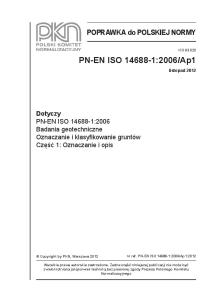
Effect of the High Glucose Concentrations and PMA on TGF-1 Promoter Activity and Localization of the Responsible cis-Regulatory Elements To study whether the two AP-1 binding sites ⫺418/⫺412 and ⫺371/⫺364 mediate the high glucose effect, we mutated both promoter regions by site-directed mutagenesis (Figure
Western Blots Nuclear extracts of mesangial cells were separated by sodium dodecyl sulfate polyacrylamide (7.5%) gel electrophoresis. Proteins were transferred to nitrocellulose by semidry electroblotting (transfer buffer, 48 mM Tris, 39 mM glycine, 0.0375% sodium dodecyl sulfate, 20% (vol/vol) methanol). Then nitrocellulose membranes were blocked with NaCl/ethylenediaminetetraacetate/Triton (NET) buffer (150 mM NaCl, 50 mM Tris/HCl [pH 7.4], 5 mM EDTA, 0.05% Triton X-100, 0.25% gelatin) and incubated with the first antibody (diluted 1:1000 in NET) overnight at 4°C. After the membranes were washed with NET, they were incubated with horseradish peroxidaseconjugated anti-rabbit or anti-goat IgG for 1 h at room temperature. Visualization of immunocomplexes was performed by enhanced chemiluminescence as described (24).
Statistical Analysis Results presented are derived from at least three independent experiments. Means ⫾ SEM were calculated and groups of data were compared using t test. Statistical significance was set at P ⬍ 0.05.
Results
Effect of Glucose and Phorbol Ester on TGF-1 and TGF-2 mRNA Levels in Mesangial Cells Previous studies, including studies with porcine mesangial cells performed in our laboratory, have shown that elevated glucose levels and particularly PMA increase TGF-1 mRNA levels (11,21). Because we found no significant effect of high
Figure 1. Effect of phorbol ester (PMA) stimulation on mesangial transforming growth factor-1 (TGF-1) and TGF-2 mRNA levels. Mesangial cells were grown in standard medium with or without 0.1 M PMA for the time indicated, and RNase protection assays were performed. The single-stranded antisense RNA probes used in RNase protection assay were obtained by run-off transcription using T7 or SP6 polymerase as described (21). The TGF-2 probe is the Aci I fragment (267 bp) starting 12 bp upstream of the coding sequence for the active peptide (38). Labeled cRNA probes for TGF-1, TGF-2, and glyceraldehyde phosphate dehydrogenase (GAPDH) were added to each sample. Arrows on the left indicate the shift of full-length cRNA probes for TGF-1 (590b), TGF-2 (310b), and GAPDH (241b) to protected cRNA probes for TGF-1 (528b), TGF-2 (267b), and GAPDH (211b) after RNase A/T1 digestion. No undigested probe is visible in the lanes of the samples. TGF-/GAPDH ratios were obtained by scanning the optical density of the bands.
2010
Journal of the American Society of Nephrology
J Am Soc Nephrol 11: 2007–2016, 2000
Table 2. Comparison of the effect of high glucose on the human TGF-1 promoter activity in porcine and human mesangial cellsa
Glucose concentration (mM) Relative luciferase activity (%)
Porcine MC
Human MC
6
6
100
30 191 ⫾ 22*
30
100 176 ⫾ 25**
a TGF-1, transforming growth factor-1. The pGL3wt plasmid was transiently transfected in porcine or human mesangial cells, and the cells were cultured for 24 h with 6 or 30 mM glucose. Luciferase activities are expressed as percentages of the activity measured with 6 mM glucose. In every independent transfection assay, a high glucose-induced activation of the TGF-1 promoter activity could be observed. Three independent experiments were performed in duplicate. Results shown are means ⫾ SEM. * P ⬍ 0.01 versus 6 mM glucose; ** P ⬍ 0.05 versus 6 mM glucose.
2B). As shown in Figure 3A, mutation in AP-1 box A or box B or in both boxes completely prevented the high glucoseinduced increase of the TGF-1 promoter activity and reduced the promoter activity in both glucose conditions below control levels. The lowest remaining promoter activity was found after mutation of both AP-1 sites (approximately 20% in normal glucose and in high glucose condition), whereas mutation of AP-1 box B had the lowest effect on promoter activity (approximately 50%). Furthermore, we examined the regulatory function of one GC-box, which represents a high-affinity stimulating protein 1 (Sp1) binding site. Mutation in this GC-box (Figure 2B) reduced the promoter activity in normal and high glucose conditions; however, the high glucose-induced stimulation was essentially unaffected (Figure 3A). Because addition of PMA increased TGF-1 mRNA levels in mesangial cells, we studied whether PMA induced an increase in TGF-1 promoter activity and whether AP-1 binding sites are involved in this activation. Stimulation with PMA led to a 2.3-fold increase of the wild-type TGF-1 promoter activity in the mesangial cells (Figure 3B). Mutation in the AP-1 box A and box B or in both boxes prevented the activation by the phorbol ester completely. These results clearly indicate an important role of both AP-1 boxes in hyperglycemia- and PMA-induced activation of the TGF-1 promoter and further emphasize that these cis-regulatory elements are responsible for an AP-1–mediated activation.
Effect of Inhibitors of AP-1 and Sp1 on the GlucoseInduced Stimulation of the TGF-1 Promoter To elucidate further the role of the transcription factors AP-1 and Sp1 in high glucose-induced TGF-1 promoter activation, transfection experiments with the known inhibitors of AP-1 and Sp1, curcumin (25) and mithramycin (26), were performed (Figure 4). In the presence of curcumin, the high glucose effect was prevented. Addition of mithramycin did not abolish the high glucose effect. These data further support the functional
Figure 2. (A) cis-Regulatory elements in the 5⬘-flanking region of the human TGF-1 gene. Positions are deduced from sequence data as described by Kim et al. (15). activating protein-1 (AP-1) binding sites are indicated by dark rhombus, filled circles show GC-boxes. Mutated binding sites are indicated by asterisks. (B) Constructs of the 5⬘flanking region of the TGF-1 gene in pGL3basic, which contains the firefly (photinus pyralis) luciferase coding region. The pGL3wt includes the wild-type (wt) TGF-1 fragment ⫺453/⫹11 fused to the luciferase gene and was the template for the site-directed mutagenesis as described in the Materials and Methods section. After an initial denaturation at 94°C for 5 min, 25 cycles of amplification were performed (50°C, 1 min; 72°C, 12 min; 94°C, 1 min). The samples contained 100 ng template, 100 pmol of each primer, 2 mM MgCl2, 0.2 mM dNTPs, OptiPerformTMBuffer III, OptiZymeTMEnhancer, and 4 U PowerScript DNA polymerase (all from PAN Systems, Aidenbach, Germany) in a total volume of 100 l. After completion of the PCR, the dam-methylated template DNA was digested by 20 U DpnI at 37°C for 1 h and used directly for transformation. The AP-1 binding sites A and B and the GC-box ⫺220/⫺211 were mutated by a change of two bases in the consensus sequence as indicated. Substituted bases are depicted by italic lowercase letters. The GC-box ⫺220/⫺211 has been proved to be a high-affinity binding site of Sp1 (16).
participation of AP-1 in the regulation of TGF-1 promoter activity by high glucose.
Effect of Elevated Glucose Concentrations on the Amount of AP-1 Proteins To study the regulation of the activity of AP-1 proteins in mesangial cells by high glucose, we determined in Western blot analysis whether high glucose alters nuclear levels of AP-1 proteins. As shown in Figure 5, A through D, no significant differences of c-Jun, JunB, JunD, and ATF-2 could be detected after incubation of mesangial cells with high glucose for 40 h. In Western blots with nuclear extracts from cells cultured with high glucose for 24 h, the same results were obtained (data not shown). In contrast, the amount of c-Fos was significantly increased in nuclear extracts after high glucose incubation for 24 h (Figure 5E) and returned to basal levels after 40 h of high glucose incubation (data not shown). Stimulation of mesangial
J Am Soc Nephrol 11: 2007–2016, 2000
High Glucose and TGF-1 Gene Activation
2011
Figure 4. Effect of inhibitors of AP-1 and Sp1 on high glucoseinduced TGF-1 promoter activity. Transfection assays of mesangial cells were performed with the wild-type promoter plasmid pGL3wt in 6 mM and 30 mM glucose in the absence or presence of the AP-1 inhibitor curcumin (4 M) or the Sp1 inhibitor mithramycin (25 nM). Luciferase activities are expressed as percentages of the activity measured with 6 mM glucose without inhibitor. Data are means ⫾ SEM of three independent experiments. ** P ⬍ 0.01 versus 6 mM glucose.
Figure 3. Effect of base mutations in the TGF-1 promoter region on the high glucose-induced (A) and PMA-induced (B) promoter activity. The wild-type and the mutated plasmids used for the transfection experiments are shown in Figure 2B. (A) Mesangial cells were transfected with equal amounts of the different plasmids and grown in 6 or 30 mM glucose for 24 h. Luciferase activities are expressed as percentages of the activity measured with the wild-type plasmid and 6 mM glucose. (B) Mesangial cells were transfected as described in (A), and 9 h before harvesting 0.5 M PMA dissolved in ethanol was added. Wild-type promoter activity determined in transfected cells incubated with ethanol as control was set as 100%. Data are means ⫾ SEM of three different experiments. * P ⬍ 0.01 versus 6 mM glucose; ** P ⬍ 0.01 versus 6 mM glucose.
cells with PMA also led to an increased level of c-Fos (Figure 5E).
High Glucose-Induced Activation of the p38 Pathway Is Inhibited by SB 203580 and Bisindolylmaleimide I In addition to enhanced gene expression, the transcriptional activity of AP-1 proteins may be regulated by site-specific phosphorylation by the stress-activated kinases JNK and p38 MAPK. Therefore, immunoblotting was performed with nuclear extracts from mesangial cells cultured with normal and high glucose for 24 h, and phosphorylated AP-1 proteins were detected using a phospho-specific c-Jun antibody and a phospho-specific ATF-2 antibody (Figure 6A). Stimulation of mesangial cells with 10 g/ml anisomycin, which is known to activate both JNK and p38 MAPK (27), resulted in an additional band of the estimated molecular mass of 46 kD corre-
sponding to the expected size of the phosphorylated c-Jun (Figure 6A). No bands with similar size were detected in nuclear extracts from normal and high glucose conditioned cells with the phospho-Jun ser-63 antibody, indicating the absence of this phosphorylated form of c-Jun. The lower band detected with the phospho-specific c-Jun antibody could be explained by less phosphorylated forms of c-Jun, but the amount of these proteins was similar in normal and high glucose conditioned cells, whereas treatment with 10 g/ml anisomycin increased the intensity of this band. In contrast, the amount of a phosphorylated form of ATF-2 was markedly increased by high glucose, indicating an activation of the p38 MAPK pathway by high glucose. Stimulation of the cells with high glucose for 40 h yielded the same results (data not shown). To elucidate the signal transduction pathways activated by high glucose potentially leading to enhanced gene activation of TGF-1, we studied the effect of the PKC inhibitor bisindolylmaleimide and p38 MAPK inhibitor SB 203580 on the activity of the p38 MAPK pathway by quantification of the phosphorylated form of the endogenous substrate ATF-2. In nuclear extracts of mesangial cells, phosphorylated ATF-2 and ATF-2 protein were detected by immunoblotting and quantified (Figure 6B). High glucose increased the level of the phosphorylated forms twofold, whereas 1 M SB 203580 and 50 and 500 nM bisindolylmaleimide prevented this increase. These data show that addition of either one of the substances is sufficient to inhibit the activation of p38 MAPK by high glucose in mesangial cells.
The High Glucose-Induced Activation of the TGF-1 Promoter Is Prevented by Inhibitors of PKC and p38 MAPK The results obtained with the phospho-specific ATF-2 antibody suggest a participation of activated p38 MAPK in the
2012
Journal of the American Society of Nephrology
Figure 5. Effect of high glucose on the amount of AP-1 proteins. (A through D) Western blot analyses of nuclear extracts from mesangial cells incubated for 24 h with 6 mM glucose (NG) or 30 mM glucose (HG) were performed as described in the Materials and Methods section. For immunodetection of AP-1 proteins, 25 g of nuclear proteins and antibodies recognizing c-Jun (A), JunB (B), JunD (C), and ATF-2 (D) were used. The antibodies against c-Jun, JunB, and JunD were specific and not cross-reactive with the other Jun proteins. The ATF-2 antibody cross reacts with other proteins besides the 68 kDa ATF-2 protein. The positions of the molecular weight markers are indicated. Arrows on the right side of each figure marks bands identified by the specific antibodies. (E) Western blot analyses with 25 g of nuclear proteins from mesangial cells stimulated with 0.5 M PMA for 6 h (PMA) or ethanol as control (c) or incubated for 24 h with 6 mM glucose (NG) or 30 mM glucose (HG). The position of the 66 kDa molecular weight marker is indicated. The arrow on the right side of the figure marks the 62 kDa protein identified by the antibody.
high glucose-stimulated TGF-1 gene expression. To study whether these signaling pathways are functionally involved in TGF-1 promoter activation, we transfected and cultured mesangial cells in the presence of the inhibitors of the p38 MAPK or the PKC pathway. Addition of 1 and 10 M SB 203580 as well as 50 and 500 nM bisindolylmaleimide I prevented the activation of the TGF-1 promoter, whereas the promoter activity in normoglycemic conditions was unaffected (Figure 7). The results clearly indicate a participation of both the p38 MAPK and the PKC pathway in the activation of the TGF-1 promoter by ambient high glucose concentrations.
J Am Soc Nephrol 11: 2007–2016, 2000
Figure 6. High glucose induces phosphorylation of ATF-2, which is prevented by protein kinase C (PKC) and MAPK inhibitors. Western blot analyses were performed as described in the Materials and Methods section. Twenty-five g of nuclear proteins from mesangial cells incubated for 24 h with 6 mM glucose (NG) or 30 mM glucose (HG) were used. (A) Control cells were cultured in 6 mM glucose and stimulated with anisomycin for 1 h before harvesting. Left: Antibody specific for c-Jun phosphorylated on serine 63; the phosphorylated c-Jun protein with a molecular mass of 46 kD is indicated by the arrow. Right: Antibody specific for ATF-2 phosphorylated on threonine 71; the different phosphorylated forms of ATF-2 are indicated by arrows. (B) p38 MAPK activity was assessed by phosphorylation of ATF-2. Mesangial cells were cultured as described in A in the presence of 1 M SB203580 or 50 and 500 nM bisindolylmaleimide I (BIS I), and phosphorylation of ATF-2 and ATF-2 protein was detected with specific antibodies. Bar graphs show densitometric quantification of the ratio of phospho-ATF-2/ATF-2 protein. The mean value of normal glucose is defined as 1. Each value is expressed as means ⫾ SEM of three independent experiments. *P ⬍ 0.05 30 mM versus 6 mM glucose without inhibitor; #P ⬍ 0.05 30 mM versus 30 mM glucose with inhibitor.
Enhanced Binding of Nuclear Proteins from Cells Cultured in High Glucose to AP-1 Boxes A and B of the TGF-1 Promoter Our data show that both AP-1 boxes are functionally involved in mediating the high glucose effect. To evaluate
J Am Soc Nephrol 11: 2007–2016, 2000
Figure 7. Effect of inhibitors of p38 MAPK and PKC on high glucose-induced TGF-1 promoter activity. Transfection assays of mesangial cells were performed with the wild-type promoter plasmid pGL3wt in normo- and hyperglycemic conditions in the presence of the different inhibitors. Cells were transfected as described in the Materials and Methods section and cultured in 6 or 30 mM glucose for 24 h with 1 and 10 M SB 203580 as p38 MAPK inhibitor or 50 and 500 nM bisindolylmaleimide I (BIS I) as PKC inhibitor. Luciferase activities are expressed as percentages of the activity measured with 6 mM glucose without inhibitor. Data are means ⫾ SEM of three independent experiments. **P ⬍ 0.01 versus 6 mM glucose.
whether this effect is mediated by increased binding of AP-1 to the corresponding DNA consensus sequences in the TGF-1 promoter region, we performed electrophoretic mobility shift assays with wild-type or mutated oligonucleotides (Table 1). In nuclear extracts from cells cultured in high glucose concentrations for 24 h, an enhanced binding to the AP-1 B and to a much lesser extent to the AP-1 A fragments was observed (Figure 8A, lanes 2 and 5). Similar results were obtained with nuclear extracts from cells cultured for 40 h with high glucose (data not shown). The specificity of the binding was assessed by using the mutated oligonucleotides. As shown in Figure 8A (lanes 3 and 6), upper bands (arrows) disappeared almost completely whereas the bands marked with a bracket remained unchanged, indicating unspecific binding of nuclear proteins. Furthermore, after addition of an excess of cold wild-type nucleotides, these bands were still apparent (AP-1 B: Figure 9B, lanes 6 and 7; AP-1 A: data not shown). Binding of nuclear proteins to the oligonucleotide containing a high affinity Sp1 binding site was not influenced by high glucose (Figure 8B, lane 2). With the mutated Sp1 oligonucleotide, only a very weak binding was detectable (Figure 8B, lane 4); the shifted band disappeared completely after addition of an excess of cold wild-type oligonucleotide. These results indicate that the response to high glucose is regulated by enhanced binding of proteins.
Identification of Nuclear Proteins Binding to AP-1 Box B of the TGF-1 Promoter by Specific Anti–AP-1 Antibodies To identify the proteins involved in high glucose-induced AP-1 activation mobility, we performed shift experiments with antibodies specific for several AP-1 proteins. In experiments
High Glucose and TGF-1 Gene Activation
2013
Figure 8. Glucose treatment activates binding of nuclear proteins of mesangial cells to AP-1 binding sites of the TGF-1 promoter. Mobility shift experiments were performed with nuclear extracts from mesangial cells cultured for 24 h in 6 (NG) or 30 mM glucose (HG). Thirteen g of nuclear proteins were incubated with 50,000 cpm of the 32P-labeled oligonucleotides. The mutated oligonucleotides (Table 1) contain the same mutation as the luciferase constructs used for the transfection assays. (A) Binding of nuclear proteins from normo- and hyperglycemic mesangial cells to the AP-1 boxes A and B (wt) and the mutated AP-1 boxes (mut). Arrows indicate specifically shifted bands. Nonspecific binding is marked by the bracket. (B) The mobility shift assays with the Sp1 binding site. To distinguish nonspecific shifted bands, 50⫻ molar excess of cold Sp1 oligonucleotide was added to the binding reaction (⫹50⫻ cold). The corresponding control with the AP-1 box B is demonstrated in Figure 9B. The insignificant specific binding of nuclear proteins to the mutated oligonucleotides shows that the mutations are suitable to test the function of these elements in the reporter gene assays.
with AP-1 box A only, the anti–AP-1 antibody attenuated the binding of nuclear proteins, characterizing the AP-1 box A as a weak AP-1 binding site (data not shown). In experiments with AP-1 box B, the presence of anti–AP-1, anti-JunD, or anti– c-Fos antibodies caused an almost complete disappearance of the specific shifted band in normal and in high glucosetreated cells (Figure 9A, arrow), whereas addition of anti-JunB antibody (Figure 9A, lanes 3 and 8) had little effect. Noteworthy is that the high glucose-induced increase in binding of AP-1 was prevented by anti–AP-1 antibody. To detect supershifted bands, we extended the run time of electrophoresis and increased the exposure time. Under these conditions, the binding of JunD and c-Fos to the AP-1 box B is obvious (Figure 9C). Because the data described above indicate an involvement of the p38 MAPK in the high glucose-induced activation of the TGF-1 promoter and the AP-1–related proteins CREB-1, ATF-1, and ATF-2 are activated by this pathway, we studied the binding of these transcription factors to the AP-1 boxes. Specific antibodies, however, could not detect any of these
2014
Journal of the American Society of Nephrology
J Am Soc Nephrol 11: 2007–2016, 2000
binding site and JunD and c-Fos as component of the complex bound by AP-1 box B.
Discussion
Figure 9. Identification of AP-1 compounds of the shifted proteinDNA complexes of AP-1 box B. Mobility shift experiments were performed with nuclear extracts from cells incubated for 24 h with 6 mM glucose (NG) and 30 mM glucose (HG), using 50,000 cpm of the 32 P-labeled oligonucleotide AP-1 B in the presence or absence of antibodies against AP-1 as indicated in the figure. (A) Mobility shift analyses with the AP-1 antibodies AP-1, which cross reacts with all Jun proteins JunB, JunD, and c-Fos. The arrow indicates the band identified as AP-1 proteins. Nonspecific bands are marked by the bracket. (B) Mobility shift analyses with antibodies specific for CREB-1, ATF-1, and ATF-2. To distinguish nonspecific shifted bands, 20⫻ and 50⫻ molar excess of cold AP-1 box B oligonucleotide was added to the binding reaction (⫹20 or 50⫻ cold). The arrow indicates the specific shifted band; nonspecific bands are marked by the bracket. (C) To obtain supershifted bands, we performed mobility shift assays with extended run-time and longer exposure up to 3 d. Antibodies against AP-1, cJun, JunB, JunD, and c-Fos were used. Supershifted bands occurred with the JunD and c-Fos antibody and are indicated by arrows on the right side; shifted bands are marked by the bracket on the left side of the figure.
proteins in the DNA binding complex derived from high glucose-stimulated mesangial cells (Figure 9B, data for AP-1 box A are not shown). In conclusion, the supershift analyses identified AP-1 box A as a weak and box B as a stronger AP-1
Previous studies indicated that many of the actions of high glucose on renal cells, particularly the matrix-stimulatory activity, are mediated by autocrine production and activation of the prosclerotic cytokine TGF-1. In the present study, we provide four lines of evidence that activation of AP-1 is involved in the high glucose-induced increase in mesangial TGF-1 gene expression. First, by using promoter reporter constructs, we found that elevated glucose concentrations stimulated the activation of the TGF-1 promoter. The stimulatory effect was mediated by the two AP-1 binding sites in the promoter region as shown by mutation of the AP-1 binding sites, whereas mutation of an Sp1 binding site was ineffective in preventing the increase of promoter activity. Second, only curcumin but not mithramycin prevented glucose-stimulated TGF-1 promoter activation. Third, presence of elevated glucose concentrations increased the binding of nuclear proteins to the AP-1 binding sites. Mutation of the AP-1 binding sites prevented both AP-1 binding and high glucose-induced TGF-1 promoter activation, indicating a causal relationship between AP-1 binding to promoter sequences and activation of TGF-1 gene expression. Fourth, high glucose stimulated the activity of the AP-1 factor c-Fos by transiently increasing the amount of the protein. Several laboratories showed an increased TGF-1 mRNA level in cultured mesangial cells when stimulated with glucose (7,8). Our present observation of a 1.9-fold increase of the TGF-1 promoter activity after 24 h of exposure to high glucose is in accordance with the elevation of the TGF-1 mRNA and protein levels after 48 h of glucose stimulation (21). Moreover, the data suggest that high glucose stimulates TGF-1 gene expression rather than influences TGF-1 mRNA stability. Furthermore, by mutation of the AP-1 binding sites, we could demonstrate the important role of both AP-1 binding sites in mediating the high glucose effect. The finding that mutations of one AP-1 site reduced TGF-1 promoter activity under control levels suggests a participation of the AP-1 binding sites in the regulation of basal promoter activity. Our results that mutation in both AP-1 binding sites diminished the promoter activity more than mutation in one AP-1 box indicate a cooperative effect of the two binding sites in the regulation of the promoter activity in basal and hyperglycemic conditions (28). The AP-1 boxes are located in the promoter region ⫺453 to ⫺323, which also is responsible for phorbol ester responsiveness and autoinduction via AP-1 proteins (16). Because AP-1 proteins are activated by a variety of stimuli, the role of these two AP-1 sites is not restricted to high glucose. Recently, Hoffman et al. (29) reported a high glucose-induced de novo synthesis of TGF-1 mRNA in murine mesangial cells and an induction of the murine TGF-1 promoter activity by high glucose after 24 h. However, the regulation of the glucose responsiveness of the murine TGF-1 promoter cannot be explained by AP-1 sites because the glucose-responsive region of the mouse promoter was localized between ⫺835 and ⫺406
High Glucose and TGF-1 Gene Activation
J Am Soc Nephrol 11: 2007–2016, 2000
upstream of the first transcriptional start site, where no AP-1 binding sites reside (30). The finding that high glucose stimulates the human TGF-1 promoter activity via AP-1 binding sites is strongly supported by the glucose-induced enhanced binding of nuclear proteins to both AP-1 sites. The AP-1 proteins JunD and c-Fos were identified in the protein complex binding to AP-1 box B, and the enhanced expression of c-Fos by high glucose suggests the involvement of this protein in the glucose-dependent activation of the TGF-1 promoter. However, both AP-1 proteins may also participate in basal promoter regulation. The observed weak DNA binding activity of AP-1 box A alone, which can be explained by the nontypical T3 A switch at position 7 of the AP-1 consensus sequence (Figure 2A), may indicate that further promoter sequences, e.g., AP-1 box B, are necessary for the function of AP-1 box A. Recently, increased binding of AP-1 to a synthetic oligonucleotide containing two PMA responsive elements in mesangial cells cultured in high glucose was detected after 3 d of high glucose conditioning (31). However, we used AP-1 binding consensus sequences of the TGF-1 promoter, which may exert a higher affinity for the high glucose-induced protein complexes. The present study supports the suggested involvement of PKC activation in the development of diabetic nephropathy (17,18). However, the detailed mechanism of the link between hyperglycemia-induced PKC activation and TGF-1 gene activation has not been reported. Three lines of evidence support a PKC involvement in TGF-1 promoter regulation by high glucose. First, activation of the TGF-1 promoter by the PKC activator PMA and glucose was mediated via the same AP-1 binding sites. Second, application of the PKC inhibitor bisindolylmaleimide I prevented the high glucose-induced increase of promoter activity. Third, high glucose and PMA led to a transient increase of c-Fos, which is in line with the reports of Kreisberg et al. (32). Particularly, the PKC  isoform seems to be involved in mediating high glucose effects on TGF-1 gene expression and deposition of matrix components (18,33). The concentration of the PKC inhibitor bisindolylmaleimide I used in the present study shows high selectivity for the PKC ␣, 1, and 2 isoforms (34). Because the PKC 2 isoform could not be detected in our mesangial cells by Western blotting (S. Facchin and E. Schleicher, unpublished results), participation of the PKC ␣ and 1 isoforms is very likely in mediating the high glucose-induced TGF-1 gene expression. The involvement of p38 MAPK in the upregulation of TGF-1 promoter activity by high glucose was shown by three independent results. p38 MAPK is activated by high glucose as demonstrated by enhanced phosphorylation of its endogenous substrate ATF-2 (35); this phosphorylation is prevented by the p38 MAPK inhibitor SB 203580 and the PKC inhibitor bisindolylmaleimide I, and the identical concentrations of these inhibitors suppress the upregulation of TGF-1 promoter activity by high glucose. The activation of p38 MAPK by high glucose was also demonstrated in mesangial-like smooth muscle cells (36). Furthermore, our data suggest that PKC is involved in the activation of p38 MAPK. The activated p38 MAPK pathway may mediate the enhanced binding of AP-1 to
2015
the high glucose-responsive AP-1 sites of the TGF-1 promoter by increasing their DNA binding activity or inducing protein–protein interactions, thereby leading to enhanced transcriptional activity (35). However, the AP-1–related transcription factors and substrates of p38 MAPK ATF-1, ATF-2, and CREB-1 could not be detected in the protein complex binding to the AP-1 sites of the TGF-1 promoter. Therefore, an as yet unidentified protein may be the p38 MAPK substrate, which is involved in the glucose response of the TGF-1 promoter, whereas the recently reported increased stabilization of mRNA by p38 MAPK (37) also provides a possible link to the enhanced binding of transcription factors to the glucose-responsive sites of the TGF-1 promoter. In conclusion, our results indicate that hyperglycemia stimulates human TGF-1 gene expression via two adjacent AP-1 binding sites in cultured mesangial cells. The increased TGF-1 promoter activity is mediated by enhanced binding activity of AP-1 and is regulated by PKC- and p38 MAPKdependent pathways. Taken together, our data link hyperglycemia and the enhanced expression of the prosclerotic cytokine TGF-1 on a molecular basis, providing an improved insight into the pathogenetic mechanism of the development of diabetic nephropathy.
Acknowledgments We gratefully acknowledge the donation of plasmids encoding the human TGF-1 promoter by Dr. S.-J. Kim. A.P. received a Hermannand-Lilly-Schilling professorship. The work was supported by the Deutsche Forschungsgemeinschaft (Schl 239-6) to E.S.
References 1. Osterby R: Glomerular structural changes in type 1 (insulindependent) diabetes mellitus: Causes, consequences, and prevention. Diabetologia 35: 803– 819, 1992 2. Mauer SM, Steffes MW, Ellis EN, Sutherland DN, Brown DM, Goetz FC: Structural-functional relationships in diabetic nephropathy. J Clin Investig 74: 1143–1155, 1984 3. Nerlich A, Schleicher E: Immunohistochemical localization of extracellular matrix components in human diabetic glomerular lesions. Am J Pathol 139: 889 – 899, 1991 4. The Diabetes Control and Complications Trial Research Group: The effects of intensive insulin treatment of diabetes on the development and progression of long-term complications in insulin-dependent diabetes mellitus. N Engl J Med 329: 977–986, 1993 5. Kreisberg JI, Ayo SH: The glomerular mesangium in diabetes mellitus. Kidney Int 43: 109 –113, 1993 6. Heilig CW, Conception LA, Riser BL, Freytag SO, Zhu M, Cordes P: Over-expression of glucose transporters in rat mesangial cells cultured in a normal glucose milieu mimics the diabetic phenotype. J Clin Investig 96: 1802–1814, 1995 7. Ziyadeh FN, Sharma K, Ericksen M, Wolf G: Stimulation of collagen gene expression and protein synthesis in murine mesangial cells by high glucose is mediated by autocrine activation of transforming growth factor-beta. J Clin Investig 93: 536 –542, 1994 8. Kolm V, Sauer U, Olgemo¨ller B, Schleicher E: High glucoseinduced TGF-1 regulates mesangial production of heparan sulfate proteoglycan. Am J Physiol 270: F812–F821, 1996
2016
Journal of the American Society of Nephrology
9. Yamamoto T, Nakamura T, Noble NA, Ruoslahti E, Border WA: Expression of transforming growth factor  is elevated in human and experimental diabetic nephropathy. Proc Natl Acad Sci USA 90: 1814 –1818, 1993 10. Iwano M, Kubo A, Nishino T, Sato H, Nishioka H, Akai Y, Kurioka H, Fujii Y, Kanavachi M, Shiki S, Dohi K: Quantification of glomerular TGF-1 mRNA in patients with diabetes mellitus. Kidney Int 49: 1120 –1126, 1996 11. Sharma K, Ziyadeh FN: Hyperglycemia and diabetic kidney disease: The case for transforming growth factor  as a key mediator. Diabetes 44: 1139 –1146, 1995 12. Sharma K, Ziyadeh FN: Renal hypertrophy is associated with up-regulation of TGF-1 gene expression in diabetic BB and NOD mouse. Am J Physiol 267: F1094 –F1101, 1994 13. Border WA, Okuda S, Languino LR, Sporn MB, Ruoslahti E: Suppression of experimental glomerulonephritis by antiserum against transforming growth factor 1. Nature 346: 371–373, 1990 14. Sharma K, Jin Y, Guo J, Ziyadeh FN: Neutralization of TGF- by anti-TGF- antibody attenuates kidney hypertrophy and the enhanced extracellular matrix gene expression in STZ-induced diabetic mice. Diabetes 45: 522–530, 1996 15. Kim SJ, Glick A, Sporn MA, Roberts AB: Characterization of the promoter region of the human transforming growth factor-1 gene. J Biol Chem 264: 402– 408, 1989 16. Kim SJ, Angel P, Lafyatis R, Hattori K, Kim KY, Sporn MB, Karin M, Roberts AB: Auto-induction of transforming growth factor 1 is mediated by the AP-1 complex. Mol Cell Biol 10: 1492–1497, 1990 17. DeRubertis FR, Craven PA: Activation of protein kinase C in glomerular cells in diabetes: Mechanisms and potential links to the pathogenesis of diabetic glomerulopathy. Diabetes 43: 1– 8, 1994 18. Koya D, Jirousek MR, Lin YW, Ishii H, Kuboki K, King GL: Characterization of protein kinase C  isoform activation on the gene expression of transforming growth factor-, extracellular matrix components, and prostanoids in the glomeruli of diabetic rats. J Clin Investig 100: 115–126, 1997 19. Ayo SH, Radnik R, Garoni JA, Troyer DA, Kreisberg JI: High glucose increases diacylglycerol mass and activates protein kinase C in mesangial cell cultures. Am J Physiol 261: F571–F577, 1991 20. Ishii H, Jirousek MR, Koya D, Takagi C, Xia P, Clermont A, Bursell SE, Kern TS, Ballas LM, Heath WF, Stramm LE, Feener EP, King GL: Amelioration of vascular dysfunctions in diabetic rats by an oral PKC beta inhibitor. Science 272: 728 –731, 1996 21. Kolm-Litty V, Sauer U, Nerlich A, Lehmann R, Schleicher ED: High glucose-induced transforming growth factor 1 production is mediated by the hexosamine pathway in porcine glomerular mesangial cells. J Clin Investig 101: 160 –169, 1998 22. Netzker R, Weigert C, Brand K: Role of the stimulatory proteins Sp1 and Sp3 in the regulation of transcription of the rat pyruvate kinase M gene. Eur J Biochem 245: 174 –181, 1997 23. Chen C, Okayama H: High-efficiency transformation of mammalian cells by plasmid DNA. Mol Cell Biol 7: 2745–2752, 1987 24. Bouaboula M, Legoux P, Pessegue B, Delpech B, Dumont X,
J Am Soc Nephrol 11: 2007–2016, 2000
25.
26.
27.
28. 29.
30.
31.
32.
33.
34.
35. 36.
37.
38.
Piechacyk M, Casellas P, Shire D: Standardization of mRNA titration using a polymerase chain reaction method involving co-amplification with a multi-specific internal control. J Biol Chem 267: 21830 –21838, 1992 Huang TS, Lee SC, Lin JK: Suppression of c-Jun/AP-1 activation by an inhibitor of tumor promotion in mouse fibroblast cells. Proc Natl Acad Sci USA 88: 5292–5296, 1991 Blume SW, Snyder RC, Ray R, Thomas S, Koller CA, Miller DM: Mithramycin inhibits Sp1 binding and selectively inhibits transcriptional activity of the dihydrofolate reductase gene in vitro and vivo. J Clin Investig 88: 1613–1621, 1991 Cano E, Hazzalin CA, Mahadevan LC: Anisomycin-activated protein kinases p45 and p55 but not mitogen-activated kinases ERK-1 and -2 are implicated in the induction of c-fos and c-jun. Mol Cell Biol 14: 7352–7362, 1994 Tjian R, Maniatis T: Transcriptional activation: A complex puzzle with few easy pieces. Cell 77: 5– 8, 1994 Hoffman BB, Sharma K, Zhu Y, Ziyadeh FN: Transcriptional activation of transforming growth factor-1 in mesangial cell culture by high glucose concentration. Kidney Int 54: 1107– 1116, 1998 Geiser AG, Kim SJ, Roberts AB, Sporn MB: Characterization of the mouse transforming growth factor-1 promoter and activation by the ha-ras oncogene. Mol Cell Biol 11: 84 –92, 1991 Wilmer WA, Cosio FG: DNA binding of activator protein-1 is increased in human mesangial cells cultured in high glucose concentrations. Kidney Int 53: 1172–1181, 1998 Kreisberg JI, Radnik RA, Ayo SH, Garoni J, Saikumar P: High glucose elevates c-fos and c-jun transcripts and proteins in mesangial cell cultures. Kidney Int 46: 105–112, 1994 Kolm-Litty V, Tippmer S, Ha¨ring HU, Schleicher E: Glucosamine induces translocation of protein kinase C in mesangial cells: Exp Clin Endocrinol Diabetes 106: 377–383, 1998 Toullec D, Pianetti P, Coste H, Bellevergue P, Grand-Perret T, Ajakane M, Baudet V, Boissin P, Boursier E, Loriolle F, Duhamel L, Charon D, Kirilovsky J: The bisindolylmaleimide GF 109203X is a potent and selective inhibitor of protein kinase C. J Biol Chem 266: 15771–15781, 1991 Karin M, Liu ZG, Zandi E: AP-1 function and regulation. Curr Opin Cell Biol 9: 240 –246, 1997 Igarashi M, Wakasaki H, Takahara N, Ishii H, Zhen YJ, Yamauchi T, Kuboki K, Meier M, Rhodes CJ, King GL: Glucose or diabetes activate p38 mitogen-activated protein kinase via different pathways. J Clin Investig 103: 185–195, 1999 Winzen R, Kracht M, Ritter B, Wilhem A, Chen CY, Shyu AB, Muller M, Gaestel M, Resch K, Holtmann H: The p38 MAP kinase pathway signals for cytokine-induced mRNA stabilization via MAP kinase-activated protein kinase 2 and an AU-rich region-targeted mechanism. EMBO (EUR MOL BIOL ORGAN) J 18: 4969 – 4980, 1999 Mulheron GW, Mulheron JG, Danielpour D, Schomberg DW: Porcine granulosa cells do not express transforming growth factor-2 (TGF-2) messenger ribonucleic acid: Molecular basis for their inability to produce TGF- activity comparable to that of rat granulosa cells. Endocrinology 131: 2609 –2614, 1992