Scientific Articles
ANESTHESIA AND ELECTROCORTICOGRAPHY FOR EPILEPSY SURGERY: A JORDANIAN EXPERIENCE Subhi S.M. AL- Ghanem*, Abdelkarim S AL-Oweidi**, Ahmad F Tamimi*** and Abdelkarim A AL-Qudah****
Abstract Objective: Electrocorticography (ECoG) may be used to guide epilepsy surgery. However, anesthetics can suppress epileptiform activity or induce confounding burst-suppression patterns and the relationship between ECoG results and seizure outcome is controversial. In this study, we evaluated the ECoG activity under several different anesthetics and examined the relationship between ECoG and outcome. Methods: We retrospectively studied 44 patients who had ECoG during epilepsy surgery. Anesthesia was with fentanyl and isoflurane (n = 19), fentanyl and sevoflurane (n = 9), remifentanil and sevoflurane (n = 5), remifentanil and propofol (n = 9), and fentanyl with propofol sedation during local anesthesia (n = 2). Pre-resection ECoG was considered satisfactory if epileptiform activity was present and there was no burst-suppression. Post-resection ECoG was graded according to residual epileptiform activity: A (none), B (mild), C (moderate). Seizure outcome was graded: I (seizure free without medication), II (seizure free with medication), III (> 50% seizure reduction) or IV (< 50% seizure reduction). Grades I-III were considered beneficial. Results: ECoG was satisfactory in 43 of the 44 surgeries, but one of the 11 recordings during propofol showed no epileptiform activity. Thirty-six of 37 patients (97%) with ECoG grade A or B and five of seven patients (71%) with ECoG grade C benefited from epilepsy surgery. Chi-squared, p > 0.05. Conclusions: Satisfactory ECoG is possible using isoflurane or sevoflurane with nitrous oxide and fentanyl or remifentanil or using propofol and remifentanil. However, one of eleven ECoGs under propofol was negative for epileptiform activity. The amount of post-resection ECoG epileptiform activity does not significantly correlate with seizure outcome. Keywords: Epilepsy surgery, anesthesia, electrocorticography. From Faculty of Med. & Jordan Univ. Hosp. Amman, Jordan. * FFARCSI, Head of Dept. Anesth. & IC. ** MD, Assist. Prof. of Anesth. & IC. *** MD, Prof. Neurosurg. **** MD, Prof. Pediatric Neurology. Correspondence: Dr. Subhi M AL-Ghanem, Head of Dept. of Anesthesia and Intensive Care, Faculty of Medicine, Jordan University Hospital, P.O. Box: 13046, 11492 Amman, Jordan. Tel: +9626-5353444, Ext. #2383, Fax: +9626-5353388, E-Mail:
[email protected]
31
M.E.J. ANESTH 20 (1), 2009
32 Introduction Despite the great advancement in medical treatment of epilepsy, quite large number of epileptic patients remain intractable to drug therapy. Epilepsy surgery can be a better choice for control of seizures in quite good number of these medication refractory patients1-4. In the United States, it is estimated that 1500 patients undergo epilepsy surgery each year5. Intraoperatively, resection of the epileptogenic zone can be guided by electrocorticography (ECoG)6,7. The epileptogenic activity may be increased or masked by the effects of anesthetic drugs7-11. In a retrospective study of twenty patients Tanaka et al7 reported the use of ECoG in 20 children undergoing epilepsy surgery under fentanyl and pancuronium with hyperventilation and found enhanced epileptiform activities in 17 patients. While Endo et al11 found that balanced neuroleptic analgesia using fentanyl and droperidol together with sevoflurane is not suitable for epilepsy surgery requiring ECoG. The relationship between intraoperative ECoG and seizure outcome is still controversial. Cascino et al12 reported that ECoG findings are not important determinants of surgical outcome in patients with non-lesional temporal lobe epilepsy. On the other hand, Stefan et al13 found that intraoperative ECoG contributes to a better delineation of epileptic activity and good postoperative outcome.
S. S. M. AL-GHANEM ET. AL
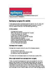
status. Preoperative data included age, sex, type, onset and duration of intractable epilepsy, psychological status and investigations like electroencephalograms (EEG), video EEG, brain computerized tomography (CT) and magnetic resonance imaging (MRI). Intraoperative data include the anesthetic techniques and monitoring, patient management during ECoG, post resection ECoG grades and type of surgical resection. Postoperative data include postoperative seizures outcome. Presurgical localization of the epileptic zones were derived from the clinical features of the epileptic foci, scalp EEG, video EEG monitoring, invasive subdural EEG monitoring, brain CT and MRI imaging studies. Epilepsy in all patients of this study, was refractory to two or more of the conventional antiepileptic drugs (AEDs), the frequency of seizures was not less than one per week and the duration of treatment was not less than 6 months before considering surgery. The selection of patients for surgery was a joint decision between the neurologist, clinical neurophysiologist and the neurosurgeon. Patients were not premedicated before anesthesia. Three patients were premedicated with diazepam before surgery and were excluded from the study. Antiepileptic drugs were discontinued at least one day before surgery. Twenty six males and 18 females were studied whose age distribution is shown in Fig. 1 and their clinical findings are summarized in Table 1.
In this study, we evaluated the presence or absence of epileptiform activity and potentially confounding burst-suppression in pre-resection ECoG under several different anesthetics during epilepsy surgery and also examined the relationship between post-resection ECoG and seizure outcome.
Fourty four patients with intractable epilepsy who were referred for epilepsy surgery at Jordan University Hospital completed their surgical management from 1994 to 2005. Data was collected on each patient by detailed review of preoperative, intraoperative and postoperative
Age Distribution of Patients
No. of Patients
Patients and Methods
Fig. 1 Age distribution of patients
20 18 16 14 12 10 8 6 4 2 0 0-5
6-10
11-20
Age (years)
>20
ANESTH. & ELOG. FOR EPILEPSY SURG.
33
Table 1 Clinical data and results of investigations of 44 patients
Table 2 Surgical procedures done on 44 patients
Age
17.34 years (Range 0.553 years)
Procedure
Male/female
26/18
Temporal lobe resection
30/44
(68.2%)
Type of seizures
Number of patients
Extra temporal lobe resection
12/44
(27.3%)
Functional hemispherectomy
2/44
(4.5%)
(%)
Complex partial
15
(34%)
Complex partial with simple partial
2
(4.5%)
Simple partial
1
(2.3%)
Complex partial with secondary generalization
18
(40.9%)
Generalized tonic clonic
7
(15.9%)
Mixed types
1
(2.3%)
Age of onset of seizure
Duration of seizure
(%)
Range (birth-45 years)
General anesthesia
42
(97%)
Local anesthesia
2
(3%)
1. Fentanyl and Isoflurane
19
(43%)
2. Fentanyl and Sevoflurane
9
(20.5%)
3. Remifentanil and Sevoflurane
5
(11%)
4. Remifentanil and Propofol during general anesthesia
9
(21%)
5. Fentanyl and Propofol during local anesthesia
2
(4.5%)
Mean = 8.75 years
Maintenance of anesthesia
Number of patients
During ECoG recording (%) (91%)
Decrease in cognitive functions
2
(4.5%)
Mentally retarded
2
(4.5%)
Number of patients
(%)
A-Focal Temporal activity
27
(61.4%)
Extra temporal activity
5
(11.3%)
12
(27.3%)
CT and MRI examination
Table 3 Anesthesia management of 44 patients Number of patients
40
B - Generalized activity
Fentanyl was used in 30 (68.2%) patients and isoflurane in 19 (43%) patients, (Table 3).
Type of anesthesia
Normal
Preoperative EEG findings
(%)
Mean =8.13 years
Range (0.5-25 years) Psychological status
Number of patients
Number of patients
(%)
Temporal structural lesions
18
(40.9%)
Extra temporal lesions
13
(29.5%)
Pre-resection and post-resection ECoG recording was used in 44 patients. Seven patients and 2 patients underwent preresection subdural and sphenoidal electrodes insertion respectively for more precise localization of the epileptic zone. Temporal lobectomy was performed in 30 (68.2%) patients, (Table 2).
The presence or absence of epileptiform activity and burst-suppression was noted and that pre-excision ECoG was defined as satisfactory when epileptiform activity was found.
General Anesthesia Management and Intraoperative ECoG Induction of general anesthesia was either inhalational with isoflurane and sevoflurane or intravenous with sodium thiopentone 2-4 mg/Kg or propofol 1-2 mg/Kg and fentanyl 2µg/Kg. Muscular relaxation was achieved by either atracurium or vecuronium and maintained by incremental doses of either drug to achieve one or two twitches on nerve stimulator. Arterial oxygen and carbon dioxide partial pressures were kept above 100 mmHg and between 30-35 mmHg respectively. Hypothermia during the M.E.J. ANESTH 20 (1), 2009
34 procedure was prevented by using warm IV fluids, water heating matrix and keeping the operating room temperature between 24-26 °C. After craniotomy and exposure of brain surface and during the period of ECoG recording before and after surgical resection, anesthesia was maintained by one of the following: = Fentanyl 0.5µg/kg incremental doses with inspired concentration of 0.5-1% isoflurane and 50% nitrous oxide in oxygen. = Fentanyl 0.5µg/kg incremental doses with inspired concentration of 1-2% sevoflurane and 50% nitrous oxide in oxygen. = Remifentanil infusion 1-4µg/kg/hour and inspired concentration of 1-2% sevoflurane and 50% nitrous oxide in oxygen. = Propofol infusion 1-4mg/kg/hour with remifentanil infusion 1-4μg/kg/hour.
Local Anesthesia Management For Awake Craniotomy Patients were initially deeply sedated with 2 mg/ kg propofol and 1 µg/kg fentanyl intravenously and positioned on the operating table in the semilateral position. Propofol infusion was subsequently administered at a rate of 1-4 mg/kg/hr and 25 μg incremental doses of fentanyl. After that, local anesthesia infiltration of the scalp down into the temporalis muscle and infiltration of the pin sites for Mayfield head holder was done. 0.25-0.5% lignocaine with adrenaline 1:200000 was used. Deep levels of sedation were employed during Mayfield head holder fixation, during scalp incision, raising the bone flap and manipulation, transection or coagulation of meningeal vessels. Light levels of sedation and sometimes discontinuation of the propofol infusion were employed during ECoG recording and during cortical speech mapping for verbal communication with the patient. ECoG recording during awake craniotomy was the same as during general anesthesia cases and electrodes were moved over the entire area of exposure. ECoG recording was done using 23 SLE microscribe EEG machine and E9 flexible plate 15 electrodes. Ad Tech 4, 6 or 8 electrode strips were used according to the case. ECoG was recorded. before resection to delineate the margins of resections
S. S. M. AL-GHANEM ET. AL
and after resection to detect any residual resectable epileptic focus. Twenty minutes was the minimum duration of ECoG recording during general and local anesthesia. Post-resection ECoG was graded according to Jay et al: (A) no residual epileptiform activity; (B) mild residual activity; (C) moderate residual activity; (D) unchanged from the pre-resection ECoG and (E) undetermined due to drug effect14. The range of follow-up period was from 6 months to 7 years. Postoperative seizure outcome was graded according to Kobayashi et al: grade I (seizure free without AED); grade II (seizure free on AED); grade III (> 50% reduction in seizure frequency); grade IV (< 50% reduction in seizure frequency); grade V (not improved) and grade VI (worse)15. A beneficial surgical result is defined as grade I-III seizure outcome. Postoperative seizure outcome and post-resection ECoG grade were compared with chi-square analysis of proportions.
Results No burst suppression or masking of epileptiform ECoG activity in 43 of 44 surgeries was observed regardless of the type of anesthesia. The epileptiform activity was absent in one patient under propofol anesthesia before resection. The was a 38 years old male patient scheduled for temporal lobectomy. The patient was suffering from long standing complex partial seizure with secondary general seizure and with frequency of 4 times/ month. The preoperative video EEG revealed left temporal focus. General anesthesia was employed using remifentanil and propofol. During ECoG monitoring before resection, epileptic activity could not be recorded, surgical resection could not be done and the patient awakened from general anesthesia. Three days later, general anesthesia was employed again for temporal lobectomy using fentanyl and isoflurane, epileptic activity before resection was recorded and surgery was completed. The histological diagnosis was normal brain tissue. Figure 2A, shows the ECoG with epileptic focus of a 21 years old male patient who was suffering from complex partial seizure of 17 years duration and
ANESTH. & ELOG. FOR EPILEPSY SURG.
35
Fig. 2A Pre-resection intraoperative ECoG showing the epileptic focus at electrodes 10 and 11 on the left anterior temporal lobe.
Fig. 2B Post-resection ECoG from the margin of the resection showing no residual epileptic activity on the same patient.
underwent left temporal lobectomy using fentanyl and isoflurane. Figure 2B, shows ECoG of the same patient after surgical resection without residual epileptiform activity. Post-resection ECoG grades showed 17 patients with grade A, 20 patients with grade B and 7 patients with grade C. Seizure outcome demonstrated that 6 (14%) patients are seizure free without AEDs, 16 (36%) are seizure free on AEDs and 19 (43%) patients had more than 50% reduction in the rate of seizures, (Table 4). The chi-square analysis of proportions for ECoG and beneficial outcome grades is 2.80, p = 0.094 which is not significant. Outcome grade ECoG grade
I-III
IV
A or B
36
1
C
5
2
Table 4 Postoperative seizure outcome according to post-resection ECoG results in 44 patients Postoperative Number of Post-resection grade* (Number of patients) seizure patients outcome grade** Grade A Grade I 4 (17) Grade II 6 Grade III 7 Grade B Grade I 2 (20) Grade II 8 Grade III 9 Grade IV 1 Grade C Grade II 2 (7) Grade III 3 Grade IV 2 * According to Jay et al [15]: A, no residual epileptiform activity ; B, miled residual epileptiform activity ; C, moderate epileptiform activity; D, unchanged from the pre-resection ECoG; E, undetermined due to drug effect. ** According to Kobayashi et al [16]: 1, seizure free without antiepileptic drugs; 2, seizure free on antiepileptic drugs; 3, more than 50% reduction in seizure frequency; 4, less than 50% reduction in seizure frequency; 5, not improved; 6, become worse. M.E.J. ANESTH 20 (1), 2009
36 Discussion The aim of epilepsy surgery is to stop seizures or to reduce their frequency by removing the epileptogenic zone or focus. This requires presurgical evaluation and selection of patients suitable for this type of surgery16,17. Routine scalp EEG usually provides interictal activity while ictal activity is usually obtained from short and long duration video EEG monitoring18. Temporal lobe is a common site for epilepsy focus and temporal lobectomy is a common procedure for intractable epilepsy patients. Epilepsy surgery can be done under local or general anesthesia. ECoG monitoring is more difficult during general anesthesia since anesthetic agent can suppress electrical brain activity and epileptogenic activity9,10. The present study has shown that satisfactory ECoG was obtained in 43 of the 44 surgeries done using different types of anesthetics and also 41 of 44 patients with post resection ECoG grade I -III benefited from epilepsy surgery. Many anesthetic drugs used in ECoG-guided epilepsy surgery may stimulate or supress epiletiform activity including alfentanil, fentanyl, remifentanil, sevoflurane, isoflurane, propofol and methohexitone1933 . Wass et al19 found increase in epileptiform activity after bolus 2.5 µg/kg intravenous remifentanil. Hisada et al21 reported increase in epileptogenic activity by 1.5 MAC sevoflurane anesthesia. Sato et al20 found epilepsy spikes decreased significantly with administration of nitrous oxide. Iijima et al22 found that the supplementation of nitrous oxide with sevoflurane suppressed epileptogenicity of sevoflurane in patients with epilepsy. The effect of propofol on epileptiform activity during epilepsy surgery is controversial and reports of both pro-convulsant and anti-convulsant effects have been published24-33. Hewitt et al26 reported increase in epileptic activity in 12 of 20 patients given boluses of propofol (range 40-200 mg) undergoing temporal epilepsy surgery and burst suppression was produced in the
S. S. M. AL-GHANEM ET. AL
ECoG in all but two patients. Samra et al30 reported no increase in epileptiform activity after subanesthetic doses of a propofol in volunteers with complex partial seizures. Drummond et al8 reported in a case of awake craniotomy given low dose propofol sedation an increase in EEG beta (β) activities that obscure the epileptogenic activity in ECoG recording. Rampil et al9 reported suppression of epileptiform activity by propofol during epilepsy surgery. Thirty three (75%) of our patients during intraoperative ECoG recording were given fentanyl and remifentanil with isoflurane and sevoflurane in 50% nitrous oxide in oxygen with adequate epileptiform activity recording. Despite reports of epileptiform activity suppression, we found satisfactory ECoG results regardless of the several types of anesthesia evaluated. In our study, we did not evaluate spike frequency or amplitude of ECoG that may be affected by some types of anesthetics more than others, and although ECoG was absent in one patient under propofol anesthesia, it is difficult to exclude other effects than propofol on ECoG since it is only one observation. Thirty patients in our study underwent temporal lobectomy and the present study has also shown that 41 (93%) patients benefited from epilepsy surgery using intraoperative ECoG. Various studies have shown favorable and encouraging seizure control after temporal lobe epilepsy surgery2,5,34-36. Drake et al2 reported 9 of 16 (56%) patients are seizure free after temporal lobectomy and 6 of 16 (38%) patients had more than 50% reduction in the frequency of seizures. Other reports have shown 90% and 80% over all improvements after temporal and extra temporal respectively36. In our study there was a slight trend to a higher proportion of surgical benefits (grade I-III seizure outcome) there was no statistically significant relationship between the amount of post-excision ECoG epileptiform activity and seizure outcome and this finding is in accordance with other studies12,37,38. In conclusion, our study has added further support to the literature that ECoG guided epilepsy surgery can be done with isoflurane or sevoflurane with nitrous oxide and fentanyl or remifentanil or using propofol and remifentanil, although, one of eleven ECoG was negative for epileptiform activity under propofol
ANESTH. & ELOG. FOR EPILEPSY SURG.
infusion. There was no statistically significant correlation between the amount of epileptiform activity on post-resection ECoG and seizure outcome, and it
37 seems that detailed analysis of post-resection ECoG beyond simple spike abundance may be required to better predict resection outcome39.
References 1. Ojemann GA: Surgical therapy for medically intractable epilepsy. J Neurosurg; 1987, 66(4):489-499. 2. Drake J, et al. Surgical management of children with temporal lobe epilepsy and mass lesion. Neurosurgery; 1987, 21:792-797. 3. Shimizu H: Our experience with pediatric epilepsy surgery focusing on corpus callosotony & hemispherectomy. Epilepsia; 2005, 46 suppl. 1:30-31. 4. Fish D, et al: Surgical treatment of children with medically intractable frontal or temporal lobe epilepsy: Results and highlights of 40 years’ experience. Epilepsia; 1993, 34:244-247. 5. Engel J: Epilepsy surgery. Curr Opin Neurol; 1994, 7:140-7. 6. Ojemann G: Different approaches to resective epilepsy surgery: "standard" and "tailored" Epilepsy Res; 1992, 5:169-74. 7. Tanaka T, et al: Intraoperative electrocorticography with medically intractable epilepsy, Neurol Med chil; Tokyo, 1996, 36:440-446. 8. Drummond JC, et al: Masking of epileptiform activity by propofol during seizure surgery. Anesthesiology; 1992, 76:652-654. 9. Rampil IJ, et al: Propofol sedation may disrupt interictal epileptiform activity from a seizure focus. Anesth Analg; 1993, 77:1071-1073. 10. Sato Y, Sato K, Shamoto H, Kato M, Yoshimoto T: Effect of nitrous oxide on spike activity during epilepsy surgery. Acta Neurochir; Wien, 2001, 143:1213-1216. 11. Endo T, et al: Effect of sevoflurane on electrocorticography in patients with intractable temporal lobe epilepsy. J Neurosurg Anesthesia; 2002, 14:59-62. 12. Cascino GD, et al: Electrocorticgraphy and temporal lobe epilepsy: relationship to quantitative MRI and operative outcome. Epilepsia; 1995 Jul, 36(7):692-6. 13. Stefan H, et al: Interictal triple ECoG characteristics of temporal lobe epilepsies: An intraoperative ECoG analysis correlated with surgical outcome. Clin Neurophysiol; 2007 Dec, 27. 14. Jay V, et al: Pathology of temporal lobectomy for refractory seizure in children. Review of 20 cases including some malformative lesions. J Neurosurg; 1993, 79:53-61. 15. Kobayashi J, et al: Surgery for intractable seizure in infants and children. Canadian J Neurol Sci; 1985, 12:178. 16. Bartolomei F, et al: The presurgical evaluation of epilepsies. Rev Neurol; Paris, 2002, 158:55-64. 17. Yagi k: Surgery for intractable epilepsy-selection and presurgical evaluation. Rinsho Ahinkeigaku; 1995, 35:1356-60. 18. Al Qudah AA, Abu sheik S, Tamimi AF: Diagnostic value of short duration out patient video electroencephalographic monitoring. Pediatric Neurology; 1999, 21(3):622-5. 19. Wass CT, et al: The effects of remifentanil on epileptiform discharges during intraoperative electrcorticography in patients undergoing epilepsy surgery. Epilepsia; 2001, 42:1340-1344. 20. Sato K, Shamoto H, Kato M: Effect of sevoflurane on electrocorticgraphy in normal brain. J Neurosurg Anesthesia; 2002, 14:63-65. 21. Hisada K, et al: Effects of sevoflurane and isoflurane on
electrocorticographic activities in patients with temporal lobe epilepsy. J Neurosurg Anesthesiology; 2001, 12(4):333-337. 22. Iijima T et al. The epileptogenic properties volatile anesthetics, sevoflurane and isoflurane in patients with epilepsy. Anesthe Analg; 2000, 91:989-995. 23. Manninen PH, et al: Intraoperative localization of an epileptogenic focus with alfentanil and fentanyl. Anesth Analg; 1999, 88:11011106. 24. Soriano SG, et al: The effect of propofol on intraoperative electrocorticography and cortical stimulation during awake craniotomies in children. Pediatric Anesthesia; 2000, 10:29-34. 25. Smith M, et al: Activation of the electrocorticogram by propofol during surgery for epilepsy. Br J Anesth; 1996, 76:499-502. 26. Hewitt PB, et al: Effect of propofol on the electrocorticogram in epileptic patients undergoing cortical resection. British J Anesth; 1999, 82:199-202. 27. Herrick IA, et al: Propofol sedation during awake craniotomy for seizures: electrocorticographic and epileptogenic effects. Anesth Analg; 1997, 84:1280-1284. 28. Finley GH, et al: Delay seizure following sedation with propofol. Can J Anesth; 1993, 40:863-865. 29. McBuray JW, Teiken PJ, Moon MR: Propofol for treating status epileptics. J Epilepsy; 1994, 7:21-22. 30. Samra SK, et al: Effect of propofol sedation on seizure and intracrauially recorded epileptiform activity in patients with partial epilepsy. Anesthesiology; 1995, 82:843-851. 31. Ebrahim ZY, et al: The effect of propofol on the electroencephalogram of patients with epilepsy. Anesth Analg; 1994, 78:275-279. 32. Geng MA, et al: Large dose of propofol alone in adult epileptic patient: electrocorticographic results. Anesth Analg; 1994, 78:275279. 33. Danks RA, et al: Patients tolerance of craniotomy performed with the patient under local anesthesia and monitored conscious sedation. Neurosurgery; 1998, 42:28-34. 34. Al Qudah AA, Tamimi AF, Ghanem S: Electrocorticography in the management of surgically treated epileptic patients. Neurosciences; 2000, 5:22-25. 35. D Barry Sinclair, et al: Pediatric Temporal lobectomy for epilepsy. Pediatric Neurosurgery; 2003, 38:195-205. 36. Mahera T, et al: Surgical treatment of children with medically intractable epilepsy. Outcome of various surgical procedures. Neurol Med Chir; Tokyo, 1996, 36:305-309. 37. Chatrian GE, Quesney LF: Intraoperative Electrocorticography. Epilepsy; 1997, 1749-65. 38. Schwartz TH, et al: The predictive value of intraoperative electrocorticography in resection for limbic epilepsy associated with mesial temporal sclerosis. Neurosurgery; 1997 Feb, 40(2):302-9. 39. Binnie CD, et al: Electrocorticography and stimulation. Acta Neurol Scand, Suppl, 1994, 152:74-82.
M.E.J. ANESTH 20 (1), 2009
38
S. S. M. AL-GHANEM ET. AL