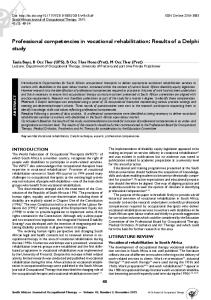
Cytometry (Communications in Clinical Cytometry) 30:166–177 (1997)
Original Articles
Analysis of Variation in Results of Flow Cytometric Lymphocyte Immunophenotyping in a Multicenter Study Jan W. Gratama,1* Jaco Kraan,1 Rene´ Van den Beemd,2 Berend Hooibrink,3 Dirk R. Van Bockstaele,4 and Herbert Hooijkaas2 1Department
of Clinical and Tumor Immunology, Daniel den Hoed Cancer Center, Rotterdam, The Netherlands of Immunology, Erasmus University and University Hospital, Rotterdam, The Netherlands 3Department of Clinical Viro-Immunology, Central Laboratory of the Dutch Red Cross Blood Transfusion Service, Amsterdam, The Netherlands 4Department of Hematology, University Hospital, Antwerp, Belgium 2Department
Fifty-five laboratories participated in a send-out study of four peripheral blood samples comparing a standard protocol vs. local protocols for flow cytometric lymphocyte immunophenotyping. The standard protocol included centrally provided reagents, instrument setup using triple-fluorescent microbeads and a three-color, whole-blood immunostaining technique based on fluorescein isothiocyanate and phycoerythrinlabeled monoclonal antibodies, erythrocyte lysis, washing, fixation, and identification of nucleated cells by the DNA/RNA stain LDS-751. Data analysis guidelines included lymphocyte selection using CD45,CD14assisted ‘‘backgating’’ on forward (FSC) and sideward (SSC) light scatter and placement of fluorescence (FL) markers on the basis of the isotype control staining. Most (i.e., 77%) of the variation in results of percentage lymphocyte subset assessments using the standard protocol was explained by laboratory, sample, background FL, and the interaction between laboratory and sample. Purity and completeness of the FSC,SSC lymphogate, background FL, flow cytometer type, and flow cytometer setup (which were either partly or entirely determined by laboratory) contributed significantly to the variation. The effect of the leukocyte differential count on the variation in absolute numbers of lymphocyte subsets was particularly large in lymphopenic This study was performed under the auspices of the Foundation for Immunophenotyping in Hemato-Oncology (SIHON), the Foundation for Quality Control in Medical Immunology (SKMI), the Foundation for Quality Control of Hospital Laboratories (SKZL), and the Belgian Association for Cytometry (BVC/ABC), with the participation of (in alphabetical order): M. Bas (Maasland Hospital, Sittard); F. Berends (Academic Medical Center, Amsterdam); F. Bergkamp (Kennemer Gasthuis, Haarlem); A. Bloem (University Hospital, Utrecht); Dr. Blomjous (St. Elisabeth Hospital, Tilburg); A. Borst (Sint Laurentius Hospital, Roermond); T. Braekevelt (General Hospital Zusters van Barmhartigheid, Ronse); C. Bridts (Universitaire Instelling, Antwerp); J. Ceuppens (University Hospital St. Raphael, Leuven); J. Damoiseaux (University Hospital, Maastricht); S. Darwood (Coulter Electronics, Luton, England); J. De JonghLeuvenink (MariaHospital and Dr. B. Verbeeten Institute, Tilburg); M. De Metz (Canisius/Wilhelmina Hospital, Nijmegen); H. De Muynck (University Hospital, Leuven); M. De Nooijer (Academic Medical Center, Amsterdam); M. De Vos (C.R.I., Zwijnaarde); J. D’Hautcourt (Clinique Saint Joseph, Mons); R. Dinkelaar (Drechtsteden Hospital, Dordrecht); Dr. Engelen (Centrum Medische Analyse, Herentals); E. Gemen (Bosch Medicentrum, Den Bosch); J. Goudswaard (Regional Laboratory ‘‘Zeeland,’’ Goes); J. Gratama (Daniel den Hoed Cancer Center, Rotterdam); J. Hoffmann (Catharina Hospital, Eindhoven); B. Hooibrink (Central Laboratory of the Dutch Red Cross Blood Transfusion Service, Amsterdam); H. Hooijkaas (Erasmus University and University Hospital, Rotterdam); W. Huisman (Westeinde Hospital, The Hague); S. IJpma (Merwede Hospital, Dordrecht); G. Janssen (St. Joseph Hospital, Veldhoven); C. Kallenberg (University Hospital, Groningen); J. Kerckhaert (De Lichtenberg Hospital, Amersfoort); L. Kestens (Institute for Tropical Medicine, Antwerp); J. Kluin-Nelemans (University Hospital, Leiden); P. Kramer (Sophia Hospital, Zwolle); A. Lammers (Regional Hospital Koningin Beatrix, Winter-
r 1997 Wiley-Liss, Inc.
swijk); S. Lauwers (Central Laboratory, Antwerp); M. Leerling (National Institute of Health and Environmental Hygiene, Bilthoven); A. Martens (Twenteborg Hospital, Almelo); P. Meeus (Onze Lieve Vrouw Hospital, Aalst); H. Messerschmidt (Carolus Hospital, Den Bosch); J. Modderman (Laboratory for Public Health, Leeuwarden); J. Molenaar (SSDZ, Delft); W. Nooijen (Anthonie van Leeuwenhoek Hospital, Amsterdam); F. Olthuis (Medisch Spectrum Twente, Enschede); D. Park (Slotervaart Hospital, Amsterdam); J. Philippe (University Hospital, Ghent); F. Preijers (University Hospital, Nijmegen); T. Rammeloo (St. Franciscus Hospital, Roosendaal); S. Rensink-Matter (St. Lucas Hospital, Amsterdam); G. Rijkers (Wilhelmina Children’s Hospital, Utrecht); K. Roozendaal (Onze Lieve Vrouwe Gasthuis, Amsterdam); J. Rummens (Virga Jesse Hospital, Hasselt); J. Sepers (Ortho Diagnostic Systems, Beerse, Belgium); K. Sintnicolaas (Red Cross Blood Bank, Rotterdam); A. Ten Haaft (University Hospital, Maastricht); B.’t Hart (Biomedical Primate Research Center, Rijswijk); R. Tiemessen (Rijnstate Hospital, Arnhem); L. Vaessen (University Hospital, Rotterdam); E. Van den Abbeele (Europa St. Michiels Clinic, Brussels); J. Van der Linden (University Hospital, Utrecht); H. Van der Veen (Holy Hospital, Vlaardingen); J. Van Diggelen (Interconfessional Hospital De Baronie, Breda); Dr. Van Erum (Henri Serruys Hospital, Oostende); H. Van Helden (St. Antonius Hospital, Nieuwegein); A. Van Houte (Medical Center Alkmaar, Alkmaar); H. Vanhouteghem (M. Middelares, Ghent); M. Van Poucke (St. Pieters Hospital, Brussels); M. Van Tol (University Hospital, Leiden); E. Van Voorst tot Voorst (De Weezenlanden Hospital, Zwolle); J. Van Wersch (De Wever Hospital, Heerlen); B. Von Blomberg (Academic Hospital of the Free University, Amsterdam). *Correspondence to: Jan W. Gratama, MD, Department of Clinical and Tumor Immunology, Daniel den Hoed Cancer Center, P.O. Box 5201, 3008 AE Rotterdam, The Netherlands. E-mail:
[email protected] Received 2 January 1997; Accepted 28 April 1997
MULTICENTER STUDY ON FLOW CYTOMETRIC IMMUNOPHENOTYPING
167
samples. The use of this standard protocol vs. local protocols did not reduce the interlaboratory variation. Instrument incompatibility with the standard protocol (e.g., incompatible filter combinations for LDS-751 detection) and lack of experience of many participants with three-color flow cytometry (in particular with the use of LDS-751) may have contributed to that result. We suggest that training and experience in a universally applicable standard protocol are critical for minimization of interlaboratory variation in flow cytometric immunophenotyping. Cytometry 30:166–177, 1997. Key words: flow cytometry; lymphocyte immunophenotyping; multicenter study; variation; standardization
Flow cytometric enumeration of leukocyte subsets is a valuable research and clinical tool, in particular for the diagnosis and monitoring of cellular immunodeficiency diseases (28), leukemias, and lymphomas (8). Accurate enumeration of lymphocyte subsets is of critical importance if diagnostic classifications or therapeutic decisions are based on such quantifications. A case in point is the role of the absolute number of CD41 lymphocytes in the clinical management of patients infected with the human immunodeficiency virus (HIV; 5). Whereas each laboratory is responsible for keeping its intralaboratory variation of such assays as low as possible, variation between laboratories must be kept to a minimum in multicenter studies. For that reason, several external quality assurance surveys have been set up to address the variation in (CD41) lymphocyte immunophenotyping (7,9–11,14,20– 22,27). These studies showed that a plethora of methodologic features can affect the results of flow cytometric immunophenotyping: blood sample (characteristics, integrity), sample preparation (leukocyte isolation, staining technique, washing, fixation), staining reagents, flow cytometer (instrument characteristics, setup, and performance), data acquisition and analysis (selection of cells of interest, classification of fluorescence [FL] signals as positive or negative), and absolute cell count assessment. Thus, the interlaboratory coefficient of variation (CV) of percentage CD41 lymphocytes in HIV2 donors ranged between 10% and 40% without standardization of the technique and was reduced to between 2% and 10% after implementation of highly standardized procedures. We report here the results of a multicenter study held in The Netherlands and Belgium as an introduction to a biannual external quality assurance survey. The participants were requested to perform a standard protocol in parallel with their own protocols on centrally distributed blood samples. The combined effects of several methodologic features were addressed by the standard procedure in order to achieve a significant reduction of interlaboratory variation. The participants were provided with fluorescein isothiocyanate (FITC)- and phycoerythrin (PE)-labeled monoclonal antibody (mAb) cocktails, ready to use in a whole blood immunostaining assay according to published guidelines for lymphocyte immunophenotyping (4), because different pairings of conjugated mAb and labeling with different fluorochromes reportedly contribute to interlaboratory variation (9,20). Triple-labeled microbeads for flow cytometer setup and erythrocyte lysis and gating reagents were also centrally supplied. As gating reagent,
we used the DNA and RNA stain LDS-751 (excitation at 488 nm, peak emission at 670 nm; 25) to allow the selection of nucleated cells during data acquisition using the third FL parameter, because incomplete erythrocyte lysis can significantly add to interlaboratory variation (10). LDS-751 is as effective in resolving lymphocytes from unlysed red cells as the widely used third-color CD45 mAb (18,19) but does so at a much lower cost (per staining, a third-color mAb typically costs several $US vs. ,,1¢ for LDS-751). MATERIALS AND METHODS Study Design The study was organized by the Flow Cytometry Working Party under the coordinated auspices of the Foundation for Immunophenotyping in Hemato-Oncology (SIHON), the Foundation for Quality Control in Medical Immunology (SKMI), the Foundation for Quality Control of Hospital Laboratories (SKZL), all in The Netherlands, and the Belgian Association for Cytometry (BVC/ABC) and was designed as a single send-out study to all Dutch and Flemish laboratories that routinely performed flow cytometric lymphocyte immunophenotyping as an introduction to a biannual external quality assessment scheme. The aims of the study were to analyze the sources of variation in results of flow cytometric immunophenotyping and to investigate whether the use of a standard protocol, designed by the Flow Cytometry Working Party, vs. local protocols would reduce that variation. The participants were provided with the standard protocol for lymphocyte immunophenotyping and the prescribed reagents and were requested to process samples of 2 ml of sodium heparin-anticoagulated peripheral blood of each of four patients on the day of receipt according to both standard protocol and their local protocols. The coordinating laboratory (Daniel den Hoed Cancer Center) obtained and aliquotted the blood samples and had them dispatched by overnight express mail at ambient temperature (i.e., 15– 25°C) to arrive at the participating laboratories the following day at 9 am, i.e., within 24 h after venipuncture. In this way, the study design complied with NIAID (4) and NCCLS (17) guidelines for specimen collection and handling. After reporting of the results to the coordinating laboratory and completion of the analyses by that laboratory, each participant was sent a confidential report with comments on his own results in comparison to those of the anonymous other participants. In addition, the results of the study were presented and discussed at a plenary
GRATAMA ET AL.
168
meeting held 2 months after the deadline for data submission. Standard Protocol Flow cytometer setup. Each participant was provided with 1 QC Windows kit of microbeads (Flow Cytometry Standards Corp., San Juan, Puerto Rico), consisting of microbeads triple-labeled with FITC; PE; PE-Cy5, named QC3; and nonfluorescent microbeads, named Certified Blank. In addition, centrally prepared suspensions of mononuclear cells from a healthy donor that were unstained or had been stained with CD4/FITC, CD8/PE, or LDS-751 were supplied for adjustment of electronic compensation for spectral overlap and adjustment of forward light scatter (FSC) photodiode and sideward light scatter (SSC) photomultiplier settings. Flow cytometers had to be set up as previously described (12). In brief, standardized positioning in ‘‘sample space’’ (i.e., the characteristics of the sample and their relationships, which are independent of the instrument) of the flow cytometer’s ‘‘window of analysis’’ (i.e., the portion of sample space analyzed by the flow cytometer), had to be achieved for each parameter by placement of the respective FL signals of CD41 lymphocytes (FSC and SSC parameters) or QC3 microbeads (all FL parameters) into predefined target channels. Thereafter, electronic compensation for spectral overlap was adjusted and activated. Sample preparation. Each participant was provided with ready-to-use mAb mixtures (SimulTEST series; CD45/ FITC 1 CD14/PE for setting an FSC,SSC lymphocyte gate; IgG1/FITC 1 IgG1/PE for isotype control; CD3/FITC 1 CD16156/PE; CD3/FITC 1 CD4/PE; CD3/FITC 1 CD8/ PE, and CD5/FITC 1 CD19/PE, all from Becton Dickinson Immunocytometry Systems [BDIS], San Jose, CA), FACS lysing solution (103 stock; BDIS) and 1 µg/ml LDS-751 stock solution (Exciton, Dayton, OH). The blood samples had to be kept at room temperature and processed on the day of receipt. A leukocyte differential count had to be performed according to local protocols. If the sample contained between 0.5 and 10 3 109/liter leukocytes, 100 µl undiluted blood had to be added to 12 3 75 mm tubes each containing 20 µl mAb mixture, incubated for 20 min at room temperature (RT), followed by erythrocyte lysis during 10 min at RT and a single washing step using phosphate-buffered saline (PBS) supplemented with 1% bovine serum albumin. If the sample contained .10 3 109 leukocytes/liter, the blood sample volume had to be decreased to add a maximum of 1 3 106 leukocytes to the mAb mixture. After the washing step, the cell pellet was resuspended in 0.5 ml PBS containing 1% paraformaldehyde and 0.04 µg/ml LDS-751. Samples containing ,0.5 3 109 leukocytes/liter were considered unsuitable for processing according to this procedure. Flow cytometry had to be performed within 2 h after completion of sample preparation. For data acquisition, a live gate had to be set on the nucleated cells (LDS-7511) to acquire list mode data on 10,000 events. If the differential count revealed ,10% lymphocytes, the number of acquired events had to be increased proportionally so that at
minimum 1,000 lymphocytes were measured. For data analysis of each specimen, an FSC,SSC gate had to be set around the lymphocytes (hereafter referred to as lymphogate) in the CD45/FITC 1 CD14/PE-stained sample to contain .95% of the lymphocytes (CD451,142) with the minimal possible number of nonlymphoid cells (16). The subsequent stainings of that specimen had to be analyzed after activation of that lymphogate. Markers to distinguish between positive and negative FITC and PE signals had to be set on the isotype control staining in such a way that the FITC2,PE2 quadrant contained $98% of the gated events. These marker settings had to be used for the analysis of the remaining stainings of that specimen. Data reporting. The participants were requested to submit printed output of list mode data analyses and to report on a questionnaire form for each specimen: the type(s) of instruments used for absolute cell count and immunophenotype assessments and the results of 1) the leukocyte differential count; 2) the completeness and purity of the lymphogate; and 3) for all stainings other than CD45/FITC 1 CD14/PE, the proportions (hereafter referred to as %) of events per quadrant (i.e., FITC1,PE2; FITC1,PE1; FITC2,PE2 and FITC2,PE1) within the lymphogate. Local Protocols The participants were requested to report on a questionnaire 1) the results of assessments of the % and absolute numbers (hereafter referred to as #) of NK lymphocytes, CD31, CD41, and CD81 T lymphocytes, and B lymphocytes; the coexpression of CD16 and/or CD56 by T lymphocytes; and the coexpression of CD5 by B lymphocytes, according to their own methods with respect to flow cytometer setup, immunostaining, and flow cytometry; and 2) a summary of the methodology used. Data Processing and Statistical Analysis Data submitted on questionnaires were checked for inconsistencies and entered into a computer database by the coordinating laboratory. If necessary, comparisons with printed output of list mode data analyses were made, and incorrect data entries were corrected after consultation with the submitting laboratory. Data processing and statistical analyses were performed using SAS-PC (Statistical Analysis Systems, Cary, NC) and Stata (Stata Corporation, College Station, TX) software. Standard statistical methods used are specified in Results and in the legends to the figures. For the data obtained with the standard protocol, the % and # of the following lymphocyte subsets were computed: all T lymphocytes (defined by CD31), all T lymphocytes (defined by CD192,51), all NK lymphocytes (CD161/ 561,32), T-helper/inducer lymphocytes (CD31,41), T-cytotoxic/suppressor lymphocytes (CD31,81), NK lymphocyte subset (CD32,81), and all B lymphocytes (CD191). In addition, the % coexpressions of CD16/56 by the CD31 T lymphocytes and of CD51 by the CD191 B lymphocytes were calculated. The % lymphocyte subsets were not
MULTICENTER STUDY ON FLOW CYTOMETRIC IMMUNOPHENOTYPING
corrected for contamination of the lymphogate with residual erythrocytes, because these had already been eliminated from the list mode data by live gating on the nucleated (LDS-7511) events. Background FL was not subtracted from the experimental results because marker setting as per protocol had been aimed at obtaining low % background FL (i.e., a total of #2% in the FITC1,PE2, FITC1,PE1, and FITC2,PE1 quadrants). For the analysis of the effects of laboratory, sample, flow cytometer type and setup, lymphogate, and FITC,PE marker setting on the variation in results obtained with the standard protocol (Fig. 4), the data were summarized using the average residual (ARijk), expressed in percentage positive cells (i.e., fraction of lymphocytes), defined as follows:
AR ijk 5
Œo 1
4
4 q51
(Pijkq 2 Mjkq )2
in which i 5 1 . . . 55 for laboratory; j 5 1 . . . 4 for sample; k 5 1 . . . 4 for staining (i.e., 1 5 CD3/FITC 1 CD16156/ PE; 2 5 CD3/FITC 1 CD4/PE; 3 5 CD3/FITC 1 CD8/PE; and 4 5 CD5/FITC 1 CD19/PE); q 5 1 . . . 4 for quadrant (i.e., 1 5 FITC1,PE2; 2 5 FITC1,PE1; 3 5 FITC2,PE2; and 4 5 FITC2,PE1); Pijkq 5 percentage positive cells (expressed as fraction of lymphocytes) reported by laboratory i on sample j, staining k and quadrant q; M jkq 5
1 55
55
oP
ijkq
.
i51
For the analysis of variation in absolute numbers of CD41 lymphocytes and the parameters from which this count is derived (Fig. 5), and in % lymphocyte subsets observed with standard vs. local protocols (Fig. 6), the data were summarized using the residual (Rijm), expressed as percentage of the mean result of all laboratories, defined as follows: R ijm 5
0 Pijm 2 M jm 0 M jm
3 100%
in which i 5 1 . . . 55 for laboratory; j 5 1 . . . 4 for sample; m 5 1 . . . 10 for immunological parameter (i.e., 1–4 for those shown in Fig. 5 and 5–10 for those shown in Fig. 6);
169
Pijm 5 result of immunological parameter m reported by laboratory i on sample j; M jkq 5
1
55
P 55 o
ijkq
.
i51
RESULTS Sent-Out Specimens and Response Rate Sixty-nine laboratories (listed in the footnote at the beginning) registered for the workshop at a cost of $US125.00 per laboratory. Four patients agreed, after informed consent, to donate 150 ml venous blood per person. The results obtained with the standard protocol have been summarized in Figures 1 (%) and 2 (#). Patient 1, a 39-year-old female, had received an allogeneic bone marrow transplant (BMT) for acute lymphoblastic leukemia in second remission 13 years ago. She still had mild chronic graft-vs.-host disease (GVHD). Her blood specimen had normal % and # T lymphocytes and subsets, normal % and # NK lymphocytes, but elevated % and # of B lymphocytes (featuring polyclonal sIgk and sIgl expression [data not shown]). Patient 2 was a 56-year-old female with metastatic breast cancer, 1 week after standard-dose chemotherapy. Patient 3 was a 45-year-old male with B non-Hodgkin’s lymphoma, 13 days after autologous peripheral blood stem cell reinfusion. Their blood specimens had very low # of lymphocytes, with normal subset distributions (%). Patient 4 was a 45-year-old male 4 months after allogeneic BMT for chronic B-lymphocytic leukemia (BCLL). His clinical course was complicated by active cytomegalovirus infection and mild chronic GVHD. His blood showed increased % and # of CD31,81 T lymphocytes and normal % and # of CD31,41 T, NK, and B lymphocytes. However, the % CD191 lymphocytes coexpressing CD5 was increased, suggesting in his case the presence of residual B-CLL. The specimens from patients 1–4 are referred to as samples 1–4, respectively. Results were submitted by 55 laboratories (80%) after processing the samples on the day of arrival (n 5 50) or 1 day later (n 5 5). The remaining 14 laboratories were unable to participate because of lack of personnel at the time of the send-out or lack of experience with flow cytometric three-color FL assays. Results of Flow Cytometer Setup, Placement of Lymphogates, and Setting of FITC and PE Markers per Standard Protocol Of the 55 responding laboratories, 34 operated a FACScan, 6 a FACStar, 3 a FACSort, and 2 a FACStrak flow cytometer (all from BDIS); 5 operated an XL and 1 a Profile (both from Coulter, Hialeah, FL) and 4 a CytoronAbsolute flow cytometer (Ortho Diagnostics, Raritan, NJ). FACScan and FACSort instruments were grouped together in the subsequent analyses on the basis of their similar technical design and equipment. Flow cytometer setup per the standard protocol was performed entirely correctly by 39 laboratories. Thirteen laboratories had deficiencies in their flow cytometer setup. Three of these thirteen laboratories failed to meet the requested initial target channels for one
170
GRATAMA ET AL.
FIG. 1. Percentages of cells expressing a given immunophenotype (shown at the top of each panel) within (subsets of) lymphocytes in samples from the four patients assessed using the standard protocol. The boxes extend from the 25th (p25) to the 75th (p75) percentile; the line in the middle represents the median. The vertical lines extend to the upper
and lower adjacent values, which are defined as 1.5 3 (p75 2 p25), rolled back to where there are data. Outliers, more extreme than the adjacent values, have been individually plotted. The width of the boxes is proportional to the number of observations, i.e., 30 on sample 3, 53 on sample 2, and 55 each on samples 1 and 4.
or two of the three FL parameters by 5–80 channels on a linear scale of 1,024 histogram channels. Two of these three plus the remaining 10 laboratories obtained suboptimal fluorescence compensation (i.e., either undercompensation or overcompensation). Furthermore, two laboratories operating FACStrak instruments were unable to set up their flow cytometers per the standard protocol owing to software limitations. Finally, one laboratory reported incomplete data on its flow cytometer setup. Correct flow cytometer settings were more frequently achieved by laboratories operating a FACScan or FACSort instrument (34 of 36 [94%]) than those operating other instruments (i.e., FACStar, XL, Profile, or CytoronAbsolute; 5 of 16 [31%]; Fisher’s two-sided exact P , 0.0001). Discussions at the plenary meeting revealed that the optical filter combinations on the FACStar, Profile, and XL instruments were inadequate for detection of LDS-751 fluorescence, and the instructions per the standard protocol had been difficult to perform on CytoronAbsolute instruments because of their large sampling volumes.
A lymphogate including .95% of all lymphocytes, as per the protocol, was set by 33 of 46 (72%) and 24 of 47 (51%) laboratories submitting such data on samples 1 and 4 with normal or supranormal absolute lymphocyte counts, respectively. Typically, most of these lymphogates had only minor contamination with nonlymphoid cells (i.e., .90% purity; Fig. 3). However, only 12 of 45 (27%) and 8 of 26 (31%) laboratories placed a .95% complete lymphogate on the lymphopenic samples 2 and 3, respectively. The difficulty in setting appropriate lymphogates on these samples is further illustrated by the fact that the purity of these gates was also lower than on samples 1 and 4 (Fig. 3). However, there was no straightforward correlation between completeness and purity of suboptimally, i.e., ,95% complete, lymphogates (Fig. 3). Lymphogates were placed with significantly greater purity on FACScan and FACSort instruments than on other instruments (median [range], 93% [10–100%] vs. 87% [3–100%], respectively; P 5 0.001 using Wilcoxon’s rank test). The effects of flow cytometer type on lymphogate completeness and of flow
MULTICENTER STUDY ON FLOW CYTOMETRIC IMMUNOPHENOTYPING
171
FIG. 2. Absolute numbers of lymphocyte subsets in samples from the four patients assessed using the standard protocol. See legend to Figure 1.
cytometer setup (i.e., correct vs. incorrect) on lymphogate completeness and purity were not significant. Placement of FITC and PE markers on the isotype control staining yielding .98% double-negative events, as per protocol, was done by 49 of 54 (91%) and 48 of 54 (89%) laboratories submitting such data on samples 1 and 4, respectively. Setting the FITC and PE markers in such a way was more difficult on the lymphopenic samples 2 and 3: Only 38 of 50 (76%) and 18 of 29 (62%) laboratories did so, respectively. The sum of % background fluorescence in the FITC1,PE2, FITC1,PE1, and FITC2,PE1 quadrants was lower for the FACScan and FACSort instruments than for the other instruments (0.6% [0–16.3%] vs. 1.6% [0–19.2%], respectively; P 5 0.02). Effects of Laboratory, Sample, Flow Cytometer Type and Setup, Placement of Lymphogates, and Setting of FITC and PE Markers on Variation of Immunophenotyping Results Obtained With the Standard Protocol For each laboratory, the average residual, i.e., the average deviation from the mean result per staining and
sample, expressed as % of cells within the lymphogate, was used as a parameter for the variation of results obtained with the standard protocol. One-way analyses of variance (ANOVA) revealed that laboratory (coded 1–55) accounted for 36% of the variation; sample (i.e., 1–4) for 27%; lymphogate purity (i.e., $90% vs. ,90%) for 15%; lymphogate completeness (i.e., $95% vs. ,95%) for 9% (Fig. 4); instrument type (i.e., FACScan or FACSort vs. other), instrument setup (i.e, correct vs. incorrect), and background FL (i.e., #2% vs. .2%) each for 3%; and staining (i.e., CD3/FITC 1 CD16156/PE; CD3/FITC 1 CD4/PE; CD3/FITC 1 CD8/PE; and CD5/FITC 1 CD19/ PE) for ,0.5% of the variation (not shown). Because several of these parameters were highly correlated (see above), we also studied their combined effects on the average residual in a multivariate ANOVA. The best fitting model explained 77% of the variation. Laboratory (32%), sample (29%), background FL (7%), and effect of laboratory as a function of sample (9%) contributed significantly (P , 0.0001) to the variation in this model, whereas the contributions of lymphogate completeness and purity
172
GRATAMA ET AL.
FIG. 3. Correlation plots of completeness vs. purity of the FSC,SSC lymphogates set on samples 1–4 assessed according to the standard protocol. The line y 5 x is shown as a reference in each panel.
(each ,0.1%) were nonsignificant. Lymphogate completeness and lymphogate purity were largely determined by laboratory (not shown); hence, their effects in the multivariate model were decreased compared to the univariate analyses. Flow cytometer type and setup were entirely determined by laboratory and were therefore not included in the model. Inspection of the results of individual laboratories revealed that the median value of the average residual exceeded 5% for 19 laboratories and 10% for four (Labs 16, 36, 42, and 52; Fig. 4, upper left panel). A 1 day delay in performing the assays did not cause significant variation; only one of five labs that had such a delay (Lab 16) was an outlier. A coincidence of problems may have caused the aberrant results in these four cases. Labs 16, 36, and 52 detected too low a % FITC1,PE2, FITC1,PE1, and FITC2,PE1 cells in all four stainings of all four samples, whereas Lab 42 detected consistently too high a % T cells (CD31 or CD51,192) in sample 1 and too low a % T cells in sample 3. All four labs used instruments other than FACScan/ FACSort. Two flow cytometers (36 and 52) had been set up incorrectly. In some cases, lymphogate completeness
was too low (Labs 16 and 52) or not analyzed (Labs 36 and 42). FITC,PE marker settings by Labs 16 and 42 were inappropriate. In addition to these factors, inadequate sample preparation, poor flow cytometer performance, problems with data acquisition (such as live gating on LDS-7511 events), and/or deterioration of blood samples prior to processing might have caused these aberrant results. Using local protocols, Labs 16 and 42 obtained results similar to those of the majority of participants, excluding sample deterioration and poor flow cytometer performance in their cases. Lab 36 obtained too low a % positive cells using both protocols, and Lab 52 did not report on its local protocol, which left (combinations of) any of the above-mentioned problems open as additional causes. Analysis of Interlaboratory Variation in Absolute Numbers of Lymphocyte Subsets We traced back the variation in absolute numbers of lymphocyte subsets to the parameters from which they were derived per the standard protocol: # leukocytes, %
MULTICENTER STUDY ON FLOW CYTOMETRIC IMMUNOPHENOTYPING
173
FIG. 4. Effects of laboratory (upper left), sample (upper right), completeness of FSC,SSC lymphogate (lower left), and purity of FSC,SSC lymphogate (lower right) on variation of immunophenotyping results obtained with the standard protocol. The average residual has been used as a
parameter to quantify variation (for computation, see Materials and Methods). Upper left: The median values of the average residual for each laboratory have been connected with a line; the horizontal lines at 5% and 10% serve as references. For other panels, see legend to Figure 1.
lymphocytes (fraction of leukocytes) and % subsets (fraction of events within the lymphogate). We took the CD31,41 subset as a case in point (Fig. 5). The # leukocytes (Fig. 5, upper row) contributed relatively little; overall, the median value of the residual was 6%. Sample 3 was exceptional in its low leukocyte count (,0.5 3 109/liter) and yielded most of the outliers and, hence, the largest residuals. The variation in % lymphocytes (Fig. 5, second row) was slightly larger (i.e., median residual 9%); most of the variation was observed in the two samples with relative lymphopenia (sample 2, ,7%, and sample 3, ,16% of leukocytes). The residual of the % CD31,41 lymphocytes (Fig. 5, third row) was again larger (median 13%), but there were few outliers, indicating that the effect of low proportions of lymphocytes in the samples on the variation in flow cytometric assessment of % CD31,41 lymphocytes was relatively minor. The median residual of the absolute number of CD31,41 lymphocytes was largest (i.e., 19%; Fig. 5, lower row) as a result of the combined effect of the variation in the three parameters discussed above. As expected, samples 2 and 3 showed most variation.
We also analyzed the effect of type of instrument for absolute cell count assessment (for short, hematology analyzer) on the variation in # leukocytes, % lymphocytes, and # CD31,41 lymphocytes (Fig. 5). Fifteen of the fifty-five responding laboratories used a hematology analyzer from Coulter, 22 from Technicon, 12 from Sysmex, and the remaining 6 laboratories either an Argos analyzer (n 5 1) or only manual enumeration (n 5 5). The effect of hematology analyzer on the variation in # leukocytes and % lymphocytes was significant only for the lymphopenic samples (i.e., samples 2 and 3; P 5 0.01 and 0.0001, respectively, using the Kruskal-Wallis test). Most variation in # leukocytes and % lymphocytes was seen in the ‘‘other’’ group (i.e., Argos or manual). However, the effect of hematology analyzer on the variation in # CD31,41 lymphocytes was nonsignificant regardless of lymphopenia of the samples. Comparison of Variation in Results Obtained With Standard and Local Protocols For flow cytometric data analysis per local protocols, 25 of the 55 responding laboratories used SimulSET software
174
GRATAMA ET AL.
FIG. 5. Interlaboratory variation in results of assessments of # leukocytes (upper row), % lymphocytes within leukocytes (second row), % CD31,41 cells within lymphocytes (third row), and # CD31,41 lymphocytes (lower row) as a function of sample and type of instrument for absolute cell count assessment. Abbreviations: Clt: Coulter; Tcn: Techni-
con; Ssx: Sysmex; Oth: other. The residuals have been used as a parameter to quantify variation (for computation, see Materials and Methods). The median values of the residuals for each parameter (i.e., # leukocytes, % lymphocytes, % CD31,41 lymphocytes, and # CD31,41 lymphocytes) are indicated with horizontal lines. See legend to Figure 1.
(BDIS), providing automated placement of a lymphogate on the basis of backgating on the CD451,142 population (16). Thirteen laboratories performed this procedure manually. Four laboratories used CD45,SSC lymphogating in a three-color technique (18), and the remaining 13 laboratories placed a lymphogate on the basis of FSC and SSC only or followed other gating routines. The effects of local gating strategies on the results of % lymphocyte subset assessments were not significant (data not shown). NK lymphocytes were defined as CD161/561,32 by 39 of the 55 laboratories; as CD161,32 by 2, as CD561,32 by 2, as CD161 by 2, and as CD561,TCRab2 by 1 laboratory. The remaining 9 laboratories had no local protocol for NK lymphocyte enumerations. The effect of immunophenotypic definition of NK lymphocytes on the results of % NK lymphocyte assessments was not significant (data not shown). The variation in results of % lymphocyte subset assessments according to the quality of performance of the
standard protocol (i.e., fulfilling the criteria for completeness of the lymphogate and for % FITC,PE background fluorescence on the isotype control as per protocol) and according to local protocols is shown in Figure 6. For all lymphocyte subsets, the median values of the residuals obtained using the standard protocol with .95% complete lymphogates and #2% background fluorescence (LpMp in Fig. 6) were lower than those obtained with incorrectly performed standard protocols (LfMf and LfMp) or with incompletely reported standard protocols (L?M?). The median values of the residuals obtained with a correctly performed standard protocol were also smaller than those obtained with local protocols for all subsets except % CD31 and % CD(31,)41. Comparison of the residuals of the five groups (i.e., LpMp, LfMf, LfMp, L?M?, and local) revealed significant differences for all six subsets (P values ranging between 0.0001 and 0.05 using the Kruskal-Wallis test). However, the residuals obtained with a correctly performed standard protocol were significantly smaller
MULTICENTER STUDY ON FLOW CYTOMETRIC IMMUNOPHENOTYPING
FIG. 6. Comparison of variation in results of assessments of % CD161/561(,32) cells (upper left), % CD31 cells (upper right), % CD(31,)41 cells (middle right), % CD(31,)81 cells (lower left), and % CD191 cells (upper right) among lymphocytes and % CD161 and/or 561 coexpression by CD31 lymphocytes (middle left) as a function of protocol (i.e., standard vs. local) and performance of the standard protocol (i.e., incorrect [LfMf], partly correct [LfMp], correct [LpMp], or not reported [L?M?]). L: lymphogate; M: FITC,PE marker setting; f: failed (i.e., #95% completeness or .2% background FL); p: passed (i.e., .95% completeness or #2% background FL, respectively). (,32) or CD(31,) indicates that CD16/CD56, CD4 and CD8 were not counterstained with CD3 in some local protocols. The residual, computed per sample and laboratory, has been used as a parameter to quantify variation (for computation, see Materials and Methods). See legend to Figure 1.
than those obtained with local protocols for only two subsets (i.e., % CD161 and/or 561 within the CD31 lymphocytes, P 5 0.05; % CD191, P 5 0.0006), and they were even significantly larger for % CD(31),41 (P 5 0.004 using Wilcoxon’s test). Thus, the introduction of a standard protocol for lymphocyte immunophenotyping in this study did not lead to a consistent reduction of the variation in results compared to local protocols. All laboratories except the four operating CytoronAbsolute instruments used a single leukocyte differential count for calculation of the # lymphocyte subsets as per standard and local protocols; hence, conclusions in this respect were similar for # and % lymphocyte subsets (data not shown). DISCUSSION The aims of this study were to analyze, in a multicenter study, the variation in results of flow cytometric lymphocyte immunophenotyping and to investigate whether the
175
use of a standard protocol vs. local protocols would reduce that variation. Most (i.e., 77%) of the variation in results of % lymphocyte subset assessments using the standard protocol was explained by laboratory, sample, background FL, and interaction between laboratory and sample; i.e., the effect of laboratory on variation differed as a function of sample. Univariate analyses revealed that sample, laboratory, lymphogate purity, lymphogate completeness, background FL, flow cytometer type, and flow cytometer setup (which were partly or entirely determined by laboratory) contributed significantly to the variation. The use of a standard protocol vs. local protocols did not reduce the interlaboratory variation. The variation of results of % CD(31,)41 lymphocytes obtained with standard and local protocols in our study was of the same order of magnitude as in other surveys without extensive standardization between participants, i.e., between 10% and 40% depending on sample (11,13,14,26,27). Intensive training and quality control of 8–13 participants in the setting of standardized procedures has indeed been shown to reduce interlaboratory variation significantly (22,23), whereas longitudinal quality assurance surveys without the compulsory implementation of such procedures do not result in reduction of interlaboratory variation over time (14). We discuss the following technical problems as sources of variation in results of flow cytometric immunophenotyping according to the standard protocol, ranked in order of importance as apparent in this study. Unreliable Leukocyte Differential Counts on Lymphopenic Samples Our CVs of # CD31,41 lymphocytes ranged between 15% and 30% for samples with (supra)normal lymphocyte counts and between 40% and 60% for lymphopenic samples. The largest source of variation in # CD31,41 lymphocytes was, in samples with relative lymphopenia, the % lymphocytes derived from the leukocyte differential count. That situation might have been caused by rejection of such samples by automated hematology analyzers, requiring manual differentiation on relatively low numbers of leukocytes (100–500 vs. 10,000 for an automated differential). The need to reduce the contribution of the leukocyte differential count to the interlaboratory variation of absolute numbers of immunologically defined lymphocyte subsets has been widely recognized (1,10,11,20,23,26,27) and has led to the development of ‘‘single-platform’’ assays, in which absolute numbers of lymphocyte subsets are derived from a single flow cytometric assessment. One successful approach consists of the measurement of calibrated sample volumes on the CytoronAbsolute instrument from Ortho Diagnostic Systems (6). Alternatively, internal calibrators in the form of predefined amounts of reference beads are added to blood samples (Flowcount from Coulter Electronics) or vice versa (TruCount from BDIS); the effectiveness of these approaches in reducing interlaboratory variation has not yet been established.
GRATAMA ET AL.
176 Suboptimal Selection of Cells of Interest
The failure to remove all erythrocytes from the samples was a major source of variation in the Multicenter AIDS Cohort Survey (10); the light scatter characteristics of the residual erythrocytes overlapped with those of the lymphocytes, leading to erroneously reduced percentages of positive lymphocytes. To circumvent that problem, we have included selection of nucleated cells (LDS-7511) during data acquisition in the standard protocol. However, participants operating FACStar, Profile, or XL instruments reported weak LDS-751 signals because of incompatible filter combinations, whereas FACStrak users could not set LDS-751 live gates because of software limitations. Residual erythrocytes may have been gated out insufficiently in such cases, leading to decreased purity of the lymphogates; laboratories using such flow cytometers indeed reported lower lymphogate purities than those using FACScan or FACSort instruments. In addition, most of our participants did not routinely use three-color flow cytometry and were unfamiliar with the use of the DNA and RNA stain LDS-751 to select nucleated cells during data acquisition. That situation might have added to the interlaboratory variation. Selection of Lymphocytes During List Mode Data Analysis Selection of lymphocytes during list mode data analysis had a significant impact on the interlaboratory variation in our study. Setting restricted FSC,SSC gates on lymphocytes from normal donors may skew against lymphocyte subsets (16). Consequently, we included instructions to set .95% complete lymphogates using backgating on CD451,142 events in line with widely accepted guidelines (4). The variation in immunophenotyping results was, indeed, significantly smaller with relatively complete and/or pure lymphogates than in the case of relatively restricted and/or contaminated lymphogates. However, setting appropriately complete and pure lymphogates was problematic on samples with low absolute lymphocyte counts. In this context it is relevant that selection of a cell population of interest solely on the basis of FSC,SSC characteristics never can be successful when its FSC,SSC characteristics substantially overlap with those of other cell populations. In addition, completeness and purity of the cell population of interest only partially control placement of FSC,SSC lymphogates. Hence, novel, highly standardized procedures for selection of cells of interest in flow cytometric analyses are needed. Instrument Incompatibility With Standard Procedure Flow cytometer type (i.e., FACScan or FACSort vs. other) was highly correlated with flow cytometer setup (i.e., correct vs. incorrect), lymphogate completeness (i.e., .95% vs. #95%) and purity (i.e., .90% vs. #90%), and background FL (i.e., ,2% vs. $2%). That situation was due to instrument incompatibility with the standard procedure, precluding appropriate detection of LDS-751 signals
by FACStar, FACStrak, Profile, and XL instruments (see above) and hampering flow cytometer setup for CytoronAbsolute instruments. However, these results do not bear relevance for instrument performance. The performance of a flow cytometer is evaluated by calculation of a calibration regression line between predefined FL intensities of calibration standards labeled with spectrally matching fluorochromes in different intensities and the instrument’s response to their FL signals in histogram channels (24). Using that approach, the Italian Group for Cytometry found a negative correlation between the flow cytometers’ FITC detection threshold and their ability to detect dim FITC signals (3). The performance of 39 of the 55 flow cytometers was evaluated in such a way in our previous year’s survey, and only one of them functioned poorly (12). That instrument (Lab 36) yielded, indeed, outlying results in the current study. Variation in Sample Processing The participants were provided with a detailed protocol for sample preparation, ready-to-use mAb mixtures, and stock lysing solution and gating reagent (LDS-751). Any variation arising from sample processing in this study has therefore resulted from differences in performing the staining, lysing, washing, and fixation procedures or and storage of the stained cells by the various laboratories. Sample Deterioration Central processing and aliquotting of the blood samples prior to transport excluded these factors as sources of variation. However, varying degrees of sample deterioration during transport and storage may have occurred, which is inherent to this type of send-out study (2). A time lapse exceeding 30 h between venipuncture and assay significantly affected results in the Transfusion Safety Study (20). Such delays were reported by 5 of the 55 laboratories but did not contribute significantly to the observed variation (data not shown). The use of preserved samples would have minimized the risk of deterioration. However, leukocyte suspensions that have been viably cryopreserved (15), lyophilized (Cytotrol; Coulter Electronics), or otherwise stabilized (CD-Chex; BDIS) do not allow full process control and, therefore, are not representative for lymphocyte subset enumerations based on erythrocyte lysis. A recent alternative is the proprietary technique to stabilize whole blood as used in the U.K. NEQAS surveys (2). The commercial product, AbsoluteControl (Ortho Diagnostics), was not yet available at the time of this study. Currently, AbsoluteControl is available only as a mixture of blood specimens from apparently healthy donors and, therefore, has only limited value for quality assurance of enumerations of abnormal, i.e., lymphopenic, specimens. The technology of flow cytometric lymphocyte immunophenotyping is rapidly advancing. Triple- and quadruplecolor stainings can now be analyzed with single- and dual-laser benchtop flow cytometers. Commercial manufacturers of flow cytometric technology are incorporating such advances into assay kits using specialized calibrators, reagent combinations, and software. That policy is aimed
MULTICENTER STUDY ON FLOW CYTOMETRIC IMMUNOPHENOTYPING
at optimal robustness of such assays, but the divergence of technologies between manufacturers makes cross-calibration of such assays increasingly complex. An indication of this problem is provided by the difficulties of several participants in this study in performing the standard protocol because of incompatibilies with their flow cytometric hardware and software. Faced with the advancing but diverging technologies in the field of flow cytometric immunophenotyping, organizations involved in external quality assurance should coordinate the formulation, implementation, and periodic revision of consensus protocols at national and international levels. According to our experience and that of others (22,23), training and supervision of the participants are critical to the success of such an endeavor. ACKNOWLEDGMENTS We are grateful to Drs. W.L.J. van Putten and B. van der Holt for their statistical advice. LITERATURE CITED 1. Aboulker JP, Autran B, Beldjord K, Touraine F, Debre´ P: Consistence of routine measurements of CD41, CD81 peripheral blood lymphocytes. J Immunol Methods 154:155–161, 1992. 2. Barnett D, Granger V, Mayr P, Storie I, Wilson GA, Reilly JT: Evaluation of a novel stable whole blood quality control material for lymphocyte subset analysis: Results from the UK NEQAS Immune Monitoring Scheme. Cytometry 26:216–222, 1996. 3. Brando B, Sommaruga E: Nationwide quality control trial on lymphocyte immunophenotyping and flow cytometer performance in Italy. Cytometry 14:294–306, 1993. 4. Calvelli T, Denny TN, Paxton H, Gelman R, Kagan J: Guideline for flow cytometric immunophenotyping: A report from the National Institute of Allergy and Infectious Diseases, Division of AIDS. Cytometry 14:702–715, 1993. 5. Centers for Disease Control and Prevention: 1993 Revised classification system for HIV infection and expanded surveillance case definition for AIDS among adolescents and adults. MMWR 41(RR-17):1–19, 1992. 6. Connelly MC, Knight M, Giorgi JV, Kagan J, Landay AL, Parker JW, Page E, Spino C, Wilkening C, Mercolino TJ: Standardization of absolute CD41 lymphocyte counts across laboratories: An evaluation of the Ortho CytoronAbsolute flow cytometry system on normal donors. Cytometry 22:200–210, 1995. 7. Fletcher MA, Azen SP, Adelsberg B, Gjerset G, Hassett J, Kaplan J, Niland JC, Odom-Maryon T, Parker JW, Stites DP, Mosley JW, and the Transfusion Safety Study Group: Immunophenotyping in a multicenter study: The Transfusion Safety Study experience. Clin Immunol Immunopathol 52:38–47, 1989. 8. Foon KA, Todd RF III: Immunological classification of leukemia and lymphoma. Blood 68:1–31, 1990. 9. Gelman R, Cheng SC, Kidd P, Waxdal M, Kagan J: Assessment of the effects of instrumentation, monoclonal antibody, and fluorochrome on flow cytometric immunophenotyping: A report based on 2 years of the NIAID DAIDS flow cytometry quality assessment program. Clin Immunol Immunopathol 66:150–162, 1993. 10. Giorgi JV, Cheng HL, Margolick JB, Bauer KD, Ferbas J, Waxdal M, Schmid I, Hultin LE, Jackson AL, Park L, Taylor JMG, and the Multicenter AIDS Cohort Study Group: Quality control in the flow cytometric measurement of T-lymphocyte subsets: The multicenter AIDS cohort study experience. Clin Immunol Immunopathol 55:173– 186, 1990.
177
11. Goguel AF, Crainic K, Ducailar A, Ouin M: Interlaboratory quality assessment of lymphocyte phenotyping. Etalonorme 1990–1992 surveys. Biol Cell 78:79–84, 1993. 12. Gratama JW, Kraan J, Adriaansen H, Hooibrink B, Levering W, Reinders P, Van den Beemd MWM, Van der Holt B, Bolhuis RLH: Reduction of interlaboratory variability in flow cytometric immunophenotyping by standardization of instrument setup and calibration, and standard list mode data analysis. Cytometry 30:10–22, 1997. 13. Green WF, Stelzer GT: Interlaboratory comparison of flow cytometric lymphocyte phenotyping analyses: Implications for standardization and quality control. Cytometry 9(Suppl 3):23–28, 1988. 14. Homburger HA, Rosenstock W, Paxton H, Paton ML, Landay AL: Assessment of interlaboratory variability of immunophenotyping: Results of the College of American Pathologists Flow Cytometry Survey. Ann NY Acad Sci 677:43–49, 1993. 15. Kluin-Nelemans JC, Van Wering ER, Van’t Veer MB, Van der Schoot CE, Adriaansen HJ, Van der Burgh FJ, Gratama JW: Pitfalls in immunophenotyping of leukaemia and leukaemic lymphomas. Br J Haematol 95:692–699, 1996. 16. Loken MR, Brosnan JM, Bach BA, Ault KA: Establishing optimal lymphocyte gates for immunophenotyping by flow cytometry. Cytometry 11:453–459, 1990. 17. National Committee for Clinical Laboratory Standards: Clinical applications of flow cytometry: Quality assurance and immunophenotyping of peripheral blood lymphocytes; tentative guideline. NCCLS Document H42-T (ISBN 1-56238-155-5). NCCLS, Villanova, PA, Vol. 12, No. 6, 1992. 18. Nicholson JKA, Jones BM, Hubbard M: CD4 T-lymphocyte determination on whole blood specimens using a single-tube three-color assay. Cytometry 14:685–689, 1993. 19. Nicholson J, Kidd P, Mandy F, Livnat D, Kagan J: Three-color supplement to the NIAID DAIDS guideline for flow cytometric immunophenotyping. Cytometry 26:227–230, 1996. 20. Parker JW, Adelsberg B, Azen SP, Boone D, Fletcher MA, Gjerset GF, Hassett J, Kaplan J, Niland JC, Odom-Maryon T, Operskalski EA, Prince H, Scott D, Stites DP, Mosley JW, and the Transfusion Safety Study Group: Leukocyte immunophenotyping by flow cytometry in a multisite study: Standardization, quality control, and normal values in the Transfusion Safety Study. Clin Immunol Immunopathol 55:187– 220, 1990. 21. Paxton H, Kidd P, Landay A, Giorgi J, Flomenberg N, Walker E, Valentine F, Fahey J, Gelman R: Results of the flow cytometry ACTG quality control program: Analysis and findings. Clin Immunol Immunopathol 52:68–84, 1989. 22. Rickman WJ, Waxdal MJ, Monical C, Damato JD, Burke DS: Department of Army Lymphocyte Immunophenotyping Quality Assurance Program. Clin Immunol Immunopathol 52:85–95, 1988. 23. Rickman WJ, Monical C, Waxdal MJ: Improved precision in the enumeration of absolute numbers of lymphocyte phenotypes with long-term monthly proficiency testing. Ann NY Acad Sci 677:53–58, 1993. 24. Schwartz A, Ferna´ndez Repollet E, Vogt R, Gratama JW: Standardizing flow cytometry: Construction of a standardized fluorescence calibration plot using matching spectral calibrators. Cytometry 26:22–31, 1996. 25. Terstappen LWMM, Shah VO, Conrad MP, Recktenwald D, Loken MR: Discriminating between damaged and intact cells in fixed flow cytometric samples. Cytometry 9:477–484, 1988. 26. Von Ru ¨ cker A, Nebe T: External quality assessment of flow cytometry: Immunophenotyping. Klin Lab 39:321–328, 1993. 27. Waxdal MJ, Monical MC, Fleisher TA, Marti GE: Interlaboratory survey of lymphocyte immunophenotyping. Pathol Immunopathol Res 7:345– 356, 1988. 28. Wilson CB: The cellular immune system and its role in host defense. In Principles and Practice of Infectious Diseases, 3rd Ed, Mandell GL, Douglas RG, Bennett JE (eds). Churchill Livingstone, New York, 1990, pp 101–138.