Analysis of lipid uptake and processing in cultured cells (Lipid-Aufnahme und Stoffwechsel in kultivierten Zellen)
DISSERTATION zur Erlangung des Doktorgrades (Dr. rer. nat.) der Mathematisch-Naturwissenschaftlichen Fakultät der Rheinischen Friedrich-Wilhelms-Universität Bonn
vorgelegt von Radwan Mohammed Ali Hameed aus Bagdad, Irak
Bonn 2012
Angefertigt mit der Genehmigung der Mathematisch-Naturwissenschaftlichen Fakultät der Rheinischen Friedrich-Wilhelms-Universität Bonn
Promotionskommission 1. Erstgutachter (Betreuer):
Priv. Doz. Dr. Thomas Kolter
2. Zweitgutachter:
Prof. Dr. Arne Lützen
Tag der Promotion:
14.01.2013
Erscheinungsjahr:
2013
Die vorliegende Arbeit wurde in der Zeit vom Oktober 2008 bis Oktober 2012 unter der Leitung von Herrn Priv. Doz. Dr. Thomas Kolter am Life and Medical Sciences Center (LIMES), Program Unit Membrane Biology and Lipid Biochemistry, der Rheinischen Friedrich-Wilhelms-Universität Bonn angefertigt. Die Arbeit wurde ermöglicht durch ein Stipendium des Deutschen Akademischen Austauschdienstes (DAAD).
Hiermit versichere ich, dass ich diese Arbeit selbstständig und ohne jede unerlaubte Hilfe angefertigt habe, dass diese oder eine ähnliche Arbeit noch keiner anderen Stelle zur Prüfung vorgelegen hat.
---------------------------Ort, Datum
------------------------Radwan Hameed
ACKNOWLEDGEMENTS This work was performed at the Limes-Institute, Program Unit Membrane Biology & Lipid Biochemistry, Laboratory of Lipid Biochemistry / Kekulé-Institut für Organische Chemie und Biochemie, University of Bonn, from Oct. 2008 - Oct. 2012. I will like to thank all the people that have helped and supported me during this period to complete my PhD study in Germany. In particular, I would like to express my warm and sincere gratitude to Dr. Thomas Kolter, my enthusiastic supervisor, for welcoming me, introducing, teaching, guiding and helping me in Biochemistry, for expert advice and constructive criticism, for valuable suggestions, for his support and for providing excellent research facilities and creating a productive environment. Also, my unalloyed gratitude goes to Prof. Dr. Konrad Sandhoff, for giving me the apportunity to carry out my PhD research in his group and for providing efficient guidance and energetic research environment during the whole programme. I would like to thank Prof. Dr. A. Lützen, Prof. Dr. J. Piel and Dr. Hans-Hubert Kirch for being part of my Graduate Committee. I am highly indebted to Dr. Günter Schwarzmann (for helping me and advising me during my research); to Frau Andrea Raths (for her help in cell culture), Jenny Mainzer (for her technical support) Dr. Jennifer Wirtz (for her help) and Dr. Bernadette Breiden (for her powerful support and help). Many special thanks to my colleagues Dr. Alhamdani, University of Heidelberg; Dr. Baqi, University of Bonn, and my colleagues in the same research group Vincent Oninla and Martin Gantner, who have shared the time with me and for their friendship, kindness, helpful advice and cooperation. Also, immense gratitude to the present and past Co-workers in the lab, for a pleasant environment, friendship and support. I would like also to express my sincere appreciation to DAAD (Deutscher Akademischer Austauschdienst), for the financial support I have received since I started my research and for the opportunity given to me to be able to realize my dream. I want to appreciate my parent-in-law, and their entire family for being close to me at all time. A big ‘thank you’ to all my family, especially my mother (for her continuous assistance and support), my father (for his love, support and advice with regards to my life education). Finally, I want to acknowledge my darling wife, Sazan Talat Arf, for her love, support and patience, and for sharing her life with me, and to my lovely son Rami, for bringing joy to my life.
Content
Page
1.
Summary
1
2.
Introduction
4
2.1 Cell culture
4
2.1.1
A macrophage-like cell line
5
2.1.2
Human skin fibroblasts
6
2.1.3
A human epidermoid carcinoma cell line (A431 cells)
6
2.1.4
Human liver hepatocellular carcinoma cells (HepG2 cells)
7
2.1.5
Niemann-Pick type A (NPA)
7
2.2 The biological membrane
8
2.2.1
Lipid bilayers
11
2.2.2
Transmembrane lipid asymmetry
12
2.2.3
Membrane phospholipids
13
2.3 Endocytosis
16
2.3.1
Entry pathways into cells
16
2.3.2
Lysosomes
18
2.4 Lipoproteins
20
2.4.1
Low density lipoprotein (LDL)
22
2.4.2
Lipoprotein uptake and trafficking
23
2.4.3
Regulation of LDL receptors
24
2.4.4
Bovine serum albumin (BSA)
25
2.4.5
Interaction between BSA or LDL and lipids
27
2.5 Lipid transfer proteins
28
2.5.1
Cholesterol ester transfer protein
28
2.5.2
Phospholipid-transfer proteins
28
2.6 Fatty acid functions
29
2.6.1
Fatty acid biosynthesis
30
2.6.2
Fatty acid uptake and trafficking
31
2.7 Cholesterol
34
2.7.1
35
Cholesterol and cholesteryl ester biosynthesis
2.8 Di- and tri-acylglycerols 2.8.1
Di- and tri-acylglycerol biosynthesis
2.9 Phospholipids
37 37 38
2.9.1
Glycerophospholipids
39
2.9.2
Phosphatidic acid biosynthesis
39
2.9.3
Phospholipid biosynthesis
39
2.9.4
Sphingophospholipids
42
2.9.5
Sphingolipid biosynthesis
43
Cationic amphiphilic drugs (CADs)
45
2.10 3.
Aim and concept of the present study
48
4.
Results
49
4.1 Uptake and processing of C-18 fatty acids in cultured cells
49
4.1.1
Fatty acid uptake and processing
49
4.1.2
Differences between fatty acids uptake of different structure
50
4.1.3
Differences between cell types
50
4.1.4
Utilization of fatty acids
52
4.1.5
FA processing in macrophages
53
4.1.6
FA processing in HepG2 cells
53
4.1.7
FAs processing in fibroblasts
54
4.1.8
FA processing in A431 cells
54
4.1.9
FA processing in different cell types
56
4.1.10 Pulse-chase studies
57
4.1.11 The effect of cationic amphiphilic drug, desipramine on FAs uptake and processing
4.2 Analysis of two lipid delivery methods towards lipid uptake and processing
59 64
4.2.1
Cholesteryl ester uptake and processing
64
4.2.2
Cholesterol uptake and processing
66
4.2.3
Triacylglycerol uptake and processing
67
4.3 Analysis of lipid processing in Niemann-Pick disease, type A cells compared to normal fibroblasts
70
4.4 Effect of cationic amphiphilic drugs on uptake and processing of choline-containing phospholipids by different types of cultured cells
71
4.4.1
Differences between cell types
72
4.4.2
Impact of desipramine on SM and PC uptake
73
4.4.3
Effect of desipramine on SM and PC processing
73
4.4.4
Impact of chlorpromazine on SM and PC uptake
75
4.4.5
Effect of chlorpromazine on SM and PC processing
76
4.4.6
Impact of imipramine on SM and PC uptake
78
4.4.7
Effect of imipramine on SM and PC processing
78
4.4.8
Impact of chloroquine on PC and SM uptake
80
4.4.9
5.
Effect of chloroquine on PC and SM processing
80
4.4.10 Impact of FTY720 on PC and SM uptake
82
4.4.11 Effect of FTY720 on PC and SM processing
82
4.4.12 Analysis of cell viability
84
4.4.13 Analysis of lipid binding to LDL
84
Discussion
86
5.1 Uptake and processing of C-18 fatty acid in cultured cells
86
5.1.1
FA uptake in cultured cells
87
5.1.2
FA processing in HepG2 cells
88
5.1.3
FA processing in macrophages
89
5.1.4
FA processing in A431 cells
90
5.1.5
FA processing in fibroblasts
90
5.1.6
The effect of the cationic amphiphilic drug, desipramine, on FA uptake and processing
5.2 Analysis of two lipid delivery methods towards lipid uptake and processing
92 95
5.2.1
Cholesterol
96
5.2.2
Cholesteryloleate
97
5.2.3
Triacylglycerol
98
5.3 Analysis of lipid processing in Niemann-Pick A cells compared to normal fibroblasts
100
5.4 Effect of cationic amphiphilic drugs on uptake and processing of choline-containing
6.
phospholipids by cultured cells
100
5.4.1
Effect of CADs on SM uptake and processing
100
5.4.2
Effect of CADs on PC uptake and processing
103
Material and Methods
107
6.1 Material
107
6.1.1
Analytical equipment
107
6.1.2
Consumables and chemicals
108
6.1.3
Radiolabeled markers
109
6.2 Biological materials
110
6.2.1
Cells and additives
110
6.2.2
Kits
110
6.3 Methods
110
6.3.1
Cell culture
110
6.3.2
Protein determination
111
6.3.3
Lipid extraction and analysis
111
6.3.4
Anion-exchange chromatography
112
6.3.5
Alkaline hydrolysis
112
6.3.6
Reversed-phase chromatography (RP18)
113
6.3.7
Thin-layer chromatography
113
6.3.8
Feeding with Lipid-BSA complexes
114
6.3.9
Feeding with lipids-LDL complexes
114
6.3.10 Feeding with cationic amphiphilic drugs (CADs)
115
6.3.11 Cell viability assay
116
6.4 Control experiments
116
6.4.1
Gel-filtration chromatography
116
6.4.2
Pulse-chase experiment
117
6.4.3
Feeding with radiolabeled desipramine
117
7.
References
118
8.
Abbreviations
141
1
1. Summary To characterize lipid uptake pathways in cultured cells, uptake and metabolism of different isotope-labeled lipid probes were investigated in human skin fibroblasts, a mouse macrophage-like cell line (RAW264.7), human hepatocellular carcinoma cells (HepG2 cells) and a human squamous carcinoma cell line (A431 cells). As lipid probes, we used different fatty acids, cholesterol, a cholesteryl ester, a triacylglycerol, and the phospholipids phosphatidylcholine and sphingomyelin. The uptake and metabolism of exogenously added lipid probes differed with cell type, lipid structure, and mode of delivery. Cationic amphiphilic drugs (CADs) are widely used drugs that are known to interfere with lipid metabolism and to induce phospholipidosis in human patients. We investigated the influence of the desipramine, imipramine, chlorpromazine, chloroquine, and FTY720 as representative CADs on uptake and processing of the phospholipid probes. Desipramine was found to have drastic and cell-type specific effects on FA processing. Lipid processing was also impaired in a genetic phospholipidosis, Niemann-Pick disease, type A. Fatty acids: to study the uptake and processing of C-18 fatty acids in cultured cells, uptake and incorporation of four FA probes in membrane phospholipids and in nonpolar lipids were monitored. We used FA probes that differed in the degree of unsaturation: stearic acid (18:0), oleic acid (18:1,ω-9), linoleic acid (18:2, ω-6), and linolenic acid (18:3, ω-3). These FA were applied in complex with bovine serum albumin (BSA) to the four different types of cultured cells. Significant differences were found between uptake and metabolism of these fatty acids, when fatty acid class and cell type were varied. FA uptake by fibroblasts and macrophages was highest with 18:1, and lowest with 18:3, and 18:0, respectively. Uptake by A431 cells and HepG2 cells was lowest with 18:0, and highest with 18:2 and 18:3, respectively. In macrophages, stearic acid and oleic acid are predominantly incorporated into nonpolar lipid droplet (LD) lipids, while linoleic and linolenic acid are predominantly incorporated into polar lipids. Also in HepG2 cells, the level of FAs incorporated into polar lipids was much greater for unsaturated FAs than for saturated FAs. In fibroblasts, only a minor incorporation of FAs into neutral lipids and a major incorporation into polar lipids were observed. In A431 cells, 18:2 was best incorporated into neutral lipids, followed by 18:3, 18:1, and 18:0. The impact of a cationic amphiphilic drug (CAD, FIASMA = functional inhibitor of acid sphingomyelinase), desipramine, on this process was also analyzed. Treatment with desipramine caused a tremendous reduction of FA-incorporation into triacylglycerols of
2 macrophages and A431 cells, but only a slight decrease in HepG2 cells. Fibroblasts showed an unexpected increase in the incorporation of FAs into triacylglycerol (TAG) and diacylglycerol (DAG). We also measured the uptake of [3H]desipramine by different types of cells, which was lowest in fibroblasts. Cholesterol, cholesteryloleate, and triolein: to characterize the uptake pathways for these lipids, we investigated the effect of different lipid delivery methods. The exogenous lipid probes were applied to the four different types of cultured cells either in complex with bovine serum albumin (BSA), or as components of low density lipoprotein (LDL) particles. Significant differences in uptake and metabolism after application of these two methods were found. When incorporated into LDL, uptake of cholesterol, cholesterol ester, and triacylglycerol was 2-4-fold higher than when delivered by BSA. Furthermore, the uptake of cholesterol presented as BSA-complexes was best for A431 cells, while uptake of the other lipids presented as LDL- and BSA-complexes were higher in fibroblasts than the other cell types. Also the metabolic incorporation of cholesterol and oleate derived from cholesterol ester and triacylglycerol was higher. These findings indicate that LDL-associated lipid is incorporated into cultured cells via a pathway that differs significantly from that of BSAlipid. Cholesterol and phosphatidylcholine processing in Niemann-Pick disease, type A: to investigate the role of Niemann-Pick disease, type A (NPA), one of the lysosomal storage diseases, on the processing of [14C]cholesterol and [14C]phosphatidylcholine, we applied the methods mentioned above to human fibroblasts and to fibroblasts from patients with NPA disease. Incubation with LDL-associated [14C]phosphatidylcholine and LDL-associated [14C]cholesterol show reduced processing of [14C]phosphatidylcholine and [14C]cholesterol by 25, and 21%, respectively. This study indicates that in NPA disease, also nutrient delivery via the endolysosomal system is impaired. Phosphatidylcholine (PC) and sphingomyelin (SM) processing in drug-treated cells: to investigate the influence of cationic amphiphilic drugs on uptake and processing of exogenously added choline-containing phospholipids, the influence of five cationic amphiphilic drugs, desipramine (DMI), imipramine (IM), chlorpromazine (CPZ), chloroquine (CQ), and fingolimod (FTY720), were studied. The lipid probes were delivered as components of LDL-particles, and the metabolic fate of their isotope-labeled fatty acid moieties was monitored in the four different cell types mentioned before. Concentrations of
3 10μM had slightly to no apparent effect on [14C]-SM and [14C]-PC processing for all CADs tested. Profound changes were observed when CADs were administered in high concentration (20µM, and 40µM). In macrophages, all investigated drugs lead to an impaired processing of SM and PC. Incorporation of the fatty acids released from PC and SM into diacylglycerols, triacylglycerols, and glycerophospholipids was drastically reduced in the presence of 20µM of the drugs. Furthermore, each cell type showed a characteristic neutral lipid and phospholipid pattern. The effect of the investigated CADs on SM and PC processing in terms of pmol per μg cell protein depend on the concentration of the investigated CADs, the cell type, and the identity of the FA in lipid probes. For example, 20µM FTY720 caused a drastically reduced incorporation of sphingomyelin-derived stearic acid into triacylglycerols in macrophages, fibroblasts and A431 cells, but not in HepG2 cells. While 20µM FTY720 caused a slightly reduced incorporation of phosphatidylcholine-derived palmitic acid into triacylglycerols in macrophages. Therefore, our method is able to detect metabolic steps that are affected in the presence of CADs and to predict the potential of CADs to induce phospholipidosis.
4
2. Introduction In this work, the uptake and metabolism of different isotope-labeled lipid probes in fibroblasts (which can be obtained also from the patients), macrophages (which can eat/consum, or engulf/digest cellular debris), HepG2 cells (that are derived from the liver tissue which play a major role in lipid metabolism) and A431 cells (which have a high content of lipid droplets) were compared, to characterize lipid uptake pathways by cultured cells.
2.1.
Cell culture
The cell is the basic structural and functional unit of all known living organisms. It is the smallest unit of life that is classified as a living thing, and is often called the building block of life. Organisms can be classified as unicellular (consisting of a single cell; including most bacteria) or multicellular (including plants and animals). Humans contain about 1014 cells, for every 10 of those, about one is actually human and the remainder from bacteria and other microorganism. The cell is size ranging from 135µm in the anterior horn in the spinal cord to 4µm in granule cells in the cerebellum, but a typical cell size is 10µm and a typical cell mass is 1ng. Cell culture is a term referred to the growth and maintenance of prokaryotic or eukaryotic cells under sterile and appropriate conditions (typically, 37 °C, 5% CO2 for mammalian cells) out of their natural environment. Some cells can naturally survive in suspension cultures without being attached to a surface, such as cells that exist in the bloodstream. Adherent cells require a surface to continue to divide and fill the available area, such as a standard culture plastic dish. Sterile techniques are generally performed to avoid contamination
with
bacteria,
yeasts,
or
other
cells.
To
exclude
contaminating
microorganisms, antibiotics (e.g. penicillin and streptomycin) are added to the growth media, and the cell culture is carried out in a biosafety hood. Cell culture conditions can vary widely from one cell type to another. The growth medium is the most commonly varied factor among the culture conditions. It can vary in pH, glucose concentration, growth factors such as calf serum, and the presence of other nutrients. Cultured cells of different types serve as model systems of reduced complexity for living cells in their physiological surroundings. Although lipids of different classes have been applied to different types of cultured cells, a systematic study is missing that shows how different cells types take up and utilize lipids of different classes. In the present work, I provide a systematic comparison of four different cell
5 types towards their capacity to incorporate and metabolize different classes of lipids from the culture medium. This is part of a larger program to determine the kinetics by which exogenously applied lipids of different classes are taken up by cultured cells and processed to lipid droplets (LDs) constituents. LDs are intracellular storage organelles for nonpolar lipids (Goodman, 2008; Thiele, et al. 2008). They play a crucial role for lipid metabolism and homeostasis (Fujimoto, et al. 2008) and for diseases like obesity, metabolic syndrome, diabetes, and others (LeLay, et al. 2009). However, to make confident comparisons among studies using cells from different sources, the following cells were compared.
2.1.1. A macrophage-like cell line In 1893 Metchnikoff was the first who used the term “macrophage”. Human macrophages are about 21µm in diameter. Macrophages are derived from monocytes that circulate in the blood. The most convenient source of macrophages are CD34 positive bone marrow progenitors that shed their progeny after proliferation and differentiation to promonocytes in the bloodstream. They then further differentiate into monocytes and extravasate into tissues (Ross, et al., 2002). Macrophages develop from the division of monocytes that migrate from the blood into many tissues throughout the body, including connective tissues, liver, lung, lymph nodes, spleen, bone marrow, skin, and others. The type of macrophage that results from monocyte differentiation depends on the type(s) of cytokines that these cells encounter. Cytokines are peptides and proteins produced by immune cells that can influence cell behavior and affect interactions between cells. They act as antimicrobial mediators and play critical roles in immune regulation and wound-healing. Macrophages have a wide range of cellular functions; they engulf and destroy cellular debris, ingest foreign invaders of the body, damaged macromolecules, bacteria, and infected or dead body cells by phagocytosis. After infection, damaged tissue is repaired and the remaining macrophages and other leukocytes move out of the tissue and reenter the circulation. In atherosclerosis, macrophages migrate to inflamed areas of the vessels of the cardiovascular system, where they can endocytose substantial amounts of cholesterol from lipoproteins, and accumulate within the artery wall under some circumstances (Lucas, et al. 2001). As the imported cholesterol is converted into the esterform, they accumulate cholesteryl esters in lipid droplets. These lipid-filled macrophages are called foam cells because the lipid droplets have a foamy appearance. As macrophage foam cells accumulate in an artery wall, they initially form an early fatty streak, the first unique step in atherosclerosis. RAW 264.7 (Fig.
6 2.1B), a mouse leukaemic monocyte macrophage cell line, is the most commonly used monocyte-derived line. These cell lines show fundamental differences to the primary cells in that they grow continuously in culture due to permanent alterations in their genes that may have an effect on the signaling cascades that are activated by microbial ligands (Hartley, et al. 2008).
2.1.2. Human skin fibroblasts The skin is the largest organ system in the body, acting as the protective barrier against the damaging effects of the environment. The skin consists of two basic layers, the epidermis, which is largely composed of keratinocytes, and the dermis. Fibroblasts have a branched cytoplasm surrounding an elliptical, speckled nucleus having one or two nuclei that often locally align in parallel cluster when crowded. Fibroblasts are large and flat with elongated cells possessing processes extending out from the ends of the cell body (Fig. 2.1A). Fibroblasts produce collagen proteins in order to maintain structural framework integrity for many tissues. They also appear to play an important role in the pathophysiology of fibrotic diseases as well as in cutaneous wound-healing tissues (Cevikbas, et al. 2011; Diegelmann, et al. 2004) by migrating to the site of damage, where they deposit new collagen and facilitate the healing process. Fibroblasts are morphologically heterogeneous with diverse appearances depending on their location and activity. Fibroblasts show distinct phenotypes in different anatomical locations, such as fibroblast-like cells that are found between the cartilaginous fibers in the synovial membrane of joints and are called synoviocytes. Fibroblasts produce one type of the extracellular protein fibronectin. This fibronectin is able to adhere fibroblasts to the extracellular matrix. Fibronectins are essential for the migration and differentiation of many cell types in embryogenesis. These proteins are also important for wound-healing because they promote blood clotting and facilitate the migration of macrophages and other immune cells into the affected area.
2.1.3. A human epidermoid carcinoma cell line (A431 cells) A431 cells are a model cell line derived from a human epidermoid carcinoma from an 85 year old female patient (Giard, et al. 1973), which has been used for a variety of studies in cell biology. A431 cells (Fig. 2.1C) were found to have high levels of the epidermal growth factor receptor (EGFR) on its cell surface, nearly 3 million receptor sites per cell, and amplification of the EGF receptor gene (Merlino, et al. 1984). Therefore, they are used as a positive control
7 for EGFR expression, furthermore, they are used in studies of the cell cycle and cancerassociated cell signaling pathways. The growth of A431 cells has been shown to be inhibited by high concentrations of EGF (Kamata, et al. 1986), and paradoxically to stimulate A431 cell proliferation at low concentrations of EGF (Kawamoto, et al. 1983). This is in marked contrast to normal human fibroblasts, which have a much more reduced EGF receptor density.
2.1.4. Human liver hepatocellular carcinoma cells (HepG2 cells) The HepG2 cell line was established by D.P. Aden, (Aden, et al. 1979) and is the most commonly used cell line for a variety of biochemical and cell biological studies of hepatocyte functions (Fig. 2.1D). This cell line is derived from a 15 year old male patient with primary liver cancer with differentiated hepatocellular carcinoma. In morphology, these cells are epithelial as monolayers and in small aggregates, have a model chromosome number of 55, and are not tumorigenic in nude mice. HepG2 cells exhibit numerous functions, including the synthesis and secretion a variety of major plasma proteins (Knowles, et al. 1980) such as albumin, transferrin and the acute phase proteins fibrinogen, alpha 2-macroglobulin, alpha 1antitrypsin, and plasminogen, and enzymes of carbohydrate metabolism (Verspohl, et al. 1984). HepG2 cells appear to be a useful model of the human hepatocyte and are widely used as an in vitro model of human hepatic lipid metabolism (Javitt, et al. 1990).
2.1.5. Niemann-Pick diseases, type A (NPA) Niemann-Pick diseases, type A (NPA) is one of a group of metabolic diseases classified as lysosomal storage disorders (LSD’s) in which lipids accumulate in certain tissues, caused by mutations in the sphingomyelin phosphodiesterase 1 gene (SMPD1) encoding for acid sphingomyelinase (Ferlinz, et al. 1991). NPA is a severe neurodegenerative disorder of infancy. It usually begins in the first few months of life; symptoms are an enlarged liver and spleen, enlarged lymph glands, swelling of the skin of the face, and brain and nervous system impairment. Acid sphingomyelinase (ASM) is the lysosomal enzyme affected by the gene defects. Its function is to degrade sphingomyelin (SM) into ceramide and phosphorylcholine, and is found in every cell of the body. The enzyme defect leads to sphingomyelin and cholesterol accumulation within the lysosomal compartment inside cells, causing cell death in early childhood. NPA occurs in all races and ethnicities, but higher rates are seen in the Ashkenazi (Eastern European) Jewish population. In this study, the uptake and subsequent
8 metabolism
of
both
LDL-associated
[14C]cholesterol
and
LDL-associated
[14C]phosphatidylcholine into cultured skin fibroblasts from controls (healthy probands), and from a patient with Niemann-Pick diseases, type A was also examined.
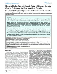
Figure 2.1: Morphological aspects of A: fibroblasts (Lysy, et al. 2007); B: RAW 264.7 macrophage (Cox, et al. 2009); C: A431 cells (Veldman, et al. 2005); and D: HepG2 cells (Reynaert, et al. 2004) that were used in the present study.
2.2.
The biological membrane
Membranes in cells typically define enclosed spaces or compartments in which cells may maintain a chemical or biochemical environment that differs from the outside. Biological membranes are asymmetric structures. Both the lipids and the proteins of membranes exhibit lateral (when lipids or proteins of particular types cluster in the plane of the membrane) and transversal asymmetries. In eukaryotic cells, also the lipid composition of the membranes of different organelles is heterogeneous. For example, the plasma membrane is highly enriched in cholesterol and glycosphingolipids, which are nearly absent from the endoplasmic reticulum (ER) (Prinz, 2002; Munro, 2003). In addition, there is a transbilayer lipid compositional asymmetry within the same membrane (Pomorski, et al. 2001), and in polarized cells, apical and basolateral membrane have different lipid and protein composition. Biological membranes and their components serve a number of essential cellular functions: They act as a selective barrier within or around a cell, where many reactions and processes occur; function as a platform for signal transduction; allow cell recognition; provide anchoring sites for cytoskeletal filaments or components of the extracellular matrix; compartmentalize cells; regulate the fusion of the membrane with other membranes in the cell and provide a passage way across the membrane for certain molecules.
9 The major components of all biological membranes are lipids, proteins and small amount of carbohydrates (as part of glycolipids and glycoproteins) of less than 10% of the mass of most membranes in variable proportion. Membranes proteins mediate and regulate transport of metabolites, macromolecules, and ions in and out of cells or subcellular organells (Shkulipa, 2006). The lipids of cell membranes play a crucial role in the function and properties of cell membranes. The membrane lipids consist of many different lipid species, classified according to head-group and backbone structures. These include glycerophospholipids, sphingolipids and cholesterol (Pomorski, et al. 2001; Edidin, et al. 2003; Holthuis, 2001; Fahy, et al. 2005). The carbohydrate moieties attached to some proteins and lipids are particularly abundant on the extracellular surface of the plasma membrane where they form the glycocalyx. The function of this layer is to prevent uncontrolled membrane fusion and to participate in recognition phenomena. In endothelial tissues, the glycocalyx serves to shield the vascular wall from the shear stresses of blood flow, impede leakage of blood constituents across the endothelial lining, and prevent adhesion of leucocytes and platelets to the endothelium (Rehm, et al. 2004). Many subcellular organelles in eukaryotes are surrounded by membranes (Voet, et al. 2011) such as nuclei, mitochondria, chloroplasts, endoplasmic reticulum, and Golgi apparatus. These organelles continually exchange biomolecules by a variety of membrane trafficking mechanisms (Sprong, et al. 2001; Mellman, 1996). In 1972, S.J. Singer, and G.L. Nicolson proposed the fluid mosaic model for membrane structure (Fig. 2.2), a widely accepted model of biological membranes. They suggested that membranes are dynamic structures composed of proteins and phospholipids. In this model, the phospholipids form a fluid bilayer (Singer, et al. 1972) in which the nonpolar regions of the lipid molecules in each layer face the core of the bilayer and their polar head groups face outward, interacting with the aqueous phase on either side. The hydrophobic nature of lipid molecules allows membranes to form spontaneously, and to act as effective barriers to polar molecules. The fluidity of the hydrocarbon core of the bilayer increases with increasing content of unsaturated or branched alkyl chains or with decreasing alkyl chain length. Membrane proteins can be embedded in the bilayer by hydrophobic interactions between the membrane lipids and hydrophobic domains of the proteins. Some proteins protrude from only one side of the membrane; others have domains exposed on both sides. The orientation of proteins in the bilayer is asymmetric, giving the membrane “sidedness”: the protein domains exposed on one side of the bilayer are different from those exposed on the other side, reflecting functional asymmetry. The individual lipid and protein units in a membrane form a
10 fluid mosaic because most of the interactions among its components are noncovalent, leaving individual lipid and protein molecules capable of rotational degrees of freedom and to move laterally in the plane of the membrane, but movement of either from one face of the bilayer to the other is restricted. Singer and Nicolson defined two classes of membrane proteins: peripheral (or extrinsic proteins) and integral proteins (or intrinsic proteins). The first includes those proteins that do not penetrate the bilayer to any significant degree and are associated with the membrane by virtue of electrostatic interactions and hydrogen bonds between the polar groups on the membrane surface and the surface of the protein. Furthermore, peripheral proteins can easily be dissociated from the membrane by treatment with salt solutions or by changes in pH. Integral proteins, in contrast, possess hydrophobic surfaces that can readily penetrate the matrix of the phospholipid bilayer itself as well as surfaces that prefer contact with the aqueous medium. In eukaryotic cells, almost all integral membrane proteins are synthesized on the surface of the rough ER (Gilmore, et al. 2012). Integral membrane proteins are strongly associated with bilayer lipids that influence specific function of certain membrane proteins. Because of these interactions, integral proteins can only be removed from the membrane by agents capable of breaking up the hydrophobic interactions within the lipid bilayer itself such as detergents and organic solvents.
Figure 2.2: The fluid-mosaic model of membrane structure proposed by (Singer, S.J. and Nicolson, G.L. 1972). In this model, a lipid bilayer is composed of phospholipids, cholesterol, glycolipids, and proteins. Peripheral proteins are embedded in either the outer or inner leaflet of the lipid bilayer, while integral proteins are firmly embedded in the lipid layers. Many of the proteins and lipids have externally exposed oligosaccharide side chains (Nelson, et al. 2005).
11
2.2.1. Lipid bilayers Amphipathic lipids spontaneously form a variety of supramolecular structures when added to an aqueous medium. They can aggregate into one of three forms: spherical micelles, liposomes, and sheetlike, two-molecule-thick bilayers (Fig. 2.3). All these structures form in ways that minimize the surface area in contact between the hydrophobic lipid chains and the aqueous milieu. For example, when small amounts of a fatty acid are mixed with an aqueous solution, a monolayer is formed at the air–water interface, with the polar head groups in contact with the water surface, shielding their hydrophobic tails from the water in contact with the air (Shkulipa, 2006). Micelles formed from an amphipathic lipid in water position the hydrophobic tails in the center of the lipid aggregate with the polar head groups facing outward. Micelles are the preferred form of aggregation in water for detergents and soaps. Phospholipids prefer to form bilayer structures in aqueous solution because their pairs of fatty acyl chains do not pack well in the interior of a micelle. Phospholipid bilayers form rapidly and spontaneously when phospholipids are added to water, and they are stable structures in aqueous solution. Extensive bilayers normally wrap around themselves and form closed vesicles. The nature and integrity of these vesicle structures are very much dependent on the lipid composition. Phospholipids can form either unilamellar vesicles (with a single lipid bilayer) known as liposomes, or multilamellar vesicles which are reminiscent of the layered structure of onions. The lipid bilayer thickness is about 3nm, 30Å thick (Heimburg, 2009), and is defined by the length, degree of saturation, and packing of the fatty acid chains. The thickness of bilayers is not a static number since thickness can vary over the surface of a membrane if microdomains of lipids are formed with different alkyl chain lengths. For example, addition of cholesterol or increasing chain length causes membrane thickening, whereas increased chain unsaturation or the strength of head group repulsions causes the bilayer to thin (Cantor, 1999). An important property of the lipid bilayer heterogeneity is that it contributes to membrane fluidity. The bilayer's fluidity allows lateral mobility within the lipid bilayer. It depends on the membrane phase and changes with the temperature. This fluidity is biologically important, influencing membrane transport. For example, the membranes of mammalian spermatozoa are composed of a complex mixture of lipids that provide the correct infrastructure and fluidity for the membrane mediated events that lead to fertilization (Ladha, 1998). Other roles for lipid diversity are the storage of precursors that can be metabolized to potent second messengers, e.g., diacylglycerol, ceramide, sphingosine, inositol trisphosphates
12 and eicosanoids. In addition, several phosphoinositides (phosphatidylinositol 3-phosphate (PI3P), phosphatidylinositol 4-phosphate (PI4P), phosphatidylinositol-4,5-biphosphate (PI45P2), phosphatidylinositol-3,4,5-triphosphate (PI345P3)) act as membrane recognition and attachment sites for protein complexes involved in protein traffic and membrane fusion events (Simonsen, et al. 2001; Barlow, et al. 2010).
2.2.2. Transmembrane lipid asymmetry The fluidity of the lipid bilayer of biological membranes has been established by biophysical studies. The lipids can rotate freely about their axis perpendicular to the plane of the membrane and diffuse readily within the lateral plane. Movement of polar lipids from one leaflet of the bilayer to the other is severely constrained and is measured in half times of hours or days. This constraint results from the requirement of free energy to move a hydrated polar moiety from the aqueous interface through the hydrocarbon interior of the structure. As a consequence of this restricted motion, an asymmetric distribution of lipids can be created and maintained across biological membranes. In many naturally occurring bilayers, the external monolayer of the mammalian cell membrane is made up almost exclusively of the neutral zwitterionic phospholipids phosphatidylcholine (PC), and sphingomyelin (SM), together with some phosphatidylethanolamine (PE). Phosphorylcholine is the most common head group accounting for about half of the phospholipids in most mammalian cells. In contrast, the internal monolayer contains anionic phospholipids as a major component which account for about 30% of cell phospholipids (Verkleij, et al. 1973; Buckland, et al. 2000; Chaurio, et al. 2009), mainly phosphatidylserine, phosphatidylethanolamine, and the phosphatidylinositols are much more abundant in the inner (cytoplasmic) leaflet (Chaurio, et al. 2009; Quinn, 2002). This is illustrated in figure 2.4, which shows the percentage distribution of the major lipid classes between the cytoplasmic and outer leaflet of the human erythrocyte membrane. Lipid asymmetry is maintained by translocases (Flippases) (Van Meer, et al. 2008). The aminophospholipid translocase is an ATPase II type enzyme that requires Mg2+ and specifically transports phosphatidylserine and phosphatidylethanolamine from the outer to the cytoplasmic leaflet of the membrane, while choline phosphatides are transported from the cytoplasmic to the outer leaflet.
13
Figure 2.3: Variety of structures of amphipathic lipid that aggregates in water. (a) In micelles, the hydrophobic fatty acid chains are sequestered at the core of the sphere with only small amounts of water in the hydrophobic interior. (b) In an open bilayer, all fatty acids acyl side chains are protected from interaction with water except those at the edges of the sheet. (c) When a bilayer folds on itself, it forms a closed bilayer (liposome) enclosing an aqueous cavity (Nelson, et al. 2005). Figure 2.4: In erythrocyte plasma membrane, percentage asymmetric distribution of the major phospholipids between the cytoplasmic and outer monolayers leaflets. This distribution is determined by treating the intact cell with phospholipase C, which removes the head groups of lipids in the outer monolayer, but cannot reach lipids in the inner monolayer (leaflet). In the outer monolayer, the proportion of each head group released provides an estimate of the fraction of each lipid (Nelson, et al. 2005).
2.2.3. Membrane phospholipids Lipids are a class of biological molecules defined by low solubility in water and high solubility in nonpolar solvents (Fahy, et al. 2005). The lipids found in biological systems are either hydrophobic or amphipathic. Phospholipids are the primary building blocks of most biological membranes. In eukaryotic cells, phospholipids are synthesized by enzymes located on the surface of the endoplasmic reticulum (ER). The membranes of mammalian cells contain more than 1,000 different phospholipid species (Vance, 2008). Phospholipids are abundant in all biological membranes and are derived from either glycerol or sphingosine, a long-chain unsaturated amino alcohol (Berg, et al. 2003). Phosphoglycerides (Fig. 2.6) consist of a glycerol backbone to which two residues (Fahy, et al. 2005), mostly fatty acids and a phosphorylated alcohol are attached in ester linkage. The fatty acid constituents are
14 usually even-numbered, most commonly of 16 or 18 carbons. Naturally occurring phospholipids contain a saturated fatty acid (such as stearic acid or palmitic acid) in position sn-1, whereas an unsaturated one (such as oleic acid, linoleic acid or arachidonic acid) in sn-2 position. The length and the degree of unsaturation of fatty acids in the membrane have an important effect on the fluidity (Chaurio, et al. 2009). The major phosphoglycerides arise from phosphatidate through the formation of an ester bond between the phosphate group of phosphatidate and the hydroxyl group of one of several alcohols. The simplest phosphoglyceride is phosphatidic acid, which is sn-1,2-diacylglycerol 3-phosphate, a key intermediate in the formation of all other phosphoglycerides. In other phosphoglycerides, the 3-phosphate is esterified to an alcohol such as ethanolamine, choline, serine, glycerol, or inositol (Fig. 2.5). The second major class of phospholipids is sphingomyelin, which contains a sphingosine backbone rather than glycerol. A fatty acid is attached by an amide linkage to the amino group of sphingosine, forming ceramide. The content of phospholipids also varies among organelles. For example, cardiolipin is a major constituent of the inner membrane of mitochondria, but is absent from other organelles (Van Meer, et al. 2008), whereas bis(monoacylglycero)phosphate is largely confined to late endosomes and lysosomes (Kolter, et al. 2010; Matsuo, et al. 2004; Kobayashi, et al. 2002). In mammalian cells, glycerophospholipids account for approximately 70% of the total membrane lipid content and thus play key roles in the structure and function of mammalian membranes; the other 30% consists of cholesterol, sphingomyelin, and glycosphingolipids (Leventis, et al. 2010). Among the phospholipids derived from glycerol, phosphatidylcholine (PC) is the most prevalent and accounts for 40-50% of the total phospholipids (Vance, 2008; Matsuo, et al. 2004). Of this amount, 76% is found in the outer monolayer, and 24% is found in the inner monolayer. Phosphatidylethanolamine (PE) is the next most abundant, which ranges from 20-45% of the total phospholipids, depending on the tissue (Vance, 2008; Murphy, et al. 2000), and is the major phospholipid in bacteria. Phosphatidylinositol (PI) (28% of the total PL), phosphatidylserine (PS) (2-10% of the total PL), phosphatidic acid (PA) (1% of the total PL), phosphatidylglycerol (PG) ( imipramine > chloroquine (Joshi, et al. 1988). The drugs may interact with either or both of the hydrophilic and lipophilic sites of dipalmitoyl PC. Chlorpromazine and imipramine showed two binding affinities of these drugs to dipalmitoyl PC, whereas chloroquine displayed a single binding affinity site to dipalmitoyl PC (Joshi, et al. 1988). In cultured macrophages, the incorporation of PC-derived palmitic acid into TAG was increased in cells pre-treated with 10µM desipramine (Fig. 4.27A). Minahk and co-worker (Minahk, et al. 2008) demonstrated that in cultured primary hepatocytes, 50% of LDL[3H]oleate-PC is converted to triacylglycerol via phospholipase C and DGAT2 rather than via lysosomal degradation. A recent study also demonstrated that PC plays a significant role to supply DAG for TAG synthesis via a PLC- mediated pathway (Robichaud, et al. 2009) , since desipramine has been demonstrated to stimulate PLC activity (Bouron, et al. 1999). Chinese hamster ovary cells can metabolize lipoprotein-associated PC to generate DAG by PLC that can be directly incorporated into TAG without prior formation of phosphatidic acid as the precursor (Igal, et al. 2001). Our results show that after 24h incubation of cultured macrophages with 40μM DMI, the total phospholipid labeling of DMI-treated macrophages was increase compared to the untreated cells (Fig. 4.27B). More specifically, a marked accumulation of label in PC, phosphatidic acid (PA) and lysoPC (LPC) occurred in a concentration-dependent manner in the DMI-treated macrophages. We also observed a significantly decrease in the incorporation into TAG by 39%, and a 56% increase into PA with 40µM DMI compared to control. Several cationic
amphiphilic
drugs
have
been
shown
to
inhibit
soluble
phosphatidate
phosphohydrolase in C6 glioma cells (Leli, et al. 1987), rat liver, and rat brain (Koul, et al. 1987). These data suggest that desipramine at 40µM may affect the activity of PA phosphohydrolase with increased formation of phosphatidyl-CMP, the intermediate for the synthesis of acidic phospholipids (F.g. 5.3). This may explain the marked increase in PA and decrease in DAG labeling, which are translated into decreased labeling of PE and TAG when treated with 40µM DMI (Fig. 4.27B). The cell viability in the presence of 40µM desipramine was reduced 28% compared to untreated cells (Fig. 4.38); the amount of cellular protein was reduced by 8%. Therefore, the reduced incorporation of FAs into TAG and DAG in the presence of 40µM desipramine in macrophages cannot be attributed only to toxicity.
105 In cells pre-treated with CPZ or DMI for 24h, levels of labeled PC were increased 3- and 2fold in the presence of 20µM CPZ or DMI, respectively. Furthermore, all drugs significantly reduced the amount of label in SM compared to the control cells and the experiments clearly show reduced processing of exogenously added PC with higher concentration of drugs. Some increase in FA was seen in macrophages treated with FTY720. In macrophages, the recovery of [14C]stearic acid derived from SM and [14C]palmitic acid derived from PC as free FAs was significantly higher in cells treated with 20µM FTY720 than in cells treated with other CADs. One hypothetical explanation is that FTY720 might cause an elevated intralysosomal pH and lead to a lower degree of protonation of the fatty acid, which, in turn, will cause the fatty acid to diffuse more slowly across the lysosomal membrane since the pKa values of FTY720 is lower (7.8) than those of the other CADs, which have pKa values of 10.4, 9.4, 9.2, and 8.1 for desipramine, imipramine, chlorpromazine, and chloroquine, respectively. Other possible explanation assumed that this might be caused by the formation of complexes between positively charged FTY720 and the negatively charged fatty acid, which become trapped in the lysosomes. Despite the difference in chain length of only two methylene groups, the saturated FAs present in our PC- and SM- probes should not necessarily undergo the same metabolic reaction. The metabolic fate of a saturated fatty acid derived from PC was significantly different from that derived from SM. For example, levels of palmitic acid derived from PC incorporated into TAG are increased in the presence of 10µM, and 20µM desipramine, while the opposite was observed for SM in the presence of desipramine. Furthermore, the incorporation of PC-derived [14C]palmitic acid into TAG decreased slightly, but not significantly in fibroblasts and A431cells pre-treated with chlorpromazine compared to controls, while there were no significant differences in the incorporation of PC-derived [14C]palmitic acid into TAG in HepG2 cells pre-treated with different concentrations of chlorpromazine and untreated control cells (data not shown). In general, CADs are lysosomotropic agents that accumulate in lysosomes, can lead to pH elevation of the acidic compartment, and to the mis-localization of soluble lysosomal enzymes (Nanoyams, et al. 2008). Some CADs induce an inhibition of phospholipid catabolism in lysosomes and increase cellular phospholipids in human and other mammalian tissues. Effects of CADs on steps downstream of lysosomal release of the fatty acid from the lipid probe depend on the applied CAD, on the identity of the fatty acid and the cell type, as indicated by previous experiments. Independent studies in cultured primary hepatocytes demonstrated that PC present in LDL is taken up not only by LDL receptors (30%) and
106 scavenger receptors (class B, type I) (20-30%), but also by additional unknown mechanisms that are responsible for the remaining (40-50%) (Minahk, et al. 2008). Therefore, CADs might impact only processing of that fraction of LDL-derived PC, which enters the acidic compartments and not on that fraction for which the interaction between LDL and its receptor proteins are irrelevant (Truong, et al. 2000). Ishikawa et al. showed that in smooth muscle cells only a fraction of LDL-PC was degraded in lysosomes. They found 25% of LDLderived PC in the lysosome-rich fraction and 25% in the cytosol-rich fraction. They concluded that LDL-derived PC was sorted equally between lysosomal and extralysosomal compartements (Ishikawa, et al. 1989) and also suggested that phospholipase A on plasma membranes, which can hydrolyze LDL-PC before LDL binds to LDL receptor, might affect the apoB epitope and the cellular uptake of LDL via the receptor. In addition, the authors found that phospholipases A1 and A2 are optimally active at neutral or alkaline pH ranges as well and might react with LDL-PC (Ishikawa, et al. 1988).
107
6. Material and Methods 6.1.
Material
6.1.1. Analytical equipment Autoclave
Systec V-150, Wettenberg, Germany
Blotter
Mini-TransBlot, BioRad, München, Germany
Centrifuges
Eppendorf, Hamburg, Germany L 8-80, mit SW-28-Ti-Rotor, Beckmann, Palo Alto, USA
Centrifuge tubes
12 mL: Costar, Cambridge, USA 15 mL: Greiner, Nürtingen, Germany 50 mL: Falcon/BectonDickinson, Bedford, USA
Glass tools
Schott-Duran, Jenaglas, Mainz, Germany
Incubator
Binder CO2, Tuttlingen, Germany
Microscope
Helmut Hund GmbH, Typ h500, Germany
pH-meter
pH 537, WTW, Weinheim, Germany
Phosphoimager
Fujix BAS 1000 Bio Imaging Analyzer, Raytest, Straubenhardt, Germany
Phosphoimaging plates
14
3
C-screen BAS MS 2040, H-Screen BAS Tr 2040, Raytest, Straubenhardt, Germany
Scintillation counter
Tri-Carb 2900TR, Packard Instruments Co., Inc, Downers Grove, USA
Shaker water bath
Gesellschaft für Labortechnik, Burgwedel, Germany
Sterile benches
LaminAirHA 2472 GS, Heraeus, Düsseldorf, Germany
TLC-tank
Desaga, Heidelberg, Germany
TLC-heater
TLC-Plate Heater III, Camag, Berlin, Germany
Ultrasound bath
Sonorex RK 100, Bandelin, Berlin, Germany
Heating-stirring module
Pierce, Therm III, Rockford
108 FLUO-STAR plate reader
BMG, Germany
Cell culture hood
Lamin Air, HB 2448, Heraeus, Hanau
Ultrasound
Cap horn, Sonifer B-12, Branson Ultrasonic Corp., Danbury, USA
Vortex
MS Minishaker, IKA-Werk, Staufen, Germany
Water purification
Millipore-Pelicon filtration device with polysulfone filter cassette, Millipore, Molsheim, France
Balance
Sartorius AG, Göttingen, Germany
Micropipette
Eppendorf
Pipette controllers
Brand, accu-jet, Wertheim, Germany
Oven
Memmert, Germany
Washer disinfectors for laboratory glassware
Miele G7783, Gütersloh, Germany
Multiskan photometer
Thermo scientific, Ascent, Vantaa, Finland
Water bath
GFI, Burgwedel, Germany 0
0
Refrigerator (4 C, -20 C)
Liebherr, Switzerland
6.1.2. Consumables and chemicals Centrifuge Falcon tube (15 mL, 50 mL)
Greiner Bio-one, Solingen
Safe-lock tube (0.5mL, 1.5mL, 2mL)
Eppendorf, Germany
Glass fiber wadding, silanized
Macherey-Nagel, Düren, Germany
Micropipette tips (20µL, 200µL, 1000µL)
Brand, Wertheim, Germany
Cell culture petri-dish
BD Falcon, USA
Cell culture flask
BD Falcon, USA
Vivaspin, 3.000 MWCO
Sartorius, Germany
96-microplates
Greiner Bio-one, Solingen
Stripette, serological pipettes
Sigma-aldrich, Germany
Pasteur pipette
VWR, Germany
Bovine serum albumin
Sigma, Taufenkirchen, Germany
Low density lipoprotein
Invitrogen
Lipoprotein deficient serum
Sigma, Germany
DEAE sephadex A-25
GE Healthcare, Uppsala, Sweden
Sepharose CL-2B
GE Healthcare, Uppsala, Sweden
109 Copper (II) sulfate solution
Sigma-Aldrich, Germany
Bicinchoninic acid solution
Sigma-Aldrich, Germany
LiChroprep® RP18 (40-63 μM)
Merck, Darmstadt, Germany
Lipid standards
Sigma, Taufkirchen, Germany
Screw cap glass
Pyrex, BibbySterlin Ltd, Stone, Great Britain
Sterile filters
Sartorius, Göttingen, Germany
Scintillation polyethylene vials (6mL, 20mL)
Perkin Elmer, USA
TLC plates (glass coated with silica gel 60)
Merck, Darmstadt, Germany
Tris-(hydroxymethyl)-aminomethan Hydrochloride (Tris-HCl)
Applichem, Darmstadt, Germany
Acetic acid 99.8%
BDH Prolabo, Darmstadt, Germany
Chloroform
Fisher chemical
Ethanol 100%
Merck
Methanol
J.T.Baker
Diethyl ether
BDH Prolabo, Darmstadt, Germany
n-Hexane
Merck
6.1.3. Radiolabeled markers [4-14C]Cholesterol
Perkin Elmer, Boston, USA
Cholesteryl [14C]-oleate
Perkin Elmer, Boston, USA
[14C]Trioleoylglycerol
Perkin Elmer, Boston, USA
[1-14C]-Stearic acid
GE Health Care, Amersham, UK
[1-14C]-Oleic acid
GE Health Care, Amersham, UK
[1-14C]-Linoleic acid
American Radio Labeled, USA
[1-14C]-Linolenic acid
Perkin Elmer, Boston, USA
Phosphatidyl ethanol amine, [1-14C-dioleoyl]
American Radio Labeled, USA
Phosphatidyl choline, [L-α-1-14Cdipalmitoyl]
Perkin Elmer, Boston, USA
110 Phosphatidyl choline, [1-14C-dioleoyl]
Perkin Elmer, Boston, USA
Sphingomyelin [Stearoyl-1-14C]
American Radio Labeled, USA
Desmethyl imipramine hydrochloride, [benzene ring, 10, 11-3H]
6.2.
Perkin Elmer, Boston, USA
Biological materials
6.2.1. Cells and additives Human skin fibroblasts
Kinderklinik St. Augustin, Germany
Mouse leukaemic monocyte macrophages, (Raw264.7)
DSMZ, Braunschweig, Germany
A431 cells
DSMZ,Braunschweig, Germany
Hep-G2 cells
DSMZ, Braunschweig, Germany
Niemann Pick A cells
Universitätsklinik, Münster, Germany
DMEM, RPMI
Gibco, Eggenstein, Germany
Foetal calf serum (FCS)
Cytogen, Berlin, Germany
Leupeptinhemi sulfate
Sigma, Taufkirchen, Germany
Streptomycin
Sigma, Deisenhofen, Germany
Trypsin
Cytogen, Berlin, Germany
N-Acetyl-L-Alanine-L-Glutamine
Seromed, Biochrom, Berlin, Germany
6.2.2. Kits CellTiter Blue (CTB) assay
6.3.
Promega, Germany
Methods
6.3.1. Cell culture Fibroblasts, macrophages, and Hep-G2 cells were grown in DMEM, A431 cells were grown in RPMI-1640, supplemented with 2mM glutamine, 10% (v/v) heat-inactivated fetal bovine serum, and 100mg/L streptomycin. Cells were collected from liquid nitrogen storage (appropriate protective equipment). The frozen screw-cap vials were thawed immediately in a water bath at 37 0C and the vials were wiped with 70% ethanol before transferring to the cell culture hood to reduce bacterial contamination. The cell suspensions in the vial were
111 transferred to a 15mL Falcon tube with 2mL FCS. The cells were centrifuged at 300g for 5min at room temperature. After the supernatant was discarded, 1mL 10% DMEM was added and mixed with the cells by pipetting. The cell mixture was transferred to 25cm2 flasks containing 1mL FCS and mixed. Growth medium (5 mL 10% DMEM) was added drop-wise and the flasks were incubated at 37 0C in a humidified atmosphere of 95% air and 5% CO2. After reaching a confluent state, the cells were sub-cultured 1:2 in 75cm2 culture flasks. For passaging of cells, the medium was removed and the cells were washed with PBS to remove the residual medium. To detach them from the flasks surface, the cells were incubated for 12min with 2mL trypsin/EDTA at 37 °C, upon gently shaking. 3mL DMEM containing 10% FBS was added to stop the digestion and pipetted to detach all cells from the flask wall. The cell suspension was transferred to a new Falcon tube and the cells were centrifuged at 300g for 5min, the supernatant was discarded, and the cells were re-suspended in growth medium. For our experiments, cells were grown until confluence in 35mm Petri-dishes or 25cm2 flasks and were seeded to a density of 1 x l06 cells/mL (average count per square*dilution factor*104). The medium was replaced with fresh culture medium every 2-3 days. Cells were examined using an inverted microscope to check confluence and confirm the absence of contaminations. All procedures with cells were carried out under sterile conditions using aseptic techniques.
6.3.2. Protein determination Cell protein was determined using the Bicinchoninic acid method with bovine serum albumin (BSA) as a standard (Smith et al., 1985). Prior to lipid extraction, cell pellets were homogenized 3 times (30sec) by ultrasonic treatment (120W) in 800μL of water, and aliquots were used for protein determination. 5μL of cell homogenate were diluted to 20μL with water in a multiwell plate (96-micro-plates). 200μL of freshly prepared reagent working solution (BicinChoninic Acid/copper (II) sulfate 50:1 by vol.) were added. After 30min incubation at 60 0C, the absorption of samples was measured at 562nm. Protein content was calculated on the basis of a standard curve, obtained by diluting BSA standard (0.25-5μg BSA/20μL).
6.3.3. Lipid extraction and analysis Cell pellets were suspended in 0.86mL water by ultrasonic treatment (120W; 3 times, 30sec). 60μL aliquots for protein determination and radioactivity measurement were issued, and the remainder was extracted by adding chloroform/methanol/water (1:2:0.8, v/v/v) to the cell homogenate. The mixture was vortexed and incubated for 12h at 40 °C in a water bath.
112 Insoluble cellular components were separated by filtration through wadding and the extract was dried under a stream of nitrogen. 1mL chloroform/methanol (1:1, v/v) was added to redissolve the dried lipids in a glass tubes, vortexed and sonicated by ultrasound bath for 5min. 10μL of cell suspension in 1mL solvent mixture were mixed with 10mL liquid scintillation cocktails in plastic scintillation vials to count the radioactivity in the scintillation counter. Incorporation of radioactivity into the lipid extract was determined using a Packard 2900 TR scintillation counter (Packard, Frankfurt) with a counter efficiency of 90-92% for 14C. Then, lipids were separated by thin layer chromatography (tlc).
6.3.4. Anion-exchange chromatography Ion exchange chromatography was carried out to separate acidic and neutral lipids according to differences in their net charge. This was done with an anion exchanger, Diethylaminoethyl (DEAE) Sephadex A-25, in which negatively charged ions bind to a positively charged resin. First, the matrix was converted from the chloride to the acetate form to obtain maximum affinity of acidic lipids. 500g of ion exchanger was first slurred with 1L methanol for 12h to allow the resin to swell. Then, it was suspended in 0.5L water and allowed to stand for 24h. Then the resin was transferred into a glass column and suspended in 0.5L 1M sodium acetate solution. To eliminate chloride ions, the resin had to be washed several times with 1M sodium acetate solution, until no chloride was detectable by silver nitrate test. Then the acetate form was washed first with 1L water and then with 1L methanol and was stored as 1:1 suspension in methanol at 4 °C before use. Small pieces of silanized glass fiber wadding were introduced into glass Pasteur pipettes and 1mL of DEAE-Sephadex A-25 (in acetate form)/methanol suspension (1:1) was added and equilibrated with 3mL of chloroform-methanol-water (3:7:1, v/v/v). Samples were dissolved in 2mL with the same solvent, sonicated for 5min and applied to the column. The column was rinsed with 6mL of the same solvent. The fractions that passed through the column contained the neutral sphingo and phospholipids. 8mL of chloroform-methanol-0.8M ammonium acetate (3:7:1, v/v/v) were applied to column to elute acidic lipids. The lipid fractions were evaporated to dryness under a nitrogen stream.
6.3.5. Alkaline hydrolysis Alkaline hydrolysis is used to remove phospholipids from lipid mixtures. Lipid samples were first dissolved in 1mL of chloroform-methanol (1:1, v/v), vortexed, and then the glass of the reaction vessel were rinsed with 1.5mL of the same solvent. After 5min ultrasonic treatment,
113 62μL of 4N NaOH were added, and further 5min ultrasound, then the solution was incubated for 2h at 37 °C in a water bath. After the reaction mixture was cooled and neutralized with 10μL glacial acetic acid, the lipid fractions were evaporated to dryness under a stream of nitrogen.
6.3.6. Reversed-phase chromatography (RP18) Desalting of lipid samples was performed according to the method described by (Williams and McCluer, 1980). In brief, small pieces of silanized glass fiber wadding were introduced into glass Pasteur pipettes and the Pasteur pipettes were clamped vertically. 1.5mL of LiChroprep RP-18 (40-63μm) (1:1) was added into the pipettes. The column was washed with solvents in the following order: 1mL methanol, 2mL water, 1mL methanol, 2mL of chloroform/methanol (1:1 (v/v)), 1mL methanol and 2mL of chloroform/methanol/0.1M KCl in water (3:48:47 (v/v/v)). After the washing process, lipid samples were dissolved in 1mL methanol, and sonicated for 5min. After further addition of 1mL ammonium acetate (300mM in water), the mixture was directly applied to the column. The glass vial was rinsed two times with 1mL ammonium acetate (200mM in methanol/water 1:1 (v/v)), and applied to the column. This was followed by washing with 6mL double distilled water to remove salt particles from the sample. 400µL methanol was added to the column. Glass tubes were placed under each column for lipid collection. Lipids were eluted with 600µL methanol followed by 8mL of chloroform/methanol (1:1 (v/v)). After the elution, the lipid fractions were evaporated to dryness under a stream of nitrogen.
6.3.7. Thin-layer chromatography The dried lipids in the glass tubes were re-dissolved with 1mL chloroform/methanol (1:1, v/v), vortexed and sonicated by ultrasound bath for 5min. An aliquot of the re-dissolved lipid was then transferred to corresponding safe-lock tubes (Eppendorf), and evaporated to dryness under a stream of nitrogen. The dried lipids inside the safe-lock tubes were re-dissolved in 40μL chloroform-methanol (1:1, v/v) and applied to silica gel 60-precoated glass plates with glass capillaries. Each of the safe-lock tubes was rinsed with 25μL of the same solvent and again applied on the TLC plates. The TLC-plates were dried in an evacuated desiccator over sodium hydroxide. The chromatography tank was lined with filter paper and filled with 200mL of mobile phase using a one-dimensional system. For the separation of lipids, two developing systems were used:
114 To separate non-polar lipids, polar lipids and free FAs, the following solvent systems were used as the mobile phase: Neutral lipids and free FAs:
n-hexane/diethyl ether/acetic acid 70:30:1 (v/v/v)
Phospholipids and sphingolipids:
chloroform/methanol/water 65:25:4 (v/v/v)
Lipid classes were assigned to visualized spots by co-chromatography with authentic standard substances (Schürer, et al. 1993). For monitoring and visualizing a radiolabeled lipid, the plate was exposed on a
14
C-Screen for 2-4 days. The radiolabeled spots were
visualized using a phosphoimager (FUJIX BAS 1000, Raytest, Straubenhardt, Germany). Lipid spots were quantified using Tina software. Standard lipids were applied to the TLC plates along with the radiolabeled lipids, and the standards were visualized by immersing the area of the plate containing the standards spots in CuSO4*5H2O/H3PO4 (85%)/H2O (15.6:9.4:75 (w/v/v)) Yao and Rastetter, 1985 followed by heating on a heater plate.
6.3.8. Feeding with Lipid-BSA complexes Stock solutions of 100nmol radiolabeled lipids in 300μL were complexed with FA-free bovine serum albumin (BSA; 7 mg) under vigorous stirring (Pütz and Schwarzmann, 1995). The lipid-BSA or FA-BSA complexes were diluted to a final concentration of 1μM for CEBSA, Chol-BSA, and TAG-BSA, and to 10μM for FA-BSA complexes in the culture medium (DMEM in the case of fibroblasts, macrophages, and Hep-G2 cells; and RPMI medium in the case of A431 cells). The molar ratio of lipid to albumin was kept at 1:1. The cells were seeded in 25cm2 plastic flasks with lipid-BSA complexes or 35mm Petri-dish with FA-BSA complexes. 1mL of radiolabeled lipid-BSA complexes were added to the cells for various incubation times as follows: CE-BSA, TAG-BSA, and Chol-BSA
1h, 6h, and 24h.
SA-BSA, OA-BSA, LA-BSA, LOA-BSA 15min, 30min, and 120min. The cells were rinsed two times with 1mL PBS, and harvested after trypsinization by a trypsin/EDTA solution. All procedures with cells were carried out under sterile conditions. All solutions were prepared freshly prior to the experiments. All experiments were conducted in duplicate and repeated two times.
6.3.9. Feeding with lipids-LDL complexes Lipid-LDL complexes were prepared according to a modified procedure based on (Brown S., and Goldstein L. 1975). The probe (100nmol) was incorporated into LDL by a solvent
115 exchange technique (Howard, 1979). The dried lipid was dissolved in 30µL of MeOH, vortexed, sonicated by ultrasound bath for 5min, and added to 4mL of LDL (750µg) in TrisBuffer. The resulting suspension was stirred for 4h at 37 0C in a water bath, and then filtered through a 3.000 MWCO Vivaspin filter (Howard, 1979). 30µL of the lipid-LDL suspension were taken before and after the filtration for scintillation counting and protein determination. The lipid-LDL complexes in suspension buffer were diluted to a final concentration with medium to yield 1μM for lipid-LDL complexes in medium containing 0.8% lipoprotein deficient serum (LPDS) in DMEM (0.8% LPDS in RPMI for A431cells). After adding 1mL of radiolabeled lipid-LDL complexes for various incubation times (1h, 6h, and 24h) to the medium, the cells were rinsed two times with 1mL PBS, and harvested after trypsinization by trypsin/EDTA solution. Although the lipid-LDL complex suspension appeared to be stable for more than a week, all lipid-LDL complexes were prepared freshly prior to the experiments.
6.3.10. Feeding with cationic amphiphilic drugs (CADs) Fibroblasts, macrophages, and HepG2 cells were maintained in dulbecco´s modified eagle medium (DMEM), A431 cells were maintained in roswell park memorial institute (RPMI) medium, cultured in 25cm2 culture flasks, and supplemented with 0.8% lipoprotein deficient serum. CADs (0, 10, 20, and 40µM), were added to the medium for 24h before the addition of the complexes. The lipid-LDL complexes were added to the medium and incubated for another 24h so that the drug was still present. Cells were maintained in an incubator containing 95% nitrogen and 5% CO2. Control cells, cultured at the same time, were treated under the same conditions. After 24h the cells were harvested and the lipids were extracted using chloroform/methanol (1:2, v/v). 60µL aliquots of harvested cells were used for protein determination and counting. Radiolabeled polar and non-polar lipids were quantified after lipid extraction and separation by tlc using the following mobile phases: First, in nhexane/diethylether/acetic acid (70:30:1, v/v/v) to separate non-polar lipids and free fatty acid, and chloroform/methanol/water (65:25:4, v/v/v) to separate polar lipids. Then, the separated lipids were visualized by phosphoimaging. Radioactivity of each lipid class was calculated from the distribution and the total radioactivity applied to each lane. Radioactivities in the homogenates were measured in a liquid scintillation counter. Lipid uptake was measured and the levels of the most prominent metabolites that did arise from incorporation of fatty acid derived from the exogenously added lipids into different lipid
116 classes were determined. A 24h incubation period was chosen for more detailed evaluation of the effects of the exogenously added lipids, because the changes during this period were more readily distinguishable.
6.3.11. Cell viability assay In order to evaluate if the addition of inhibitors affected the cells viability or induced stress resulting in apoptosis of the cells, cell viability was measured using the CellTiter Blue (CTB) assay (Evans, et al. 2001, Brien, et al. 2000), following the manufacturer's instruction (Promega). The assay is based on the ability of living cells to convert the dye resazurin into the fluorescent product resorufin. Nonviable cells rapidly lose metabolic capacity, do not reduce the indicator dye, and thus do not generate a fluorescent signal. Therefore, the fluorescence produced is proportional to the number of viable cells. In brief, cells were cultured in 100μL DMEM medium per well in 96-well microplates (10,000 cells/well) supplemented with 5% fetal calf serum and 2mM L-glutamine. Cells were grown in the presence of 0µM (control), 10µM, 20µM, and 40μM CADs for 48h. Medium was removed, and 100µL CTB medium (85µL fresh medium and 15µL CTB reagent) were added directly to the cells of each well. After 90min incubation at 37 0C, the fluorescence was determined at excitation/emission wavelengths of 544/590nm using a FLUO-STAR plate reader (BMG, Germany). For each compound, the average value of the duplicate samples was calculated. Data were processed using an Excel spreadsheet. The cell viability ranged from 70 to 112 ± 20% in the presence of inhibitors.
6.4.
Control experiments
The following experiments were used as a control.
6.4.1. Gel-filtration chromatography Gel-filteration was carried out for separation non-bound lipids from lipid incorporate into lipoproteins by passing them through Sephacryl S-300. The column was packed with Sephacryl S-300 and equilibrated with 10mM Tris-HCl (pH 7.5) containing 0.3mM NaCl, and 0.3mM EDTA as a running buffer, and the column was eluted with the same buffer. The lipid-LDL complexes were loaded to the column, and the lipoproteins were eluted with Trisbuffer. Then, 2mL fractions were collected.
117
6.4.2. Pulse-chase experiment Radiolabeled oleic acid was tracked in selected cells (fibroblasts, macrophages, A431 cells, and Hep-G2 cells) to determine the fate of incorporated radiolabeled fatty acids at various incubation times. Cells were incubated at different temperatures, either at 37 0C, or at 4 0C (which blocks the endocytosis pathways). After 2h incubation time, the radiolabeled fatty acids were removed immediately from the culture medium. The cultured cells were rinsed three times with 1mL PBS to remove unincorporated radiolabeled FA. Fresh 10% FCS in DMEM were added, and the cells were incubated again in chase medium at 37 °C. The medium was removed with five interval chase time points (24, 48, 72, 120, and 144h). At each time point, the cells were collected, washed with 1mL PBS, then trypsinized and transfered to a screw cup glass. After centrifuge for 5min, washed again with 1mL PBS. Centrifuge the cells again at 1000rpm for 5min, the pellet were store at –20°C until all time points have been completed.
6.4.3. Feeding with radiolabeled desipramine [3H]-Desipramine was used as a control to determine its uptake by different cell types. [3H]Desipramine (0.45nmol; 40µL in ethanol) was pipetted into Falcon tubes, dried to half contents under a stream of nitrogen, and then diluted with 12mL DMEM solution (containing different concentrations of unlabeled desipramine) to yield a 0.1µM [3H]-desipramine in 1.5mL medium. After incubation of the cells with 1.5mL of various concentrations of [ 3H]desipramine (10, 20, and 40µM) for 48h incubation time, the cells were washed 3 times with 1mL PBS. After addition of 1mL trypsin/EDTA, the cells were incubated for 1-2min at 37 °C. Cells were carefully detached by gently tapping and the cell suspensions were transferred into a screw cap glass. The vials were washed with 1mL PBS, and the cell suspension was collected into a screw cap glass. The cells were centrifuged at 1000rpm for 5min, and washed again with 1mL PBS. After centrifugation of the cells at 1000rpm for 5min, the pellets were stored at –20 °C until use. Cell pellets were suspended in 0.86mL water and by ultrasonic treatment (120W; 3 times, 30sec). The amounts of protein and the radiolabeled [3H]desipramine taken up were determined.
118
7. References Abdul-Hammed, M.; Breiden, B.; Adebayo, M.A.; Babalola, J.O.; Schwarzmann, G.; Sandhoff, K. (2010) The roles of endosomal membrane lipids and NPC2 in cholesterol transfer and membrane fusion. J. Lipid Res., 51: 1747-1760. Aboulaich, N.; Vainonen, J.P.; Stralfors, P.; Vener, A.V. (2004) Vectorial proteomics reveal targeting, phosphorylation and specific fragmentation of polymerase I and transcript release factor (PTRF) at the surface of caveolae in human adipocytes. Biochem. J., 383: 237–248. Aden, D.P.; Fogel, A.; Plotkin, S.; Damjanov I.; Knowles, B.B. (1979) Controlled synthesis of HBsAG in a differentiated human liver carcinomca-derived cell line. Nature, 282: 615-616. Aderem, A.; Underhill, D.M. (1999) Mechanisms of phagocytosis in macrophages. Annu. Rev. Immunol., 17: 593–623. Albouz, S.; Hauw, J.J.; Berwaldnetter, Y.; Boutry, J.M.; Bourdon, R.; Baumann, N. (1981) Tricyclic anti-depressants induce sphingomyelinase deficiency in fibroblast and neuro-blastoma cell-cultures. Biomed. Express., 35: 218-220. Anderson, N.; Borlak, J. (2006) Drug-induced phospholipidosis. FEBS Letter, 580: 5533-5540. Anderson, R.G. (1998) The caveolae membrane system. Annu. Rev. Biochem.,67: 199–225. Arikketh, D.; Nelson, R.; Vance, J.E. (2008) Defining the important of phosphatidylserine synthase-1 (PSS1). J. Biol. Chem., 283: 12888-12897. Aviram, M.; Bierman, E.L.; Chait, A. (1988) Modification of low density lipoprotein by lipoprotein lipase or hepatic lipase induces enhanced uptake and cholesterol accumulation in cells. J. Biol. Chem., 263: 15416-15422. Baillie, A.G.; Coburn, C.T.; Abumrad, N.A. (1996) Reversible binding of long-chain fatty acids to purified FAT, the adipose CD36 homolog. J. Mem. Biol., 153: 75-81. Bajjalieh, S.; Batchelor, R. (2000) Ceramide kinase. Methods Enzymol., 311: 207-215. Baluška, F.; Hlavacka, A.; Samaj, J.; Palme, K.; Robinson, D.G.; Matoh, T.; McCurdy, D.; Menzel, D.; Volkmann, D. (2002) F-actindependent endocytosis of cell wall pectins in meristematic root cells. insights from brefeldin A-induced compartments. Plant Physiol., 130: 422–431. Barlow, C.A.; Laishram, R.S.; Anderson, R.A. (2010) Nuclear phosphoinositides: a signaling enigma wrapped in a compartmental conundrum. Trends Cell Biol., 20: 25-35. Bastie, C.C.; Hajri, T.; Drover, V.A.; Grimaldi, P.A.; Abumrad, N.A. (2004) CD36 in myocytes channels fatty acids to a lipase-accessible triglyceride pool that is related to cell lipid and insulin responsiveness. Diabetes, 53: 2209-2216. Beck, M. (2010) Therapy for lysosomal storage disorders. IUBMB Life, 62: 33–40. Bendedouch, D.; Chen, S.H. (1983) Structure and interparticle interactions of bovine serum albumin in solution studied by small angle neutron scattering. J. Phys. Chem., 87: 1473-1477. Berdyshev, E.V.; Gorshkova, I.; Skobeleva, A.; Bittman, R.; Lu, X.; Dudek, S.M.; Mirzapoiazova, T.; Garcia, J.G.; Natarajan, V. (2009) FTY720 inhibits ceramide synthases and up-regulates
119 dihydrosphingosine 1-phosphate formation in human lung endothelial cells. J. Biol. Chem., 284: 5467-5477. Berger, M.; Wetzler, E.; August, T.; Tartakoff, A.M. (1994) Internalization of type I complement receptors and de novo multivesicular body formation during chemoattractant-induced endocytosis in human neutrophils. J. Clin. Invest., 94: 1113-1125. Berg, J.M.; Tymoczko, J.L.; Strayer, L. (2003) Biochemistry. 5th ed.; Spektrum, Akad. Verl.: Berlin, Germany. Berg, T.; Tolleshaug, H. (1980) The effects of ammonium ions and chloroquine on uptake and degradation of 125I-labeled asialo-fetuin in isolated rat hepatocytes. Biochem. Pharmacol., 29: 917925. Bernlohr, D.A.; Jenkins, A.E.; Bennaars, A.A. (2002) Adipose tissue and lipid metabolism. In: Biochemistry of Lipids, Lipoproteins and Membranes, Eds. D.E. Vance and J.E. Vance, 4th ed., Elsevier, Amsterdam, pp. 263-289. Bhattacharya, A.A.; Grüne, T.; Curry, S. (2000) Crystallographic analysis reveals common modes of binding of medium and long-chain fatty acids to human serum albumin. J. Mol. Biol., 303: 721–732.
Bishop-Bailey, D. (2011) PPARs and angiogenesis. Biochem. Soc. Trans., 39: 1601-1605. Blom, T.; Back, N.; Mutka, A.L.; Bittman, R.; Li, Z.; De Lera, A.; Kovanen, P.T.; Diczfalusy, U.; Ikonen, E. (2010) FTY720 stimulates 27-hydroxycholesterol production and confers atheroprotective effects in human primary macrophages. Circ. Res., 106: 720-729. Bojesen, I.N.; Bojesen, E. (1994) Binding of arachidonate and oleate to bovine serum albumin. J. Lipid Res., 35: 770-778. Botham, K.M.; Moore, E.H.; De Pascale, C.; Bejta, F. (2007) The induction of macrophage foam cell formation by chylomicron remnants. Biochem. Soc. Trans., 35: 454–458. Bouron, A., Chatton, J.Y. (1999) Acute application of the tricyclic antidepressant desipramine presynaptically stimulates the exocytosis of glutamate in the hippocampus. Neuroscience., 90: 729-36. Brien, J.O.; Wilson, I.; Orton, T.; Pognan, F. (2000) Investigation of the alamar blue (resazurin) fluorescent dye for the assessment of mammalian cell cytotoxicity. Eur. J. Biochem., 267: 5421-5426. Brindley, D.N. (1991) Metabolism of triacylglycerols. In: Biochemistry of Lipids, Lipoproteins and Membranes, Eds. D.E. Vance and J.E. Vance, Elsevier, New York, pp. 171–203. Briscoe, C.P.; Tadayyon, M.; Andrews, J.L.; Benson, W.G.; Chambers, J.K.; Eilert, M.M.; Ellis, C.; Elshourbagy, N.A.; Goetz, A.S.; Minnick, D.T.; Murdock, P.R.; Sauls, H.R.; Shabon, U.; Spinage, L.D.; Strum, J.C.; Szekeres, P.G.; Tan, K.B.; Way, J.M.; Ignar, D.M.; Wilson, S.; Muir, A.I. (2003) The orphan G protein-coupled receptor GPR40 is activated by medium and long chain fatty acids. J. Biol. Chem., 278: 11303-11311. Brousseau, M.; Schaefer, E.; Wolfe, M.; Bloedon, L.; Digenio, A.; Clark, R.W.; Mancusu, J.P.; Rader, D.J. (2004) Effects of an inhibitor of cholesteryl ester transfer protein on HDL cholesterol. N Engl. J. Med., 350: 1505–1515. Brown, M.S.; Faust, J.; Goldstein, J.L. (1975) Role of the low density lipoprotein receptor in regulating the content of free and esterified cholesterol in human fibroblasts. J. Clin. Invest., 55: 783793
120 Brown, M.S.; Goldstein, J.L. (1999) A proteolytic pathway that controls the cholesterol content of membranes, cells, and blood. Proc. Natl. Acad. Sci., 96: 11041–11048. Brown, M.S.; Goldstein, J.L. (1986) A receptor-mediated pathway for cholesterol homeostasis. Science, 232: 34–47. Brown, M.S.; Goldstein, J.L. (2004) A tribute to Akira Endo, discoverer of a “Penicillin” for cholesterol. Atherosclerosis Suppl., 5: 13–16. Brown, M.S.; Goldstein, J.L.; Krieger, M.; Ho, Y.K.; Anderson, R.G. (1979) Cholesteryl ester in macrophages incubated with acetylated lipoproteins. J. Cell. Biol., 82: 597-613. Brown, M.S.; Goldstein, J.L. (1975) Regulation of the activity of the low density lipoprotein receptor in human fibroblasts. Cell, 6: 307–3l6. Brown, S.; Dana, E.; Goldstein, L. (1975) Receptor-dependent hydrolysis of cholesteryl esters contained in plasma low density lipoprotein. Proc. Nat. Acad. Sci., 72: 2925-2929. Brunzell, J.D.; Davidson, M.; Furberg, C.D.; Goldberg, R.B.; Howard, B.V.; Stein, J.H.; Witztum, J.L. (2008) Lipoprotein management in patients with cardiometabolic risk. J. Am. Coll. Cardiol., 51: 1512-1524. Buckland, A.G.; Wilton, D.C. (2000) Anionic phospholipids, interfacial binding and the regulation of cell functions. Biochim. Biophys. Acta, 1483: 199-216. Burack, W.R.; Biltonen, R.L. (1994) Lipid bilayer heterogeneities and modulation of phospholipase A2 activity. Chem. Phys. lipids, 73: 209-222. Calder, P.C.; Bond, J.A.; Harvey, D.J.; Gordon, S.; Newsholme, E.A. (1990) Uptake and incorporation of saturated and unsaturated fatty acids into macrophage lipids and their effect upon macrophage adhesion and phagocytosis. Biochem. J., 269: 807-814. Calder, P.C. (2011) Fatty acids and inflammation: the cutting edge between food and pharma. Eur. J. pharmacol., 668: 50-58. Calder, P.C. (2010) Omega-3 fatty acids and inflammatory processes. Nutrients, 2: 355-374. Cantor, R.S. (1999) lipid composition and the lateral pressure profile in bilayers. Biophys. J., 76: 2625-2639. Carter, D.C.; He, X.; Munson, S.H.; Twigg, P.D.; Gernert, K.M.; Broom, M.B.; Miller, T.Y. (1989) Three-dimensional structure of human serum albumin. Science, 244: 1195–1198. Carter, D.C.; Ho, J.X. (1994) Structure of serum albumin. Adv. Protein Chem., 45: 153–203. Cevikbas, F.; Seeliger, S.; Fastrich, M.; Hinte, H.; Metze, D.; Kempkes, C.; Homey, B.; Steinhoff, M. (2011) Role of protease-activated receptors in human skin fibrosis and scleroderma. Exp. Dermatol., 20: 69-71. Chaiyasut, C.; Tsuda, T. (2001) Isoelectric points estimation of proteins by electroosmotic flow: PH relationship using physically adsorbed proteins on silica gel. Chromatography, 22: 91-95. Charbonneau, D.M.; Tajmir-Riahi, H.A. (2010) Study on the interaction of cationic lipids with bovine serum albumin. J. Phys. Chem. B., 114: 1148–1155. Chatelain, P.; brotelle, R. (1985) Phospholipid composition of rat lung after amiodarone treatment. Res. Commun. Chem. Pathol. Pharmacol., 50: 407-418.
121 Chaurio, R.A.; Janko, C.; Muñoz, L.E.; Frey, B.; Herrmann, M.; Gaipl, U.S. (2009) Phospholipids: key players in apoptosis and immune regulation. Molecules, 14: 4892-4914. Cheng, D.; Iqbal, J.; Devenny, J.; Chu, C.H.; Chen, L.; Dong, J.; Seethala, R.; Kelm, W.J.; Azzara, A.V.; Lawrence, R.M.; Pelleymounter, M.A.; Hussain, M.M. (2008) Acylation of acylglycerols by acyl coenzyme A: Diacylglycerol acyltransferase 1 (DGAT1). J. Biol. Chem., 283: 29802-29811. Chen, S.; Gjerde, A.U.; Holmsen, H.; Nerdal, W. (2005) Importance of polyunsaturated acyl chains in chlorpromazine interaction with phosphatidylserines: a 13C and 31P solid-state NMR study. Biophys.chem., 117: 101-109. Choi, J.K.; Ho, J.; Curry, S.; Qin, D.; Bittman, R.; Hamilton, J.A. (2002) Interactions of very longchain saturated fatty acids with serum albumin. J. Lipid Res., 43: 1000–1010. Chun, J.; Hartung, H.P. (2010) Mechanism of action of oral fingolimod (FTY720) in multiple sclerosis. Clin. Neuropharm., 33: 91-101. Connelly, M.A.; Klein, S.M.; Azhar, S.; Abumrad, N.A.; Williams, D.L. (1999) Comparison of class B scavenger receptors, CD36 and scavenger receptor BI (SR-BI), shows that both receptors mediate high density lipoprotein-cholesteryl ester selective uptake but SR-BI exhibits a unique enhancement of cholesteryl ester uptake. J. Biol. Chem., 274: 41-47. Cox, C.; Cao, S.; Lu, Y. (2009) Enhanced detection and study of murine noroviruse-I using a more efficient microglial cell line. Virol. J., 6: 196-202. Damyanova, R.; Markovska, T.; Mitev, V.; Staneva, G.; Petkova, D. (2010) Uptake of fatty acids by fibroblasts cultured in three-dimensional matrix. Comp. Rend. Acad. Bulg. Sci., 63: 999-1002. Daniel, L.W.; Kucera, L.S.; Waite, M. (1980) Metabolism of fatty acids by cultured tumor cells and their diploid precursor fibroblasts. J. Biol. Chem., 255: 5697-5702. Dawson, G.; Qin, J.; (2011) Gilenya (FTY720) inhibits acid sphingomyelinase by a mechanism similar to tricyclic antidepressants. Biochem. Biophys. Res. Commun., 404: 321-323. Deevska, G.M.; Nikolova-Karakashian, M.N. (2011) The twists and turns of sphingolipid pathway in glucose regulation. Biochim., 93: 32-38. Deevska, G.M.; Rozenova, K.A.; Giltiay, N.V.; Chambers, M.A.; White, J.; Boyanovsky, B.B.; Wei, J.; Daugherty, A.; Smart, E.J.; Reid, M.B.; Merrill, A.H.; Nikolova-Karakashian, M. (2009) Acid sphingomyelinase deficiency prevents diet-induced hepatic triacylglycerol accumulation and hyperglycemia in mice. J. Biol. Chem., 284: 8359-8368. De Filippi, L.; Fournier, M.; Cameroni, E.; Linder, P.; De Virgilio, C.; Foti, M.; Deloche, O. (2007) Membrane stress is coupled to a rapid translational control of gene expression in chlorpromazinetreated cells. Curr. Genet., 52: 171-185. Demant, E.J.F.; Richieri, G.V.; Kleinfeld, A.M. (2002) Stopped-floe kinetic analysis of long-chain fatty acid dissociation from bovine serum albumin. Biochem. J., 363: 809-815. Deng, W.; Bukiya, A.N.; Rodríguez-Menchaca, A.A.; Zhang, Z.; Baumgarten, C.M.; Logothetis, D.E.; Levitan, I.; Rosenhouse-Dantsker, A. (2012) Hypercholesterolemia induces up-regulation of KACh cardiac currents via a mechanism independent of phosphatidylinositol 4,5-bisphosphate and Gβγ. J. Biol. Chem., 287: 4925-4935.
122 Derksen, J.T.; Morselt, H.W.; Scherphof, G.L. (1987) Processing of different liposome markers after in vitro uptake of immunoglobulin-coated liposomes by rat liver macrophages. Biochim. Biophys. Acta., 931: 33-40. Diegelmann, R.F.; Evans, M.C. (2004) Wound healing: an overview of acute, fibrotic and delayed healing. Front Biosci., 9: 283–289. Dixon, J.L.; Ginsberg, H.N. (1993) Regulation of hepatic secretion of apolipoprotein B-containing lipoproteins: information obtained from cultured liver cells. J. Lipid Res., 34: 167-79. Dubois, B.W.; Cherian, S.F.; Evers, A.S. (1993) Volatile anesthetics compete for common binding sites on bovine serum albumin: A 19F-NMR study. Pharmacol., 90: 6478-6482. Doherty, G.J.; McMahon, H.T. (2009) Mechanisms of Endocytosis. Annu. Rev. Biochem., 78: 857– 902. Doherty, P.; Benedek, G.B. (1974) The effect of electric charge on the diffusion of macromolecules. J. Chem. Phys., 61: 5426-5435. Dokko, R.C.; Cho, B.H.; Chung, B.H. (1998) Cellular uptake of stearic, oleic, linoleic, and linolenic acid and their effects on synthesis and secretion of lipids in Hep-G2 cells. Inter. J. Biochem. Cell Biol., 30: 65-76. Downes, C.E.; MacPhee, C.H. (1990) Myo-inositol metabolites as cellular signals. Eur. J. Biochem., 193: 1-18. Dixon, J.L.; Furukawa, S.; Ginsberg, H.N. (1991) Oleate stimulates secretion of apolipoprotein Bcontaining lipoproteins from Hep-G2 cells by inhibiting early intracellular degradation of apolipoprotein B. J. Biol. Chem., 266: 5080-5086. Duttaroy, A.K. (2009) Transport of fatty acids across the human placenta: a review. Prog. lipid Res., 48: 52-61. Du, X.; Kumar, J.; Ferguson, C.; Schulz, T.A.; Ong, Y.S.; Hong, W.; Prinz, W.A.; Parton, R.G.; Brown, A.J.; Yang, H. (2011) A role for oxysterol-binding protein–related protein 5 in endosomal cholesterol trafficking. J. Cell biol., 192: 121-135. Edidin, M. (2003) Lipids on the frontier: a century of cell-membrane bilayers. Nat. Rev. Mol. Cell Biol., 4: 414-418. ElBawab, S.; Mao, C.; Obeid, L.M.; Hannun, Y.A. (2002) Ceramidases in the regulation of ceramide levels and function. Subcell. Biochem., 36: 187–205. Elojeimy, S.; Holman, D.H.; Liu, X.; El-Zawahry, A.; Villani, M.; Cheng, J.C.; Mahdy, A.; Zeidan, Y.; Bielwaska, A.; Hannun, Y.A.; Norris, J.S. (2006) New insights on the use of desipramine as an inhibitor for acid ceramidase. FEBS letters, 580: 4751-4756. Evans, S.M.; Casartelli, A.; Herreros, E.; Minnick, D.T.; Day, C.; George, E.; Westmorelan, C. (2001) Development of a high throughput in vitro toxicity screen predictive of high acute in vivo toxic potential. Toxicol. In Vitro., 15: 579–584. Fahy, E.; Subramaniam, S.; Brown, H.A.; Glass, C.K.; Merrill, A.H.; Murphy, R.C.; Raetz, C.R.H.; Russell, D.W.; Seyama, Y.; Shaw, W.; Shimizu, T.; Spener, F.; van Meer, G.; VanNieuwenhze, M.S.; White, S.H.; Witztum, J.L.; Dennis, E.A. (2005) A comprehensive classification system for lipids. J. Lipid Res., 46: 839-862.
123 Falcone, S.; Cocucci, E.; Podini, P.; Kirchhausen, T.; Clementi, E.; Meldolesi, J. (2006) Macropinocytosis: regulated coordination of endocytic and exocytic membrane traffic events. J. Cell Sci., 119: 4758–69. Ferlinz, K.; Hurwitz, R.; Sandhoff, K. (1991) Molecular basis of acid sphingomyelinase deficiency in a patient with Niemann-Pick disease type A. Biochem. Biophys. Res. Commun., 179: 1187–1191. Fielding, P.E.; Fielding, C.E. (2002) Dynamics of lipoprotein transport in the human circulatory system. In: Biochemistry of Lipids, Lipoproteins and Membranes, Eds. D.E. Vance and J.E. Vance, 4th ed., Elsevier, Amsterdam, pp. 527–552. Frank, P.; Lisanti, M. (2004) Caveolin-1 and caveolae in atherosclerosis: differential roles in fatty streak formation and neointimal hyperplasia. Curr. Opin. Lipidol.,15: 523–9. Fritsche, K. (2006) Fatty acids as modulators of the immune response. Annu. Rev. Nut., 26: 45-73. Fromenty, B.; Freneaux, E.; Labbe, G.; Deschamps, D.; Larrey, D.; Letteron, P.; Pessayre, D. (1989) Tianeptine, a new tricyclic antidepressant metabolized by beta-oxidation of its heptanoic side chain, inhibits the mitochondrial oxidation of medium and short chain fatty acids in mice. Biochem. Pharmacol., 38: 3743-3751. Fujimoto, T.; Ohsaki, Y.; Cheng, J.; Suzuki, M.; Shinohara, Y. (2008) Lipid droplets: a classic organelle with new outfits. Histochem. Cell Biol., 130: 263-279. Fujimoto, Y.; Onoduka, J.; Homma, K.J.; Yamaguchi, S.; Mori, M.; Higashi, Y.; Makita, M.; Kinoshita, T.; Noda, J.; Itabe, H.; Takanoa, T. (2006) Long-chain fatty acids induce lipid droplet formation in a cultured human hepatocyte in a manner dependent of Acyl-CoA synthetase. Biol. Pharm. Bulletin., 29: 2174-2180. Futerman, A.H.; van Meer, G. (2004) The cell biology of lysosomal storage disorders. Nat. Rev. Mol. Cell Biol., 5: 554-65. Garbarino, J.; Padamsee, M.; Wilcox, L.; Oelkers, P.M.; D’Ambrosio, D.; Ruggles, K.V.; Ramsey, N.; Jabado, O.; Turkish, A.; Sturley, S.L. (2009) Sterol and diacylglycerol acyltransferase deficiency triggers fatty acidmediated cell death. J. Biol. Chem., 284: 30994–31005. Giacomelli, C.E.; Norde, W. (2001) The adsorption-desorption cycle. Reversibility of the BSA-silica system. J. Colloid Interface Sci., 233: 234-240. Giard, D.J.; Aaronson, S.A.; Todaro, G.J.; Arnstein, P.; Kersey, J.H.; Dosik, H.; Parks, W.P. (1973) Invitro cultivation of human tumors: establishment of cell lines derived from a series of solid tumors. J. Natl. Cancer Inst., 51: 1417-1423. Gibellini, F.; Smith, T.K. (2010) The kenndy pathway-De phosphatidylethanolamine and phophatidylcholine. IUBMB, 62: 414-428.
novo
synthesis
of
Gilmore, R.; Mandon, E.C. (2012) Understanding integration of α-helical membrane proteins: the next steps. Trends Biochem. Sci., 37: 303-308. Gimeno, R.E.; Ortegon, A.M.; Patel, S.; Punreddy, S.; Ge, P.; Sun, Y.; Lodish, H.F.; Stahl, A. (2003) Characterization of a heart-specific fatty acid transport protein. J. Biol. Chem., 278: 16039–16044. Gjerde, A.U.; Holmsen, H.; Nerdal, W. (2004) Chlorpromazine interaction with phosphatidylserines: a (13)C and (31)P solid-state NMR study. Biochim. Biophys. Acta, 1682: 28-37. Glatz, J.F.; Luiken, J.J.; Bonen, A. (2010) Membrane fatty acid transporters as regulators of lipid metabolism: implications for metabolic disease. Physiol. Rev., 90: 367–417.
124 Goldstein, J.L.; Brown, M.S. (1987) Regulation of low-density lipoprotein receptors: implication for pathogenesis and therapy of hypercholesterolemia and atherosclerosis. Circulation., 76: 504-507. Goldstein, J.L.; Brown, M.S. (2009) The LDL receptor. Arterioscler. Thromb. Vasc. Biol., 29: 431438. Goldstein, J.L.; Brunschede, G.Y.; Brown, M.S. (1975) Inhibition of proteolytic degradation of low density lipoprotein in human fibroblasts by chloroquine, concanavalin A, and Triton WR 1339. J. Biol. Chem., 250: 7854-7862. Goni, F.M.; Alonso, A. (2002) Sphingomyelinases: enzymology and membrane activity. FEBS Letters, 531: 38-46. Goodman, J.M. (2008) The gregarious lipid droplet. J. Biol. Chem., 283: 28005-28009. Granot, E.; Tabas, I.; Tall, A.R. (1987) Human plasma cholesteryl ester transfer protein enhances the transfer of cholesteryl ester from high density lipoproteins into cultured HepG2 cells. J. Biol. Chem., 262: 3482-3487. Groener, J.E.M.; Bax, W.; Poorthuis, B.J. (1996) Metabolic fate of oleic acid derived from lysosomal degradation of cholesteryl oleate in human fibroblasts. J. Lipid Res., 37: 2271-2279. Gulbins, E. (2003) Regulation of death receptor signaling and apoptosis by ceramide. Pharmacol. Res., 47: 393-399. Haberland, M.E.; Fogelman, A.M. (1985) Scavenger receptor-mediated recognition of maleyl bovine plasma albumin and the demaleylated protein in human monocyte macrophages. Proc. Natl. Acad. Sci. USA., 82: 2693-2697. Hagberg, C.E.; Falkevall, A.; Wang, X.; Larsson, E.; Huusko, J.; Nilsson, I.; van Meeteren, L.A.; Samen, E.; Lu, L.; Vanwildemeersch, M.; Klar, J.; Genove, G.; Pietras, K.; Stone-Elander, S.; Claesson-Welsh, L.; Yla-Herttuala, S.; Lindahl, P.; Eriksson, U. (2010) Vascular endothelial growth factor B controls endothelial fatty acid uptake. Nature, 464: 917-921. Hanada, K. (2006) Discovery of the molecular machinery CERT for endoplasmic reticulum-to-Golgi trafficking of ceramide. Mol. Cell Biochem., 286: 23-31. Hanada, K. (2003) Serine palmitoyltransferase, a key enzyme of sphingolipid metabolism. Biochim Biophys Acta., 1682:16-30. Hamilton, J.A. (2007) New insights into the roles of proteins and lipids in membrane transport of fatty acids. Prostaglandins Leukot. Essent. Fatty Acids, 77: 355–361. Hartley, J.W.; Evans, L.H.; Green K.Y.; Naghashfar, Z.; Macias, A.R.; Zerfas, P.M.; Ward, J.M. (2008) Expression of infectious murine leukemia viruses by RAW264.7 cells, a potential complication for studies with a widely used mouse macrophage cell line. Retrovirol., 5: 1-6. Hatch, G.M. (1994) Cardiolipin biosynthesis in the isolated heart. Biochem. J., 297: 201–208. Haunerland, N.H.; Spener, F. (2004) Fatty acid-binding proteins--insights from genetic manipulations. Prog. lipid Res., 43: 328-349. Heimburg, T. (2009) Physical properties of biological membranes. In: Handbook of Molecular Biophysics. Ed. H.G. Bohr. Wiley-VCH, pp. 593-616. Heravi, J.; Waite, M. (1999) Transacylase formation of bis(monoacylglycerol)phosphate. Biochim. Biophys. Acta, 1437: 277-286.
125
Hess, D.; Chisholm, J.W.; Igal, R.A. (2010) Inhibition of stearoylCoA desaturase activity blocks cell cycle progression and induces programmed cell death in lung cancer cells. PloS one, 5: e11394. Hevonoja, T.; Pentikäinen, M.O.; Hyvönen, M.T.; Kovanen, P.T.; Ala-Korpela, M. (2000) Structure of low density lipoprotein (LDL) particles: Basis for understanding molecular changes in modified LDL. Biochim. Biophys. Acta, 1488: 189-210. Holland, W.L.; Summers, S.A. (2008) Sphingolipids, insulin resistance, and metabolic disease: new insights from in vivo manipulation of sphingolipid metabolism. Endocrine Rev., 29: 381-402. Holthuis, J.C.; Pomorski, T.; Raggers, R.J.; Sprong, H.; Van Meer, G. (2001) The organizing potential of sphingolipids in intracellular membrane transport. Physiol. Rev., 81: 1689–1723. Horinouchi, K.; Erlich, S.; Perl, D.P.; Ferlinz, K.; Bisgaier, C.L.; Sandhoff, K. (1995) Acid sphingomyelinase deficient mice: a model of type A and b Niemann-Pick disease. Nat. Genet., 10: 288-293. Horton, J.D.; Goldstein, J.L.; Brown, M.S. (2002) SREBPs: activators of the complete program of cholesterol and fatty acid synthesis in the liver. J. Clin. Invest., 109: 1125–31. Hostetler, K.Y.; Reasor, M.; Yazaki, P.J. (1985) Chloroquine-induced phospholipid fatty liver. Measurement of drug and lipid concentrations in rat liver lysosomes. J. Biol. Chem., 260: 215-219. Howard, B.V. (1979) Uptake of very low density lipoprotein triglyceride by bovine aortic endothelial cells in culture. J. Lipid Res., 18: 561-571. Huang, C.J.; Cheng, H.H.; Chou, C.T.; Kuo, C.C.; Lu, Y.C.; Tseng, L.L.; Chu, S.T.; Hsu, S.S.; Wang, J.L.; Lin, K.L.; Chen, I.S.; Liu, S.I.; Jan, C.R. (2007) Desipramine-induced Ca2+ movement and cytotoxicity in PC3 human prostate cancer cells. Toxicol. In Vitro, 21: 449-456. Huang, W.C.; Chen, C.L.; Lin, Y.S.; Lin, C.F. (2011) Apoptosis sphingolipid ceramide in cancer therapy. J. Lipids, 2011: 1-15. Hunter, J.E. (2006) Dietary trans fatty acids: review of recent human studies and food industry responses. Lipids, 41: 967-992. Hurwitz, R.; Ferlinz, K.; Sandhoff, K. (1994) The tricyclic antidepressant desipramine causes proteolytic degradation of lysosomal sphingomyelinase in human fibroblasts. Biol. Chem. HoppeSeyler, 375: 447-450. Ibrahimi, A.; Sfeir, Z.; Magharaie, H.; Amri, E.Z.; Grimaldi, P.; Abumrad, N.A. (1996) Expression of the CD36 homolog (FAT) in fibroblast cells: effects on fatty acid transport, Proc. Nat. Acad. Sci. U.S.A., 93: 2646-2651. Ichimura, A.; Hirasawa, A. Poulain-Godefroy, O.; Bonnefond, A.; Hara, T.; Yengo, L.; Kimura, I.; Leloire, A.; Liu, N.; Iida, K.; Choquet, H.; Besnard, P.; Lecoeur, C.; Vivequin, S.; Ayukawa, K.; Takeuchi, M.; Ozawa, K.; Tauber, M.; Maffeis, C.; Morandi, A.; Buzzetti, R.; Elliott, P.; Pouta, A.; Jarvelin, M.R.; Korner, A.; Kiess, W.; Pigeyre, M.; Caiazzo, R.; Van Hul, W.; Van Gaal, L.; Horber, F.; Balkau, B.; Levy-Marchal, C.; Rouskas, K.; Kouvatsi, A.; Hebebrand, J.; Hinney, A.; Scherag, A.; Pattou, F.; Meyre, D.; Koshimizu, T.A.; Wolowczuk, I.; Tsujimoto, G.; Froguel, P. (2012) Dysfunction of lipid sensor GPR120 leads to obesity in both mouse and human. Nature, 483: 350354. Ikushiro, H.; Hayashi, H. (2011) Mechanistic enzymology of serine palmitoyltransferase. Biochim. Biophys. Acta., 1814: 1474-1480.
126
Imokawa, G.; Yada, Y.; Higuchi, K.; Okuda, M.; Ohashi, Y.; Kawamata, A. (1994) Pseudoacylceramide with linoleic acid produces selective recovery of diminished cutaneous barrier function in essential fatty acid-deficient rats and has an inhibitory effect on epidermal hyperplasia. J. Clin. Invest., 94: 89-96. Ishikawa, Y.; Nishide, T.; Sasaki, N.; Shirai, K.; Saito, Y.; Yoshida, S. (1989) Effects of chloroquine on the metabolism of phosphatidylcholine associated with low density lipoprotein in arterial smooth muscle cells. Atherosclerosis, 80: 1-7. Ishikawa, Y.; Nishide, T.; Sasaki, N.; Shirai, K.; Saito, Y.; Yoshida, S. (1988) Hydrolysis of lowdensity lipoprotein phosphohpids in arterial smooth muscle cells. Biochim. Biophys. Acta., 961: 170176. Isola, L.M.; Zhou, S.L.; Kiang, C.L.; Stump, D.D.; Bradbury, M.W.; Berk, P.D. (1995) 3T3 fibroblasts transfected with a cDNA for mitochondrial aspartate aminotransferase express plasma membrane fatty acid-binding protein and saturable fatty acid uptake, Proc. Nat. Acad. Sci. U.S.A., 92: 9866-9870. Jachimska, B.; Wasilewska, M.; Adamczyk, Z. (2008) Characterization of globular protein solutions by dynamic light scattering, electrophoretic mobility, and viscosity measurements. Am. Chem. Soc., 24: 6866-6872. Jacobson, T.A.; Miller, M.; Schaefer, E.J. (2007) Hypertriglyceridemia and cardiovascular risk reduction. Clin. Therap., 29: 763-777. Jaffrezou, J.P.; Laurent, G., Levade, T. (2002) Ceramide in regulation of apoptosis. Implication in multitoxicant resistance. Subcell. Biochem., 36: 269–284. Jan, C.R.; Lu, Y.C.; Tseng, L.L.; Jiann, B.P.; Chang, H.T.; Wang, J.L.; Chen, W.C.; Huang, J.K. (2003) Effect of the antidepressant desipramine on cytosolic Ca(2+) movement and proliferation in human osteosarcoma cells. Pharmacol., 69: 190-196. Jansen, M.; Ohsaki, Y.; Rega, L.R.; Bittman, R.; Olkkonen, V.M.; Ikonen, E. (2011) Role of ORPs in sterol transport from plasma membrane to ER and lipid droplets in mammalian cells. Traffic, 12: 218– 231. Jauhiainen, M.; Metso, J.; Pahiman, R.; Blomqvist, S.; Tol, A.V.; Ehnhom, C. (1993) Human plasma phospholipid transfer protein causes high density lipoprotein conversion. J. Biol. Chem., 269: 40324036. Javitt, N.B. (1990) Hep-G2 cells as a resource for metabolic studies: lipoprotein, cholesterol, and bile acids. FASEB J., 4: 161-8. Jensen, J.M.; Schutze, S.; Forl, M.; Kronke, M.; Proksch, E. (1999) Roles for tumor necrosis factor receptor p55 and sphingomyelinase in repairing the cutaneous permeability barrier. J. Clin. Inves., 104: 1761-1770. Jeyakumar, M.; Williams, I.; Smith, D.; Cox, T.M.; Platt, F.M. (2009) Critical role of iron in the pathogenesis of the murine gangliosidoses. Neurobiol Dis., 34: 406–416. Johnsen, G.M.; Weedon-Fekjaer, M.S.; Tobin, K.A.; Staff, A.C.; Duttaroy, A.K. (2009) Long-chain polyunsaturated fatty acids stimulate cellular fatty acid uptake in human placental choriocarcinoma (BeWo) cells. Placenta, 30: 1037-1044.
127
Jones, A.T. (2007) Macropinocytosis: searching for an endocytic identity and role in the uptake of cell penetrating peptides. J. Cell Mol. Med., 11: 670-84. Joshi, U.M.; Kodavanti, P.R.; Coudert, B.; Dwyer, T.M.; Mehendale, H.M. (1988) Types of interaction of amphiphilic drugs with phospholipid vesicles. J. Pharmacol. Exp. Therap., 246: 150157. Jump, D.B. (2004) Fatty acid regulation of gene transcription. Crit. Rev. Clin. Lab. Sci., 41: 41-78. Kamata, N.; Chida, K.; Rikimaru, K.; Horikoshi, M.; Enomoto, S.; Duroki, R. (1986) Growthinhibitory effects of epidermal growth factor and overexpression of its receptors on human squamous cell carcinomas in culture. Cancer Res., 46: 1648-1653. Kasahara, T.; Tomita, K.; Murano, H.; Harada, T.; Tsubakimoto, K.; Ogihara, T.; Ohnishi, S.; Kakinuma, C. (2006) Establishment of an in vitro high-throughput screening assay for detecting phospholipidosis-inducing potential, Toxicol. Sci., 90: 133-141. Kawamoto, T.; Sate, J.D.; Le. A.; Polikoff, J.; Sato, G.H.; Mendelsohn, J. (1983) Growth stimulation of A431 cells by epidermal growth factor: Identification of high-affinity receptors for epidermal growth factor by an anti-receptor monoclonal antibody. Proc. Natl. Acad. Sci., 80: 1337-1341. Khoo, J.C.; Vance, J.E.; Mahoney, E.M.; Jensen, D.; Wancewicz, E.; Steinberg. D. (1984) Neutral triglyceride lipase in macrophages. Arteriosclerosis, 4: 34-40. Kleinman, Y.; Krul E.S.; Burnes, M.; Aronson, W.; Pfleger, B.; Schonfeld, G. (1988) Lipolysis of LDL with phospholipase A2 alters the expression of selected apoB-100 epitopes and the interaction of LDL with cells. J. lipid Res., 29: 729-743. Kliewer, S.A.; Sundeth, S.S.; Jones, S.A. (1997) Fatty acids and eicosanoids regulate gene expression through direct interactions with peroxisome proliferator-activated receptors α and β. Proc. Natl. Acad. Sci. USA., 94: 4318–4323. Kobayashi, T.; Beuchat, M.; Chevallier, J.; Makino, A.; Mayran, N.; Escola, J.M.; Lebrand, C.; Cosson, P.; Kobayashi, T.; Gruenberg, J. (2002) Separation and characterization of late endosomal membrane domains. J. Biol. Chem., 277: 32157–32164. Kobayashi, T.; Startchev, K.; Whitney, A.J.; Gruenberg, J. (2001) Localization of lysobisphosphatidic acid-rich membrane domains in late endosomes. Biol. Chem., 382: 483–485. Kobuna, H.; Inoue, T.; Shibata, M.; Gengyo-Ando, K.; Yamamoto, A.; Mitani, S.; Arai, H. (2010) Multivesicular body formation requires OSBP–related proteins and cholesterol. PLoS Genet., 6: e1001055. Kodavanti, U.P.; Mehendale, H.M. (1990) Cationic amphiphilic drugs and phospholipid storage disorder. Pharmacol. Rev., 42: 327-354. Kölzer, M.; Werth, N.; Sandhoff, K. (2004) Interactions of acid sphingomyelinase and lipid bilayers in the presence of the tricyclic antidepressant desipramine. FEBS letters, 559: 96-98. Kolter, T.; Sandhoff, K. (2010) Lysosomal degradation of membrane lipids. FEBS Letter, 584: 17001712. Kolter, T.; Sandhoff, K. (2005) Principles of lysosomal membrane digestion: stimulation of sphingolipid degradation by sphingolipid activator proteins and anionic lysosomal lipids. Annu. Rev. Cell Dev. Biol., 21: 81–103.
128
Kolter, T.; Sandhoff, K. (1999) Sphingolipids - their metabolic pathways and the pathobiochemistry of neurodegenerative diseases. Angew. Chem. Int. Ed. 38: 1532-1568. Kondo, A.; Oku, S.; Higashitani, K. (1991) Structural changes in protein molecules adsorbed on ultrafine silica particles. J. Colloid Interface Sci., 143: 214-221. Kornhuber, J.; Tripal, P.; Reichel, M.; Muhle, C.; Rhein, C.; Muehlbacher, M.; Groemer, T.W.; Gulbins, E. (2010) Functional Inhibitors of Acid Sphingomyelinase (FIASMAs): a novel pharmacological group of drugs with broad clinical applications. Cell. Physiol. Biochem., 26: 9-20. Koul, O.; Hauser, G. (1987) Modulation of rat brain cytosolic phosphatidate phosphohydrolase: effect of cationic amphiphilic drugs and divalent cations. Arch. Biochem. Biophys., 253: 453-461. Knowles, B.B.; Howe, C.; Aden, P. (1980) Human hepatocellular carcinoma cell lines secrete the major plasma proteins and hepatitis B surface antigen. Science, 209: 497-499. Kobilka, B.K.; Lefkowitz, R.J. (2012) Studies of G-protein-coupled receptors. The Royal Swedish Academy of Sciences, Nobel Prize in Chemistry. pp.1-15. Kozarsky, K.F.; Donahee, M.H.; Rigotti, A.; Iqbal, S.N.; Edelman E.R., Krieger M. (1997) Overexpression of the HDL receptor SR-BI alters plasma HDL and bile cholesterol levels. Nature, 387: 414–417. Kuehnel, M.P.; Goethe, R.; Habermann, A.; Mueller, E.; Rohde, M.; Griffiths, G.; Valentin-Weigand, P. (2001) Characterization of the intracellular survival of mycobacterium avium ssp. Paratuberculosis: phagosomal pH and fusogenicity in J774 macrophages compared with other mycobacteria. Cell. Microbiol., 3: 551-566. Kuehnel, W. (2003) Color Atlas of Cytology, Histology, and Microscopic Anatomy (4th ed.). Thieme. pp. 34. ISBN 1-58890-175-0. Kwak, S.M.; Myung, S.K.; Lee, Y.J.; Seo, H.G. (2012) Efficacy of omega-3 fatty acid supplements (eicosapentaenoic acid and docosahexaenoic acid) in the secondary prevention of cardiovascular disease. Arch. Intern. Med., 172: 686-694. Ladha, S. (1998) Lipid heterogeneity and membrane fluidity in a highly polarized cell, the mammalian spermatozoon. J. Membrane Biol., 165: 1-10. LaLonde, J.M.; Bernlohr, D.A.; Banaszak, L.J. (1994) The up-and-down beta-barrel proteins. FASEB J., 8: 1240-1247. Lands, W.E. (1958) Metabolism of glycerolipides: A comparison of lecithin and triglyceride synthesis. J. Biol. Chem., 231: 883–888. Lansmann, S.; Schuette, C.G.; Bartelsen, O.; Hoernschemeyer, J.; Linke, T.; Weisgerber, J.; Sandhoff, K. (2003) Human acid sphingomyelinase, assignment of the disulfide bond pattern. Eur. J. Biochem., 270: 1076-1088. Le Dinh, T.; Freneaux, E.; Labbe, G.; Letteron, P.; Degott, C.; Geneve, J.; Berson, A.; Larrey, D.; Pessayre, D. (1988) Amineptine, a tricyclic antidepressant, inhibits the mitochondrial oxidation of fatty acids and produces microvesicular steatosis of the liver in mice. J. Pharmacol. Exp. Ther., 247: 745-750. Lee, A.G. (2003) Lipid-protein interactions in biological membranes: a structural perspective. Biochim. Biophys. Acta., 1612: 1-40.
129 LeLay, S.; Dugail, I. (2009) Connecting lipid droplet biology and the metabolic syndrome. Prog. Lipid Res., 48: 191-195. Leli, U.; Hauser, G. (1987) Modifications of phospholipid-metabolism induced by chlorpromazine, desmethylimipramine and propranolol in C6 glioma-cells. Biochem. Pharmacol., 36: 31-37. Lemaitre, G.; Gonnet, F.; Vaigot, P.; Gidrol, X.; Martin, M.T.; Tortajada, J.; Waksman, G. (2005) CD98, a novel marker of transient amplifying human keratinocytes. Proteomics, 5: 3637–3645. Leventhal, A.R.; Chen, W.; Tall, A.R.; Tabas, I. (2001) Acid sphingomyelinase deficient macrophages have defective cholesterol trafficking and efflux. J. Biol. Chem., 276: 44976 – 44983. Leventhal, A.R.; Leslie, C.C.; Tabas, I. (2004) Suppression of macrophage eicosanoid synthesis by atherogenic lipoproteins is profoundly affected by cholesterol-fatty acyl esterification and the Niemann-Pick C pathway of lipid trafficking. J. Biol. Chem. 279: 8084-8092. Leventis, P.A.; Grinstein, S. (2010) The distribution and function of phosphatidylserine in cellular membranes. Annu. Rev. Biophys., 39: 407–427. Lim, J.P.; Gleeson, P.A. (2011) Macropinocytosis: an endocytic pathway for internalising large gulps. Immunol. Cell Biol., 89: 836-843. Lin, Y.; Smit, M.J.; Havinga, R.; Verkade, H.J.; Vonk, R.J.; Kuipers, F. (1995) Differential effects of eicosapentaenoic acid on glycerolipid and apolipoprotein B metabolism in primary human hepatocytes compared to HepG2 cells and primary rat hepatocytes. Biochim. Biophys. Acta., 1256: 88-96. Li, Q.M.; Fuhrman, B.; Aviram. M. (1995) Low density lipoprotein-cholesteryl ester-derived linoleic acid is mainly incorporated into the phospholipid component in macrophages. Isr. J. Med. Sci., 31: 474-478. Listenberger, L.L.; Han, X.; Lewis, S.E.; Cases, S.; Farese, R.V.; Ory, D.S.; Schaffer, J.E. (2003) Triglyceride accumulation protects against fatty acid-induced lipotoxicity. Proc. Nat. Acad. Sci. U.S.A., 100: 3077-3082. Lönnfors, M.; Doux, J.P.F.; Killian, J.A.; Nyholm, T.K.M.; Slotte, J.P. (2011) Sterols have higher affinity for sphingomyelin than for phosphatidylcholine bilayers even at equal acyl-chain order. Biophys. J., 100: 2633–2641. Lopez-Montero, I.; Rodriquez, N.; Cribier, S.; Pohl, A.; Velez, M.; Devaux, P.F. (2005) Rapid transbilayer movement of ceramides in phospholipid vesicales and in human erythrocytes. J. Biol. Chem., 280: 25811-25819. Lucas, A.D.; Greaves, D.R. (2001) Atherosclerosis: role of chemokines and macrophages. Expert. Rev. Mol. Med., 3: 1–18. Lucas, L.H.; Price K.E.; Larive, C.K. (2004) Epitope mapping and competitive binding of HSA drug site II ligands by NMR diffusion measurements. J. Am. Chem. Soc., 126: 14258–14266. Lüllmann-Rauch, R.; Scheid, D. (1975) Intra alveolar foam cells associated with lipidosis-like alterations in lung and liver of rats treated with tricyclic psychotropic drugs. Virchows Arch. B Cell path., 19: 255-268. Lüllmann, H.; Lüllmann-Rauch, R.; Wassermann, O. (1978) Lipidosis induced by amphiphilic cationic drugs. Biochem. Pharmacol., 27: 1103-1108.
130 Lysy, P.A.; Smets, F.; Sibille, C.; Najimi, M.; Sokal, E.M. (2007) Human skin fibroblasts: from mesodermal to hepatocyte-like differentiation. Hepatol., 46: 1574-1585. Macierzanka, A.; Szelag, H.; Moschakis, t.; Murray, B.S. (2006) Phase transitions and microstructure of emulsion system prepared with acylglycerols/zinc stearate emulsifier. Langmuir, 22: 2487-2497. Ma, J.; Qiu, Y.; Yang, L.; Peng, L.; Xia, Z.; Hou, L.N.; Fang, C.; Qi, H.; Chen, H.Z. (2011) Desipramine induces apoptosis in rat glioma cells via endoplasmic reticulum stress-dependent CHOP pathway. J. Neurooncol., 101: 41-48. Mandon, E. C.; Ehses, I.; Rother, J.; Van Echten, G.; Sandhoff, K. (1992) Subcellular localization and membrane topology of serine palmitoyltransferase, 3-dehydrosphinganine reductase, and sphinganine N-acyltransferase in mouse liver. J. Biol. Chem., 267: 11144-11148. Matsuo, H.; Chevallier, J.; Mayran, N.; Le Blanc, I.; Ferguson, C.; Faure´, J.; Blanc, N.S.; Matil, S.; Dubochet, J.; Sadoul, R.; Parton, R.G.; Vilbois, F.; Grunenberg, J. (2004) Role of LBPA and Alix in multivesicular liposome formation and endosome organization. Science, 303: 531–34. Matsuzawa, Y.; Hostetler, K.Y. (1980) Inhibition of lysosomal phospholipase A and phospholipase C by chloroquine and 4,4'-bis(diethylaminoethoxy) alpha, beta-diethyldiphenylethane. J. Biol. Chem., 255: 5190-5194. Mayes, P.A.; Botham, K.M. (2003) Cholesterol synthesis, transport, and extraction. In: Murray, R.K.; Granner, D.K.; Mayes, P.A.; Rodwell, V.W. Harper’s Illustratted biochemistry. 26th ed., Lange Medical Books/McGraw-Hill, U.S.A. Mayes, P.A.; Botham, K.M. (2003) Metabolism of acylglycerols and sphingolipids. In: Murray, R.K.; Granner, D.K.; Mayes, P.A.; Rodwell, V.W. Harper’s Illustratted biochemistry. 26th edition, Lange Medical Books/McGraw-Hill, U.S.A. Mayor, S.; Pagano, R.E. (2007) Pathways of clathrin-independent endocytosis. Mol. Cell Biol., 8: 603-612. Maziere, C.; Maziere, J.C.; Mora, L.; Polonovski, J. (1982) Incorporation of exogenous fatty-acids into phospholipids by cultured hamster fibroblasts - effect of Sv40 transformation. Biochim. Biophys. Acta., 712: 712-715. McArthur, M.J.; Atshaves, B.P.; Frolov, A.; Foxworth, W.D.; Kier, A.B.; Schroeder, F. (1999) Cellular uptake and intracellular trafficking of long chain fatty acids. J. Lipid Res., 40: 1371-1383. McNutt, M.C.; Lagace, T.A.; Horton, J.D. (2007) Catalytic activity is not required for secreted PCSK9 to reduce low density lipoprotein receptors in HepG2 cells. J. Biol. Chem., 282: 2079920803. Meikle, P.J.; Hopwood, J.J.; Clague, A.E.; Carey, W.F. (1999) Prevalence of lysosomal storage disorders. JAMA; 20: 249 – 254. Mellman, I. (1996) Membranes and sorting. Curr. Opin. Cell Biol., 8: 497-498. Merlino, G.T.; Xu, Y.H.; Ishii, S.; Clark, A.J.L.; Semba, K.; Toyoshima, K.; Yamamoto, T.; Pastan, I. (1984) Amplification and enhanced expression of the epidermal growth factor receptor gene in A431 human carcinoma cells. Science, 224: 417-419. Merrill, A.H.; Wang, E.; Mullins, R.E. (1988) Kinetics of long-chain (sphingoid) base biosynthesis in intact LM cells: effects of varying the extracellular concentrations of serine and fatty acid precursors of this pathway. Biochem., 27: 340-345.
131 Miles, P.R.; Bowman, L.; Tucker, J.; Reasor, M.J.; Wright, J.R. (1986) Alteration in rat alveolar surfactant phospholipids and proteins induced by adminstration of chlorphentermine. Biochim, Biophys. Acta, 877: 167-178. Miller, M.; Motevalli, M.; Westphal, D.; Kwiterovich, P.O. (1993) Incorporation of oleic acid and eicosapentaenoic acid into glycerolipids of cultured normal human fibroblasts. Lipids, 28: 1-5. Minahk, C.; Kim, K.W.; Nelson, R.; Trigatti, B.; Lehner, R.; Vance, D.E. (2008) Conversion of low density lipoprotein-associated phosphatidylcholine to triacylglycerol by primary hepatocytes. J. Biol. Chem., 283: 6449-6458. Möbius, W.; van Donselaar, E.; Ohno-Iwashita, Y.; Shimada, Y.; Heijnen, H.F.; Slot, J.W.; Geuze, H.J. (2003) Recycling compartments and the internalvesicles of multivesicular bodies harbor most of the cholesterol found in theendocytic pathway. Traffic, 4: 222–231.
Montagner, A.; Rando, G.; Degueurce, G.; Leuenberger, N.; Michalik, L.; Wahli, W. (2011) New insights into the role of PPARs. Prostaglandins Leukot. Essent. Fatty Acids, 85: 235– 243. Morishita, S.; Watanabe, S. (1997) Effect of the tricyclic antidepressant desipramine on protein kinase C in rat brain and rabbit platelets in vitro. Psychiatry Clin. Neurosci., 51: 249-252. Munro, S. (2003) Lipid rafts: elusive or illusive?. Cell, 115: 377-388. Murphy, E.J.; Prows, D.R.; Jefferson, J.R.; Schroeder, F. (1996) Liver fatty acid-binding protein expression in transfected fibroblasts stimulates fatty acid uptake and metabolism. Biochim. Biophys. Acta., 1301: 191-198. Murphy, E.J.; Schapiro, M.B.; Schapiro, M.B.; Shetty, H.U. (2000) Phospholipid composition and levels are altered in down syndrome brain. Brain Res., 867: 9–18. Nawa, M.; Takasaki, T.; Yamada, K.; Kurane, I.; Akatsuka, T. (2003) Interference in Japanese encephalitis virus infection of Vero cells by a cationic amphiphilic drug, chlorpromazine. J. Gen. Virol., 84: 1737-1741. Nelson, D.L.; Cox, M.M. (2005) Lehninger’s principles of biochemistry, 4th ed., W. H. Freeman and Company, New York. Nioi, P.; Pardo, I.D.; Snyder, R.D. (2008) Monitoring the accumulation of fluorescently labeled phospholipids in cell cultures provides an accurate screen for drugs that induce phospholipidosis, Drug Chem. Toxicol., 31: 515-528. Nonoyama, T.; Fukuda, R. (2008) Drug-induced phospholipidosis-pathological aspects and its prediction. J. Toxical. Pathol., 21: 9-24. Norde, W.; Favier, J.P. (1992) Structure of adsorbed and desorbed proteins, Colloids Surf., 64: 87-93. Oh, D.Y.; Talukdar, S.; Bae, E.J.; Imamura, T.; Morinaga, H.; Fan, W.; Li, P.; Lu, W.J.; Watkins, S.M.; Olefsky, J.M. (2010) GPR120 is an omega-3 fatty acid receptor mediating potent antiinflammatory and insulin-sensitizing effects. Cell, 142: 687-698. Okwu, A.K.; Xu, X.X.; Shiratori, Y.; Tabas, I. (1994) Regulation of the threshold for lipoproteininduced acyl-CoA:cholesterol 0-acyltransferase stimulation in macrophages by cellular sphingomyelin content. J. Lipid Res.. 35: 644-655.
132 Orso, E.; Grandl, M.; Schmitz, G. (2011) Oxidized LDL-induced endolysosomal phospholipidosis and enzymatically modified LDL-induced foam cell formation determine specific lipid species modulation in human macrophages. Chem. Phys. Lipids, 164: 479-487. Papp, A.C.; Pinsonneault, J.K., Wang, D.; Newman, L.C.; Gong, Y.; Johnson, J.A.; Pepine, C.J. Kumari, M.; Hingorani, A.D.; Talmud, P.J.; Shah, S.; Humphries, S.E.; Sadee, W. (2012) Cholesteryl ester transfer protein (CETP ) polymorphisms affect mRNA splicing, HDL levels, and sex-dependent cardiovascular risk. PLoS ONE, 7: e31930. Pappu, A.; Hostetler, K.Y. (1984) Effect of cationic amphiphilic drugs on the hydrolysis of acidic and neutral phospholipids by liver lysosomal phospholipase A. Biochem. Pharmacol., 33: 1639-1644. Pastores, G.M. (2006) Miglustat: substrate reduction therapy for lysosomal storage disorders associated with primary central nervous system involvement. Recent Pat. CNS Drug Discov., 1: 7782. Passeggio, J.; Liscum, L. (2005) Flux of Fatty Acids through NPC1 Lysosomes. J. Biol. Chem., 280: 10333-10339. Paugh, S.W.; Cassidy, M.P.; He, H.; Milstien, S.; Sim-Selley, L.J.; Spiegel, S.; Selley, D.E. (2006) Sphingosine and its analog, the immunosuppressant 2-amino-2-(2-[4-octylphenyl]ethyl)-1,3propanediol, interact with the CB1 cannabinoid receptor. Mol. Pharmacol., 70: 41-50. Payne, S.G.; Oskeritzian, C.A.; Griffiths, R.; Subramanian, P.; Barbour, S.E.; Chalfant, C.E.; Milstien, S.; Spiegel, S. (2007) The immunosuppressant drug FTY720 inhibits cytosolic phospholipase A2 independently of sphingosine-1-phosphate receptors. Blood, 109: 1077-1085. Pelkmans, L. (2005) Secrets of caveolae- and lipid raft-mediated endocytosis revealed by mammalian viruses. Biochim. Biophys. Acta, 1746: 295–304.
Pelech, S.L.; Vance, D.E. (1984) Trifluoperazine and chlorpromazine inhibit phosphatidylcholine biosynthesis and CTP:phosphocholine cytidylyltransferase in HeLa cells. Biochim. Biophys. Acta, 795: 441–446 Perry, D.K.; Hand, W.L.; Edmondson, D.E.; Lambeth, J.D. (1992) Role of phospholipase D-derived diradylglycerol in the activation of the human neutrophil respiratory burst oxidase. Inhibition by phosphatidic acid phosphohydrolase inhibitors. J. Immunol., 149: 2749-2758. Peters, T. (1985) Serum albumin. Adv. Protein Chem., 37: 161-245. Pewzner-Jung, Y.; Ben-Dor, S.; Futerman, A.H. (2006) When do lasses (longevity assurance genes) become CerS (ceramide synthases)?: Insights into the regulation of ceramide synthesis. J Biol Chem., 281: 25001-25005. Pittman, R.C.; Carew, T.E.; Attie, A.D.; Witztum, J.L.; Watanabe, Y.; Steinberg, D. (1982) Receptordependent and receptor-independent degradation of low density lipoprotein in normal rabbits and in receptor-deficient mutant rabbits. J. Biol. Chem., 257: 7994-8000. Pohl, J.; Fitscher, B.A.; Ring, A.; Ihl-Vahl; R.; Strasser, R.H.; Stremmel; W. (2000) Fatty acid transporters in plasma membranes of cardiomyocytes in patients with dilated cardiomyopathy. Eur. J. Med. Res., 5: 438–442. Pohl, J.; Ring, A.; Stremmel, W. (2002) Uptake of long-chain fatty acids in HepG2 cells involves caveolae: analysis of a novel pathway. J. Lipid Res., 43: 1390-1399. Pomorski, T.; Hrafnsdottir, S.; Devaux, P.F.; Van Meer, G. (2001) Lipid distribution and transport across cellular membranes. Semin Cell Dev. Biol., 12: 139-148.
133
Prinz, W. (2002) Cholesterol trafficking in the secretory and endocytic systems. Semin Cell Dev. Biol., 13: 197-203. Pütz, U.; Schwarzmann, G. (1995) Golgi staining by two fluorescent ceramide analogues in cultured fibroblasts requires metabolism. Eur. J. Cell Biol., 68: 113-121. Pussinen, P.J.; Jauhinainen, M.; Metso, J.; Pyle, L.E.; Marcel, Y.L.; Fidge, N.H.; Ehnholm, C. (1998) Binding of phospholipid transfer protein (PLTP) to apolipoproteins A-I and A-II: location of a PLTP binding domain in the amino terminal region of apoA-I. J. Lipid Res., 39: 152-161. Puzio-Kuter, A.M. (2011) The role of p53 in metabolic regulation. Gen. Can., 4: 385-391. Quinn, P.J. (2002) Plasma membrane phospholipid asymmetry. Subcell. Biochem., 36: 39–60. Radin, N.S. (2001) Killing cancer cells by poly-drug elevation of ceramide levels: a hypothesis whose time has come?. Eur. J. Biochem., 268: 193-204. Reasor, M.J.; Hastings, K.L.; Ulrich, R.G. (2006) Drug-induced phospholipidosis: issues and future directions. Exp. Opin. Drug Saf., 5: 567-583. Rehm, M.; Zahler, S.; Lötsch, M.; Welsch, U.; Conzen P.; Jacob M.; Becker B.F. (2004) Endothelial glycocalyx as an additional barrier determining extravasation of 6% hydroxyethylstarch or 5% albumin solutions in the coronary vascular bed. Anesthesiology, 100: 1211–1223. Renooij, W.; Snyder, F. (1981) biosynthesis of 1-alkyl-2-acetyl-sn-glycero-3-phosphocholine (platelet activating factor and a hypotension lipid) by cholinephosphotransferase in various rat tissues. Biochim. Biophys. Acta, 663: 545-556. Reynaert, H.; Rombouts, K.; Vandermonde, A.; Urbain, D.; Kumar, U.; Bioulac-Sage, P.; Pinzani, M.; Rosenbaum, J.; Geerts, A. (2004) Expression of somatostatin receptors in normal and cirrhotic human liver and in hepatocellular carcinoma. Gut, 53: 1180-1189. Rhainds, D.; Brodeur, M.; Lapointe, J.; Charpentier, D.; Falstrault, L.; Brissette, L. (2003) The role of human and mouse hepatic scavenger receptor class B type I (SR-BI) in the selective uptake of lowdensity lipoprotein-cholesteryl esters. Biochem., 42: 7527-7538. Richieri, G.V.; Anel, A.; Kleinfeld, A.M. (1993) Interactions of long-chain fatty acids and albumin: determination of free fatty acid levels using the fluorescent probe ADIFAB. Biochem., 32: 7574– 7580. Richieri, G.V.; Ogata, R.T.; Kleinfeld, A.M. (1994) Equilibrium constants for the binding of fatty acids with fatty acid-binding proteins from adipocyte, intestine, heart, and liver measured with the fluorescent probe ADIFAB. J. Biol. Chem., 269: 23918–23930. Richmond, G.S.; Smith, T.K. (2011) Phospholipases A1. Int. J. Mol. Sci., 12: 588-612. Ring, A.; Le Lay, S.; Pohl, J.; Verkade, P.; Stremmel, W. (2006) Caveolin-1 is required for fatty acid translocase (FAT/CD36) localization and function at the plasma membrane of mouse embryonic fibroblasts. Biochim. Biophys. Acta., 1761: 416-423. Roda, S.K.; Skretting, G.; Garred, Q.; Vilhardt, F.; Deurs, B.V.; Sandvig, K. (1999) Extraction of cholesterol with methyl-β-cyclodextrin perturbs formation of clathrin coated endocytosis vesicles. Mol. Biol. Cell., 10: 961-974.
134 Roeber, B.M.; Pical, C. (2002) Inositol phospholipid metabolism in arabidopsis. Characterized and putative isoforms of inositol phospholipid kinase and phosphoinositide-specific phospholipase C1. Plant Physiol., 130: 22–46. Rock, C.O. (2008) Fatty acid and phospholipid metabolism in prokaryotes. In: Biochemistry of lipids, lipoproteins and membrane, Eds. Vance, D.E; Vance, J.E, 5th ed. Elsevier, pp. 59-95. Rohwedder, A.; Zhang, Q.; Rudge, S.; Wakelam, M. (2011) LipidomicNet Steering Committ Huh-7 Cells. Rosenson, R.S. (2004) Statins in atherosclerosis: lipid-lowering agents with antioxidant capabilities. Atherosclerosis, 173: 1–12. Rosenthal, M.D. (1981) Accumulation of neutral lipids by human skin fibroblasts: differential effects of saturated and unsaturated fatty acids. Lipids, 16: 173-182. Rosenthal, M.D. (1987) Fatty acid metabolism of isolated mammalian cells. Prog. Lipid Res., 26: 87124. Rosenthal, M.D. (1980) Selectivity in incorporation, utilization and retention of oleic and linoleic acids by human skin fibroblasts. Lipids, 15: 838-848. Ross, J.A.; Auger, M.J.; Burke, B.; Lewis, C.E. (2002) The biology of the macrophage. In: The Macrophage, Eds. Burke, B.; Lewis, C.E., 2nd ed., Oxford Medical Publication, pp. 1-72. Rother, J.; Van Echten, G.; Schwarzmann, G.; Sandhoff, K. (1992) Biosynthesis of sphingolipids: dihydroceramide and not sphinganine is desaturated by cultured cells. Biochem. Biophys. Res. Commun., 189, 14-20. Rudenko, G.; Henry, L.; Henderson, K.; Ichtchenko, K.; Brown, M.S.; Goldstein, J.L.; Deisenhofer, J. (2002) Structure of the LDL receptor extracellular domain at endosomal pH. Science, 298: 2353– 2358. Ryser, H.J. (1967) A membrane effect of basic polymers dependent on molecular size. Nature, 215: 934-936. Saito, H.; Lund-Katz, S.; Phillips, M.C. (2004) Contributions of domain structure and lipid interaction to the functionality of exchangeable human apolipoproteins. Prog. Lipid Res., 43: 350–380. Samaj, J.; Read, N.D.; Volkmann, D.; Menzel, D.; Baluska, F. (2005) The endocytic network in plants. Trends Cell Biol.,15: 425–33. Sandhoff, K.; Linke, T.; Wilkening, G.; Sadeghlar, F.; Moczall, H.; Bernardo, K.; Schuchman, E. (2001) Interfacial regulation of acid ceramidase activity: stimulation of ceramide degradation by lysosomal lipids and sphingolipid activator proteins. J. Biol. Chem., 276: 5760-5768. Saqr, H.E.; Pearl, D.K.; Yates, A.J. (1993) A review and predictive of ganglioside uptake by biological membrane. J. Neurochem., 61: 395-411. Schaap, F.G.; Van der Vusse, G.J.; Glatz, J.F.C. (1998) Fatty acid-binding proteins in the heart. Mol. Cell. Biochem., 180: 43-51. Scherer, M.; Schmitz, G.; Liebisch, G. (2010) Simultaneous quantification of cardiolipin, bis(monoacylglycero)phosphate and their precursors by hydrophilic interaction LC-MS/MS including correction of isotopic overlap. Anal. Chem., 82: 8794-8799.
135 Schuchman, E.H. (2010) Acid sphingomyelinase, cell membranes and human disease: lessons from Niemann-Pick disease. FEBS letters, 584: 1895-1900. Schürer, N.Y.; Köhne, A.; Schliep, V.; Barlag, K.; Goerz, G. (1993) Lipid composition and synthesis of HacaT cells, an immortalized human keratinocyte line, in comparison to normal human adult keratinocytes. Exp. Dermatol., 2: 179-185. Schürer, N.Y.; Monger, D.J.; Hincenbergs, M.; Williams, M.L. (1988) Fatty acid metabolism in human keratinocytes cultured at an air-medium interface. J. Invest. Derma., 92: 196-202. Schürer, N.Y.; Stremmel, W.; Grundmann, J.U.; Schliep, V.; Kleinert, H.; Bass, N.M.; Williams, M.L. (1994) Evidence for a novel keratinocyte fatty acid uptake mechanism with preference for linoleic acid: comparison of oleic and linoleic acid uptake by cultured human keratinocytes, fibroblasts and a human hepatoma cell line. Biochim. Biophys. Acta., 1211: 51-60. Schwendener, R.A. (1988) Incorporation of chlorpromazine into bilayer liposomes for protection against microsomal metabolism and liver absorption. Eur. J. Drug Metab. Pharmacokinet., 13: 135141. Schwenk, R.W.; Holloway, G.P.; Luiken, J.J.; Bonen, A.; Glatz, J.F. (2010) Fatty acid transport across the cell membrane: regulation by fatty acid transporters. Prostaglandins Leukot. Essent. Fatty Acids, 82: 149-154. Scott, J. (2004) Pathophysiology and biochemistry of cardiovascular disease. Curr. Opin. Genet. Dev., 14: 271–279. Shcharbin, D.; Janicka, M.; Wasiak, M.; Palecz, B.; Przybyszewska, M.; Zaborski, M.; Bryszewska, M. (2007) Serum albumins have five sites for binding of cationic dendrimers. Biochim. Biophys. Acta, 1774: 946–951. Shcharbin, D.; Klajnert, B.; Mazhul, V.; Bryszewska, M. (2005) Dendrimer interactions with hydrophobic fluorescent probes and human serum albumin. J. Fluoresc., 15: 21–28. Shen, M.M.S.; Krauss, R.M.; Lindgren, F.T.; Forte, T.M. (1981) Heterogeneity of serum low density lipoproteins in normal human subjects. J. Lipid Res., 22: 236-244. Shields, D.J.; Altarejos, J.Y.; Wang, X.; Agellon, L.B.; Vance, D.E. (2003) Molecular dissection of the S-Adenosylmethionine-binding site of phosphatidylethanoamine N-Methyltransferase. J. Biol. Chem., 278: 35826-35836. Shkulipa, S.A. (2006) Computer simulations of lipid bilayer dynamics. Thesis. University of Twente, Enschede, pp. 1-139. ISBN: 90.365.2408.3. Silver, D.L.; Wang, N.; Xiao, X.; Tall, A.R. (2001) High density lipoprotein (HDL) particle uptake mediated by scavenger receptor class B type 1 results in selective sorting of HDL cholesterol from protein and polarized cholesterol secretion. J. Biol. Chem., 276: 25287–25293. Simonsen, A.; Wurmser, A.E.; Emr, S.D.; Stenmark, H. (2001) The role of phosphoinositides in membrane transport. Curr. Opin. Cell Biol., 13: 485-492. Simons, K.; Ikonen, E. (1997) Functional rafts in cell membranes. Nature, 387: 569–572. Singer, S.J.; Nicolson, G.L. (1972) The fluid mosaic model of the structure of cell membranes. Science, 175: 720-731.
136 Simard, J.R.; Zunszain, P.A.; Ha, C.E.; Yang, J.S.; Bhagavan, N.V.; Petitpas, I.; Curry, S.; Hamilton, J.A. (2005) Locating high-affinity fatty acid-binding sites on albumin by X-ray crystallography and NMR spectroscopy. Proc. Natl. Acad. Sci., 102: 17958–17963. Smith, E.L.; Schuchman, E.H. (2008) The unexpected role of acid sphingomyelinase in cell death and the pathophysiology of common diseases. FASEB J., 22: 3419-3431. Smith, P.K.; Krohn, R.I.; Hermanson, G.T.; Mallia, A.K.; Gartner, F.H.; Provenzano, M.D.; fujimoto, E.K.; Goeke, N.M.; Olson, B.J.; Klenk, D.C. (1985) Measurement of protein using bicinchoninic acid. Anal. Biochem., 150: 76-85. Sorkin, A. (2004) Cargo recognition during clathrin-mediated endocytosis: a team effort. Curr. Opin. Cell Biol., 16: 392-399. Sorrentino, D.; Robinson, R.B.; Kiang, C.L.; Berk, P.D. (1989) At physiologic albumin/oleate concentrations oleate uptake by isolated hepatocytes, cardiac myocytes, and adipocytes is a saturable function of the unbound oleate concentration. Uptake kinetics are consistent with the conventional theory. J. Clin. Invest., 84: 1325-1333. Sovic, A.; Balazs, Z.; Hrzenjak, A.; Reicher, H.; Panzenboeck, U.; Malle, E.; Sattler, W. (2004) Scavenger receptor class B, type I mediates uptake of lipoprotein-associated phosphatidylcholine by primary porcine cerebrovascular endothelial cells. Neurosci. letters, 368: 11-14. Spady, D.K.; Bilheimer, D.W.; Dietschy, J.M. (1983) Rates of receptor-dependent and -independent low density lipoprotein uptake in the hamster. Proc. Natl. Acad. Sci. USA., 80: 3499-3503. Spector, A.A.; Kiser, R.E.; Denning, G.M.; Koh, S.W.; DeBault, L.E. (1979) Modification of the fatty acid composition of cultured human fibroblasts. J. Lipid Res., 20: 536-547. Spector, A.A. (1986) Structure and lipid binding properties of serum albumin. Methods Enzymol., 128: 320-339. Sprenger, R.R.; Speijer, D.; Back, J.W.; De Koster, C.G.; Pannekoek, H.; Horrevoets, A.J. (2004) Comparative proteomics of human endothelial cell caveolae and rafts using two dimensional gel electrophoresis and mass spectrometry. Electrophoresis, 25: 156–172. Sprong, H.; Van der Sluijs, P.; Van Meer, G. (2001) How proteins move lipids and lipids move proteins. Nat. Rev. Mol. Cell Biol., 2: 504-513. Stathopoulos, V.M.; Coco-Maroney, A.; Wei, C.W.; Goth, M.; Zaricznyj, C.; Macara, I.G. (1990) Identification of two cytosolic diacylglycerol kinase isoforms in rat brain, and in NIH-3T3 and rastransformed fibroblasts. Biochem. J., 272: 569-575. Stiban, J.; Caputo, L.; Colombini, M. (2008) Ceramide synthesis in the endoplasmic reticulum can permeabilize mitochondria to proapoptotic protein. J. lipid Res., 49: 625-634. Storch, J.; Thumser, A.E. (2000) The fatty acid transport function of fatty acid-binding proteins. Biochim. Biophys. Acta, 1486: 28-44. Strelow, A.; Bernardo, K.; Adam-Klages, S.; Linke, T.; Sandhoff, K.; Krönke, M. (2000) Overexpression of acid ceramidase protects from tumor necrosis factor-induced cell death. J. Exp. Med., 192: 601-611. Stremmel, W.; Diede, H.E. (1989) Fatty acid uptake by human hepatoma cell lines represents a carrier-mediated uptake process. Biochim. Biophys. Acta., 1013: 218-222.
137 Stremmel, W.; Pohl, L.; Ring, A.; Herrmann, T. (2001) A new concept of cellular uptake and intracellular trafficking of long-chain fatty acids. Lipids, 36: 981–989. Stremmel, W.; Potter, B.J.; Berk, P.D. (1983) Studies of albumin binding to rat liver plasma membranes. Implications for the albumin receptor hypothesis. Biochim. Biophys. Acta., 756: 20-27. Stromhaug, P.E.; Berg, T.O.; Gjoen, T.; Seglen P.O. (1997) Differences between fluid-phase endocytosis (pinocytosis) and receptor-mediated endocytosis in isolated rat hepatocytes. Eur. J. Cell Biol., 73: 28-39. Stubhaug, I.; Tocher, D.R.; Bell, J.G.; Dick, J.R.; Torstensen, B.E. (2005) Fatty acid metabolism in Atlantic salmon (Salmo salar L.) hepatocytes and influence of dietary vegetable oil. Biochim. Biophys. Acta., 1734: 277-288. Stuhne-Sekalec, L.; Chudzik, J.; Stanacev, N.Z. (1987) Effect of chlorpromazine on the synthesis, hydrolysis, and transfer of microsomal cytidine liponucleotides and mitochondrial polyglycerophosphatides. Can. J. Physiol. Pharmacol., 65: 377-384. Swarnakar, S.; Temel, R.E.; Connelly, M.A.; Azhar, S.; Williams, D.L. (1999) Scavenger receptor class B, type I, mediates selective uptake of low density lipoprotein cholesteryl ester. J. Biol. Chem., 274: 29733–29739. Synnes, M.; Prydz, K.; Lovdal, T.; Brech, A.; Berg, T. (1999) Fluid phase endocytosis and galactosyl receptor-mediated endocytosis employ different early endosomes. Biochim. Biophys. Acta, 1421: 317328. Tafesse, F.G.; Huitema, K.; Hermansson, M.; Van der Poel, S.; Van den Dikkenberg, J.; Uphoff, A.; Somerharju, P.; Holthuis, J.C. (2007) Both sphingomyelin synthases SMS1 and SMS2 are required for sphingomyelin homostasis and growth in human HeLa cells. J. Biol. Chem., 282: 17537-17547. Takahashi, E.; Inanami, O.; Asanuma, T.; Kuwabara, M. (2006) Effect of ceramide inhibition on radiation-induced apoptosis in human leukemia MOLT-4 cells. J. Radiat. Res., 47: 19-25. Tanaka, A. (1995) Acid lipase deficiency: Wolman disease and cholesteryl ester storage disease. Nihon. Rinsho., 53: 3004-3008. Tanford, C. (1980) The hydrophobic effect: formation of micelles and biological membranes. 2nd ed. John Wiley and Sons, New York. Tayeh, N.; Rungassamy, T.; Albani, J.R. (2009) Fluorescence spectral resolution of tryptophan residues in bovine and human serum albumins. J. Pharm. Biomed. Anal., 50: 107–116. Temel, R.E.; Trigatti, B.; De Mattos, R.B.; Azhar, S.; Krieger, M.; Williams, D.L. (1997) Scavenger receptor class B, type I (SR-BI) is the major route for the delivery of high density lipoprotein cholesterol to the steroidogenic pathway in cultured mouse adrenocortical cells. Proc. Natl. Acad. Sci. U.S.A. 94: 13600–13605. Teruya, J.; Cluette-Brown, J.; Szczepiorkowski, Z.M.; Laposata, M. (1995) Mode of transport of fatty acid to endothelial cells influences intracellular fatty acid metabolism. J. Lipid Res., 36: 266-276. Teufel, A.; Maass, T.; Galle, P.R.; Malik, N. (2009) The longevity assurance homologue of yeast lag1 (Lass) gene family (review). Int. J. Mol. Med., 23: 135-140. Thiele, C.; Spandl, J. (2008) Cell biology of lipid droplets. Curr. Opin. Cell Biol., 20: 378-385.
138 Tollefson, J.H.; Ravnik, S.; Albers, J.J. (1988) Isolation and characterization of a phospholipid transfer protein (LTP-II) from human plasma. J. Lipid Res., 29:1593–1602. Tolleshaug, H.; Berg, T. (1979) Chloroquine reduces the number of asialo-glycoprotein receptors in the hepatocyte plasma membrane. Biochem. Pharmacol., 28: 2919-2922. Trigatti, B.L.; Krieger, M.; Rigotti, A. (2003) Influence of the HDL receptor SR-BI on lipoprotein metabolism and atherosclerosis. Arterioscler. Thromb. Vasc. Biol., 23: 1732-1738. Truong, T.Q.; Auger, A.; Denizeau, F.; Brissette, L. (2000) Analysis of low-density lipoprotein catabolism by primary cultures of hepatic cells from normal and low-density lipoprotein receptor knockout mice. Biochim. Biophys. Acta., 1484: 307-315. Ueda, Y.; Royer, L.; Gong, E.; Zhang, J.; Cooper, P.N.; Francone, O.; Rubin, E.M. (1999) Lower plasma levels and accelerated clearance of high density lipoprotein (HDL) and non-HDL cholesterol in scavenger receptor class B type I transgenic mice. J. Biol. Chem., 274: 7165-7171. Urban; S.; Zieseniss, S.; Werder, M.; Hauser, H.; Budzinski, R.; Engelmann, B. (2000) Scavenger receptor BI transfers major lipoprotein-associated phospholipids into the cells. J. Biol. Chem. 275: 33409–33415. Vance, D.E. (2002) Phospholipid biosynthesis in eukaryotes. In: biochemistry of lipids, lipoprotein and membrane, Eds. Vance, D.E.; Vance, J.E., 4th ed. Elsevier Science. Vance, J.E. (2008) Phosphatidylserine and phosphatidylethanolamine in mammalian cells: two metabolically related aminophospholipids. J. Lipid. Res., 49: 1377–1387. Van Meer, G. (2000), Cellular organelles: How lipids get there, and back. Trends Cell. Biol., 10: 550– 552. Van Meer G.; Voelker, D.R.; Feigenson, G.W. (2008) Membrane lipids: where they are and how they behave. Nature, 9: 112-124.
Varga, T.; Czimmerer, Z.; Nagy, L. (2011) PPARs are a unique set of fatty acid regulated transcription factors controlling both lipid metabolism and inflammation. Biochim. Biophy. Acta, 1812: 1007-1022. Veldman, R.J.; Koning, G.A.; Hell, A.V.; Zerp, S.; Vink, S.R.; Storm, G.; Verheij, M., Van Blitterswijik, W.J. (2005) Conformulated N-octanoyl-glucosylceramide improves cellular delivery and cytotoxicity of liposomal doxorubicin. J. Pharmacol. Exp. The., 315: 704-710. Verkleij, A.J.; Zwaal, R.F.; Roelofsen, B.; Comfurius, P.; Kastelijn, D.; Van Deenen, L.L. (1973) The asymmetric distribution of phospholipids in the human red cell membrane. A combined study using phospholipases and freeze-etch electron microscopy. Biochim. Biophys. Acta, 323: 178–193. Verspohl, E.J.; Roth, R.A.; Vigneri, R.; Goldfine, I.D. (I984) Dual regulation of glycogene metabolism by insulin and insulin-like growth factors in human hepatoma cells (HEP-G2). J. Clin. Invest., 74: 1436-1443. Vigh, L.; Maresca, B.; Harwood, J.L. (1998) Does the membrane's physical state control the expression of heat shock and other genes?. Trends Biochem. Sci., 23: 369-374. Voet, D.; Voet, J.G. (2011) Biochemistry, 4th ed., United States of America. John Wiley and Sons. ISBN 978-0470570951.
139 Vogel, S.; Piantedosi, R.; Frank, J.; Lalazar, A.; Rockey, D.C.; Friedman, S.L.; Blaner, W.S. (2000) An immortalized rat liver stellate cell line (HSC-T6): a new cell model for the study of retinoid metabolism in vitro. J. Lipid Res., 41: 882–893. Vork, M.M.; Glatz, J.F.; Van Der Vusse, G.J. (1993) On the mechanism of long chain fatty acid transport in cardiomyocytes as facilitated by cytoplasmic fatty acid- binding protein. J. Theor. Biol., 160: 207–222. Vorum, H.; Brodersen, R.; Kragh-Hansen, U.; Pedersen, A.O. (1992) Solubility of long- chain fatty acids in phosphate buffer at pH 7.4. Biochim. Biophys. Acta, 1126: 135–142. Wakelam, M.; Rohwedder, A; Zhang, Q.; Rudge, S. (2012) LipidomicNet 5 October, Regulation of lipid droplet formation by oleate in Huh7 cells. Wang, L.H.; Rothberg, K.G.; Anderson, R.G. (1993) Mis-assembly of clathrin lattices on endosomes reveals a regulatory switch for coated pit formation. J. Cell Biol., 123: 1107-1117. Wang, X.; Devaiaha, S.P.; Zhang, W.; Welti, R. (2006) Signaling functions of phosphatidic acid. Prog. Lipid Res., 45: 250–278. Wang, Y.; Oram, J.F. (2007) Unsaturated fatty acids phosphorylate and destabilize ABCA1 through a protein kinase C delta pathway. J. lipid Res., 48: 1062-1068.
Weisiger, R.A.; Ma, W.L. (1987) Uptake of oleate from albumine solution by rat liver. J. Clin. Invest., 79: 1070-1077. Wijk, N.V.; Watkins, C.L.; Hageman, R.J.J.; Sijben, J.W.C.; Kamphuis, P.G.; Wurtman, R.J.; Broersen, L.M. (2012) Combined dietary folate, vitamin B-12, and docosahexaenoic acid concentration in rats. Nut. Metab., 9: 1-8. Wileman, T.; Harding, C.; Stahl, P. (1985) Receptor mediated endocytosis. Biochem. J., 262: 19404– 19413. Williams, M.A.; Mc Cluer, R.H. (1980) The use of Sep-Pak C18 cartridges during the isolation of gangliosides. J. Neurochem., 35: 266-269. Wirtz, K.A.W. (2006) Phospholipid transfer proteins in perspective. FEBS Letters, 580: 5436-5441. Woldseth, B.; Christensen, E.; Christophersen, B.O. (1993) Incorporation of stearic acid (18:0) and palmitic acid (16:0) in phospholipid molecular species studied in isolated rat liver cells. Biochim. Biophys. Acta., 1167: 296-302. Wolfrum, C.; Buhlmann, C.; Rolf, B.; Borchers, T.; Spener, F. (1999) Variation of liver-type fatty acid binding protein content in the human hepatoma cell line HepG2 by peroxisome proliferators and antisense RNA affects the rate of fatty acid uptake. Biochim. Biophys. Acta., 1437: 194-201. Wong, H.; Hashimoto, S. (1987) Accumulation of cholesteryl ester and lipid droplets in macrophages after uptake of cholesterol-rich necrotic products. Arterioscler. Thromb. Vasc. Biol., 7:185-190 Wüstner, D.; Mondal, M.; Tabas, I.; Maxfield, F.R. (2005) Direct observation of rapid internalization and intracellular transport of sterol by macrophage foam cells. Traffic, 6: 396–412. Yao, D.; Ehrlich, M.; Henis, Y.I.; Leof, E.B. (2002) Transforming growth factor-beta receptors interact with AP2 by direct binding to beta 2 subunit. Mol. Biol. Cell, 13: 4001-4012.
140 Yao, J.K.; Rastetter, G.M. (1985) Microanalysis of complex tissue lipids by high-performance thinlayer chromatography. Anal. Biochem., 150: 111-116. Yoshida, Y.; Arimoto, K.; Sato, M.; Sakuragawa, N.; Arima, M.; Satoyoshi, E. (1985) Reduction of acid sphingomyelinase activity in human fibroblasts induced by AY-9944 and other cationic amphiphilic drugs. J. Biochem., 98: 1669-1679. Zeidan, Y.H.; Pettus, B.J.; Elojeimy, S.; Taha, T.; Obeid, L.M.; Kawamori, T.; Norris, J.S.; Hannun, Y.A. (2006) Acid ceramidase but not acid sphingomyelinase is required for tumor necrosis factor{alpha}-induced PGE2 production. J. Biol. Chem., 281: 24695-24703. Zhan, Y.; Ginanni, N.; Tota, M.R.; Wu, M.; Bays, N.W.; Richon, V.M.; Kohl, N.E.; Bachman, E.S.; Strack, P.R.; Krauss, S. (2008) Control of cell growth and survival by enzymes of the fatty acid synthesis pathway in HCT-116 colon cancer cells. Clin. Can. Res., 14: 5735-5742. Zhou, J.; Stubhaug, I.; Torstensen, B.E. (2010) Trans-membrane uptake and intracellular metabolism of fatty acids in Atlantic salmon (Salmo salar L.) hepatocytes. Lipids, 45: 301-311. Zinser, E.; Sperka-Gottlieb, C.D.; Fasch, E.V.; Kohlwein, S.D.; Paltauf, f.; Daum, G. (1991) Phospholipid synthesis and lipid composition of subcellular membranes in the unicellular eukaryote saccharomyces cerevisiae. J. Bacteriol., 173: 2026-2034.
141
8. ABBREVIATIONS ACSL
acyl-CoA synthetase
BSA
bovine serum albumin
PBS
phosphate buffered saline
DMEM RPMI TLC
dulbecco’s modified eagle’s medium roswell park memorial institute thin layer chromatography
LDL
low density lipoprotein
LPDS
lipoprotein deficient serum
CAD
cationic amphiphilic drug
CE
cholesteryl ester
DAG
diacylglycerol
TAG
triacylglycerol
Chol
cholesterol
LD
lipid droplet
FA
fatty acid
FABP
fatty acid binding protein
LCPUFA
long chain polyunsaturated fatty acid
PL
polar lipid
SA
stearic acid
OA
oleic acid
LA
linoleic acid
LOA
linolenic acid
PC
phosphatidylcholine
PG
phosphatidylglycerol
PA
phosphatidic acid
PE
phosphatidylethanolamine
BMP
bis(monoacylglycero)phosphate
PI
phosphatidylinositol
SM
sphingomyelin
Cer
ceramide
GlcCer LacCer
glucosylceramide lactosylceramide