55
Research
5
10
15
Q2
20
Q3
25
30
Q4 35
Q5
40
Preliminary Report
Adipose Derived Stromal Cell (ADSC) Injections for Pain Management of Osteoarthritis in the Human Knee Joint
Aesthetic Surgery Journal 2015, 1–8 © 2015 The American Society for Aesthetic Plastic Surgery, Inc. Reprints and permission:
[email protected] DOI: 10.1093/asj/sjv135 www.aestheticsurgeryjournal.com
65
Peter B. Fodor, MD, FACS; and Stephen G. Paulseth, PT, MS, DPT, SCS, ATC
70
Abstract Background: This safety and feasibility study used autologous adipose-derived stromal vascular cells (the stromal vascular fraction [SVF]), to treat 8 osteoarthritic knees in 6 patients of grade I to III (K-L scale) with initial pain of 4 or greater on a 10-point Visual Analog Scale (VAS). Objectives: The primary objective of the study was evaluation of the safety of intra-articular injection of SVF. The secondary objective was to assess initial feasibility for reduction of pain in osteoarthritic knees. Methods: Adipose-derived SVF cells were obtained through enzymatic disaggregation of lipoaspirate, resuspension in 3 mL of Lactated Ringer’s Solution, and injection directly into the intra-articular space of the knee, with a mean of 14.1 million viable, nucleated SVF cells per knee. Metrics included monitoring of adverse events and preoperative to postoperative changes in the Western Ontario and McMaster Universities Arthritis Index (WOMAC), the VAS pain scale, range of motion (ROM), timed up-and-go (TUG), and MRI. Results: No infections, acute pain flares, or other adverse events were reported. At 3-months postoperative, there was a statistically significant improvement in WOMAC and VAS scores (P < .02 and P < .001, respectively), which was maintained at 1 year. Physical therapy measurements for ROM and TUG both improved from preoperative to 3-months postoperative. Standard MRI assessment from preoperative to 3-months postoperative showed no detectable structural differences. All patients attained full activity with decreased knee pain. Conclusions: Autologous SVF was shown to be safe and to present a new potential therapy for reduction of pain for osteoarthritis of the knee. Level of Evidence: 4
Accepted for publication June 12, 2015.
60
75
80
85
90
Therapeutic 95
Q7 45Q8
50
Osteoarthritis (OA) of the knee results from degeneration of the cartilage in the knee and is the most common musculoskeletal disorder.1 Causes of OA include age, heredity, injury, excessive exercise, obesity, and disease, and occurrence is expected to increase exponentially as the world population ages and obesity increases.2 Currently, treatments for OA include medication to control pain, injection of corticosteroids to reduce inflammation, or injection of viscosupplements based on hyaluronic acid. None of these treatment types reverse or repair the degenerative nature of the disease.3 Regenerative cell therapy uses the antiinflammatory and healing properties of a patient’s own
cells to treat inflamed and painful tissues. Recent studies on animals and humans have shown the efficacy of Dr Fodor is an Associate Clinical Professor of Plastic Surgery, UCLA Medical Center, Los Angeles, California. Dr Paulseth is an Adjunct Instructor of Clinical Physical Therapy, Division of Biokinesiology and Physical Therapy, University of Southern California, Los Angeles, California. Corresponding Author: Dr Peter B. Fodor, 2080 Century Park East, Suite 710, Los Angeles, CA 90067, USA. E-mail:
[email protected]
100
105
Aesthetic Surgery Journal
2
adipose-derived stem and stromal vascular fraction (SVF) cells to decrease inflammation and pain and to increase range of motion (ROM) in joints.4-11 Regenerative cells that may be derived from adipose tissue include the SVF cells, which are a heterogeneous reparative cell population.12-18 The adipose-derived SVF cells 115 are readily obtained from human lipoaspirate samples using enzymatic digestion to separate the SVF cells from the extracellular matrix and the adipocytes. The SVF obtained from adipose tissue has been characterized by flow cytometry and contains a mesenchymal stem cell compartment (6.7%), an endothelial precursor cell compartment (2%), and a mono120 cyte/macrophage compartment (10%).19-21 Differences in cytometric assessment result from different isolation techniques, different cell surface markers, and/or different gating strategies for the flow cytometer. The SVF does not include any mature adipocytes (floating cells). Only non125 floating mono-nucleated cells are counted in the SVF, and the counting method used to assay the SVF needs to be Q9 capable of accurately excluding red blood cells (RBCs), other Q10 nonviable small debris fragments, and oil droplets. In this article, we describe autologous adipose-derived 130 SVF to treat OA in 8 knees of 6 patients (2 patients with bilateral OA and 4 patients with unilateral disease) with initial pain evaluated at 4 or greater on a 10-point Visual Analog Scale (VAS), a validated pain scale instrument. Our primary objective with this pilot study was to evaluate the safety of 135 Q11 SVF injection for OA of the knee and initial potential clinical changes in knee pain resulting from SVF injection. 110
140
METHODS
Study participants voluntarily provided written informed consent to participate in the study and signed the Health Insurance Portability and Accountability Act (HIPAA) authorization before any study procedures were performed. This clinical study received institutional review board 145 review and approval (IntegReview, Austin, TX) and was conducted in accordance with the guidelines set forth in Q12 the Declaration of Helsinki. The study is listed on the clinical trials.gov website (number NCT02357485). 150
Study Inclusion/Exclusion Criteria Inclusion criteria were men and women, aged 20 to 70 years,
Q13 grades I to III radiologically documented OA of 1 or both 155Q14 knees, ASA class I to II, body mass index (BMI) less than 35 Q15 kg/m2, knee pain graded as greater than 4 out of 10 on screen-
160
ing questionnaire, having previously tried a regimen of antiinflammatory systemic medicines and/or physical therapy and/or injections (corticosteroids or viscosupplements), and the ability to speak, read, and understand English. Exclusion criteria were any patient parameters falling outside of the inclusion criteria parameters, any current oral or parenteral
steroid or blood thinner use, any hyaluronic acid-based injection to the affected knee joint within the previous 6 months, or any corticosteroid injection to the affected knee joint within the previous 3 months. End stage (Grade IV) OA was excluded. The patient screening questionnaire assessed the participant’s ability to avoid steroids or strong pain medications during the study.
165
170
Adipose Harvest Because some of the patients enrolled in the study presented with low BMI and limited donor tissue that necessitated harvest from multiple sites, it was elected, for standardiza175 tion of the approach, to utilize LMA general anesthetic for all Q16 patients. Adipose tissue was harvested from the abdomen, flanks, and or lateral thighs. The SuperWet technique was used with approximately 1 cc of infusate for each cc of estimated aspirate. Wetting solution (1 L Lactated Ringer’s [LR], 180 50 mg 1% lidocaine, and 1 cc of 1:1000 epinephrine) was administered to donor subcutaneous fat through infiltration prior to suction. Fat was harvested with the standard Suctioned-Assisted Lipoplasty (SAL) method with a 3.7 mm 185 blunt Mercedes cannula. A target volume of approximately 150 to 250 cc of lipoaspirate was harvested directly into a sterile tissue-processing container (GID SVF-1, Louisville, CO). The harvested adipose was processed completely within Q17 the tissue-processing container to produce the SVF. The SVF dose was a direct result of the SVF generated from the 190 adipose harvest. For the 2 patients with bilateral OA, the SVF was divided between the 2 knees. Just before the patient emerged from general anesthetic, the affected knee(s) was injected subcutaneously at the planned intra-articular injection site with 1% Marcaine 195 (bupivacaine HCI; Hospira Inc.,). The injection of the local Q18 anesthetic was superficial with extreme care taken not to enter the intra-articular space.
Adipose-Processing Method The entire harvest and tissue processing for the SVF was accomplished within the SVF-1 sterile disposable device, which contains the washing mechanism, the mesh filter, and the centrifuge capability. After harvest, the lipoaspirate was washed 3 times with 37°C LR solution and the fluid portion was removed using the mesh filter system, leaving washed adipose tissue inside the canister. The washed adipose was disaggregated using Type I collagenase (Worthington, Lakewood, NJ) at a concentration of 200 CDU/mL of total catalytic volume in which total catalytic volume is the volume of the adipose tissue plus an equal volume of 37°C LR solution. The collagenase was injected into the canister through a sterile 0.22-µm sterilizing filter (Millex-MP, Millipore, Cork, Ireland). The device with adipose, LR solution, and collagenase was then placed into an incubated
200
205
210
215
Fodor and Paulseth
Q19 shaker for 40 minutes at 38°C at 150 revolutions per minute. After disaggregation, human albumin solution was added to achieve a concentration of 2.5% and to reduce further collage220Q20 nase activity. The device was then centrifuged at 800g for 10 minutes (Sorvall ST-40, ThermoScientific, Asheville, NC) to Q21 concentrate the cell pellet (Figure 1). The supernatant, including all floating cells and debris and the aqueous phase, was removed using a port on the top of the device and discarded. 225 The SVF pellet at the bottom was resuspended in 3 mL (single knee) or 6 mL (bilateral knees) of sterile LR solution accessed via the central port on the device using a 14 gauge 5.5-in spinal needle (Abbocath-T, Hospira, Sligo, Ireland). A 0.5 mL sample of the resuspension was collected in a 1.5 mL Eppendorf tube to be used for cell counting and assay. The 6 230 mL of resuspended SVF cells was divided between two 3 mL syringes for patients with bilateral OA.
3
evaluated for the presence of effusion using diagnostic ultrasonography (Sonosite, Bothell, WA). Next, the knee joint(s) were circumferentially re-prepped with Technicare solution (Aplicare Inc., Meridian, CT) and draped in the usual sterile fashion. If an effusion was found at the ultrasonography, up to 5 mL of fluid in the knee joint was aspirated using an 18 gauge 1.5-in needle. All of the 3 cc of SVF suspension was then slowly injected into the intraarticular space through the same 18 gauge/1.5-inch needle. The needle was then removed and direct pressure applied over the injection site for approximately 10 seconds. Hemostasis after injection was confirmed and the injection site was cleaned with an alcohol wipe and covered with a sterile bandage.
235
240
245
The SVF cell count and viability was assessed using a ChemoMetec NC-200 image cytometry system (ChemoMetec, Allerod, Denmark). The NC-200 uses acridine orange and DAPI staining to count viable and nonviable nucleated cells. A 100 µL aliquot of the assay sample was diluted with a 1:5 ratio (1 part sample to 5 parts sterile LR solution) to adjust the sample within the operating limits of the NC-200. The total volume of the resuspension in the syringe was multiplied by the concentration to give the total number of resuspended mononucleated cells (no adipose cells, no RBCs, and no fragments included in the counting process).
Injection 250
Once the patient emerged from general anesthetic, the patient was transferred from the operating table on to a gurney and kept in the supine position. The knee joint was
255
280
285
Postoperative Instructions Cell Counting
275
The patient was given crutches and instructed to be nonweight bearing on the injected knee for 2 days. The patient was allowed to bend and flex the knee as long as nonweight bearing was observed.
Pain and Mobility Assessment Knee pain and daily function was assessed using the Western Ontario and McMaster Universities Arthritis Index (WOMAC) patient questionnaire, a validated pain and function scale developed for assessment of OA (a blank copy of the WOMAC questionnaire is available as Supplementary Material at www.aestheticsurgeryjournal.com). The WOMAC questionnaire consists of 24 questions: 5 for pain (20 total points), 2 for stiffness (8 total points), and 17 for daily activities (68 total points). Patient responses were converted to numeric values in a validated scale with a range from 0 (minimum) to 96 (maximum). Additionally, each patient was administered a VAS 10-point validated pain scale questionnaire with 0 indicating no pain and 10 indicating worst possible pain (a blank copy of the VAS scale questionnaire is available as Supplementary Material at www.aestheticsurgery journal.com). Both instruments were administered preoperatively and postoperatively at 3 months and 1 year.
Q22
290
295
300
305
310
260
Orthopedic Evaluation
265
Q1 270Q6
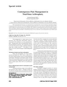
Figure 1. GID SVF-1 device (Louisville, CO) with concentrated cell pellet.
The patient’s orthopedic status was evaluated by one of the investigators (S. P., a doctor of physical therapy) prior to injection and again at 3 months post-SVF injection. ROM and timed up-and-go (TUG) measurements, as described by Podsiadlo and Richardson,22 were recorded. The TUG evaluation assessed the patient’s ability to rapidly rise from a chair, move rapidly 2 m from the chair, turn, and return and sit in the chair.
315
320
Aesthetic Surgery Journal
4
325
Statistical Analysis A t test for paired data (2-tailed) was used to determine statistical significance.
330
RESULTS
350
380
Radiologic Evaluation
Magnetic Resonance Imaging (MRI) of the patient’s knee (s) preoperatively and postoperatively at 3 months was completed using a Philips Achieva 1.5 Tesla MRI device (Andover, MA), with sequencing as follows: coronal T1, Q23 335 STIR; axial T2 fat sat; sagittal T1, PD, fat sat. MRI was only conducted preoperatively and postoperatively at 3 months; no additional images were obtained. The study’s protocol Q24 did not call for further MRIs. A comparative assessment of the preoperative and 3-month postoperative images was 340 completed by a single, independent, experienced orthopedic radiologist.
345
for small-volume lipoplasty. The patients did not experience any deformity related to adipose harvest and were uniformly content with the aesthetic quality of the outcomes. There were no adverse events (including pain and infection) related to the knee injection.
All 6 patients were followed for 1 year (April 2014 to May 2015), with no loss to follow-up. Five patients were women and 1 was a man, with an age range of 51 to 69 years (mean, 59 years). Patient demographics and procedural data are shown in Table 1.
Adverse Events Donor site postoperative course was uneventful other than minimal discomfort, edema, and ecchymosis characteristic 355
385
SVF Processing Average lipoaspirate harvest was 173.5 mL, with an average viable SVF cell count of 14.1 million (Table 1). Tissue processing time from the end of harvest to delivery of the SVF in a syringe for injection into the knee was 60 to 70 minutes. The completion of the liposuction procedure and preparation of the patient for the knee injection was completed during this processing time.
390
395
WOMAC and VAS Scores A decreasing WOMAC score and/or a decreasing VAS score represents decreasing pain (ie, an improving outcome). The WOMAC score decreased from a preoperative mean of 32.9 to a postoperative mean of 10.8 at 3 months and 9.4 at 1 year, respectively (Table 2 and Figure 2). Results were statistically significant at α = 0.05 for both the 3-month and 1-year postoperative time points using the t test for paired data (2-tailed) (Table 2). The VAS score decreased from a preoperative mean of 5.9 to a postoperative mean of 1.8 at 3 months and 2.1 at 1 year, respectively (Table 2 and Figure 3). Results were statistically significant at α = 0.05 for both the 3-month and
400
405
410
Table 1. Patient and Procedural Data Patient
Knee No.
Side
K-L Score
Age (years)
Gender
Viable SVF Cells Injected (millions)
Total Lipoaspirate Harvest (mL)
1
1
Left
II
63
Woman
41.0
215
2
2
Right
II
51
Woman
7.0
160
3
Left
III
3
4
Left
III
51
Woman
17.1
150
4
5
Left
I
64
Woman
17.6
146
5
6
Right
I
58
Woman
7.6
247
7
Left
III
8
Right
III
69
Man
7.9
Mean
NA
NA
59
NA
14.1
173.5
STD DEV
NA
NA
7.3
NA
11.8
47.3
SEM
NA
NA
2.6
NA
4.2
16.7
95% Conf. Int.
NA
NA
5.1
NA
8.2
32.7
360
365
415
7.0
420
7.6
370 6
375
NA, not applicable.
123
425
430
Fodor and Paulseth
5
Table 2. Preoperative, 3-Month Postoperative, and 1-Year Postoperative WOMAC and VAS Scores Knee
WOMAC Preoperative
WOMAC 3 Months
WOMAC 1 Year
VAS Preoperative
VAS 3 Months
VAS 1 Year
435
440
1
29
0
0
6
0
0
2
37
0
4
5
0
1
3
43
4
15
7
1
3
4
18
10
5
6
2
5
5
9
22
7
5
1
3
6
31
5
6
4
1
0
7
41
38
32
8
8
3
8
55
7
6
6
1
1
Mean
32.9
10.8
9.4
5.9
1.8
2.0
STD DEV
14.6
13.1
10.1
1.2
2.6
1.8
5.1
4.6
3.6
0.4
0.9
0.6
10.1
9.0
7.0
0.9
1.8
1.2
490
495
445
450
SEM
500
505 95% CI T test (p) from preoperative 455
.020
.003
NA
.001
.0003
NA, not applicable; VAS, visual analog scale; WOMAC, Western Ontario and McMaster Universities Arthritis Index. 510
460 515
465 520
470 525
475
Figure 2. Preoperative, 3-month postoperative, and 1-year postoperative WOMAC Scores. 530
1-year postoperative time points using the t test for paired data (2-tailed) (Table 2). 480
485
Physical Therapy Measurements The results for ROM and TUG testing preoperatively and postoperatively at 3 months are shown in Table 3. ROM increased an average of 10 degrees but was not statistically significant at α = 0.05. The average time for the TUG test decreased by 2.6 seconds and was statistically significant at α = 0.05.
Radiologic Evaluation No significant observations of differences were detectable between 0 and 3 months in the MRI images as read by the same independent, experienced orthopedic radiologist.
535
DISCUSSION Similar to previous pilot studies, this study confirmed that the use of autologous adipose-derived SVF for treatment of
540
Aesthetic Surgery Journal
6
595
545 600
550 605
555 610
Figure 3. Preoperative, 3-month postoperative, and 1-year postoperative VAS Scores. 560 615
Table 3. Preoperative and 3 Month Postoperative Knee Flexion ROM and TUG Results Knee 565
570
575
580
585
ROM Preoperative (degrees)
ROM 3 Months (degrees)
TUG Preoperative (seconds)
TUG 3 Months (seconds)
1
135
156
6.0
2.5
2
135
142
6.0
2.5
3
139
144
3.0
3.0
4
138
145
3.0
3.0
5
132
145
7.0
3.0
6
130
140
7.0
3.0
7
153
145
5.0
2.5
8
131
132
6.0
3.0
Mean
136.6
143.6
5.4
2.8
10.0
NA
−2.6
Change in Mean STD DEV
7.3
6.7
1.6
0.3
SEM
2.6
2.4
0.6
0.1
95% Conf. Int.
5.1
4.6
1.1
0.2
T test (p)
NA
.053
NA
.003
NA, not applicable; ROM, range of motion; TUG, timed up-and-go. 590
OA pain was safe and feasible for treatment of pain.6,8,10,23 This pilot study clearly confirmed that the harvest of donor adipose tissue, isolation of the SVF, and then use of the SVF for an intra-articular knee injection is a relatively
simple procedure to perform by a surgeon functioning with a team limited to what he is accustomed to in the course of performing lipoplasty procedures, with the addition of approximately 60 to 70 minutes of time spent on processing and injecting 1 or 2 knees. At 1-year postoperative, the WOMAC scores decreased (indicating improvement) in 8 of 8 knees in this study. Similarly, at 1-year postoperative the VAS scores decreased (indicating improvement) in 8 of 8 knees in this study. There was no statistically significant change in the mean WOMAC score from 3 months to 1 year. Similarly, there was no statistically significant change in the mean VAS score from 3 months to 1 year, while 2 of the knees did show an increase between 3 months and 1 year. One knee of patient 5 (knee no. 7) did not respond to the SVF injection at 3 months, with only a small change on the WOMAC score and no change in the VAS score. At 1 year this patient reported a modest improvement in the WOMAC score and a more significant improvement in the VAS score (Table 2). The amount of SVF yield varied; however there was no dose response to the amount of SVF injected. The yield of SVF did not seem to be dependent on the amount of adipose because the highest SVF yield was not due to the greatest amount of adipose harvested (Table 1). The patient with the highest SVF yield was, however, the only patient to report no pain on the WOMAC and VAS at both 3 months and 1 year (Table 2). It is unknown why this patient had a higher SVF yield. ROM increased by an average of 10 degrees but was not statistically significant at α = 0.05. TUG decreased by 2.6 seconds, which was statistically significant, a reduction of 48% from an initial value of 5.4 seconds, demonstrating increased functionality.
620
625
630
635
640
645
Fodor and Paulseth
650
655
660
665
This study shows initial longer-term benefits compared with standard-of-care treatments. Studies have shown standard-of-care treatments such as viscosupplementation and corticoidsteroid injections last for a few weeks to 6 months,24 whereas the procedure in our study showed results were maintained for 1 year. The decrease in WOMAC and VAS scores at 3 months and the further maintenance of these scores to 1 year is indicative of the potential for this therapy. OA is a chronic inflammatory condition of the tissues of the knee, having both inflammation and degradation of knee tissues, resulting in pain. While not specifically investigated in this study, a proposed mechanism of action for the adipose-derived SVF on tissues of the OA knee is reduction of pain due to the anti-inflammatory properties of the SVF cells.4,6,10,25 The inflammation-degradation cycle is difficult to interrupt, as evidenced by the relatively short-lived results of standard-of-care treatments for OA of the knee, including use of corticosteroids and viscosupplementation. Decreasing or breaking the inflammation cycle potentially gives the knee tissues a reparative period with reduced pain and slowed degradation.
670
Limitations
675
680
685
690
695
700
MRI imaging using standard a MRI device, and sequencing showed no observable changes between the preoperative and 3-month evaluations. This was not surprising, especially in light of the postoperative MRIs being performed only 3 months after treatment. Another MRI evaluation at least 6-months postoperative would be desirable using a different, significantly improved MRI sequencing. We learned that the critical sequencing parameter for the MRI is the step size. For the current study, a standard orthopedic 1 mm step size was used, which is not sufficient to capture small changes on the order of 0.01 or 0.1 mm. Should future studies wish to determine these changes, imaging should be conducted with a higher Tesla magnetic field and a decreased step size with advanced image assessment software. However, such imaging will take much more time than standard MRI sequencing. At screening, patient 4 reported moderate or greater pain with a VAS score of 5 out of a possible 10 points. At the preoperative assessment, the same patient’s WOMAC questionnaire score was 9 out of a possible score of 96, thus essentially documenting a minimally symptomatic knee. At 3-months postoperative, this patient reported a VAS score of 1, demonstrating decreased and minimal pain, however the WOMAC score, at the same time period, indicated that the patient’s knee was more symptomatic than before treatment. It is not known why the patient reported such divergent scores, but in the future, studies using multiple questionnaires with similar measurement parameters should be interpreted as soon as possible to investigate the reason for such a dichotomy.
7
Placing the SVF precisely into the intra-articular space is essential for obvious reasons. Aspiration of joint fluid prior to injection is reassuring as to the correct needle placement for SVF installation into the articular space. At this point in time, the majority of the evidence for cellular therapies comes from small studies with low numbers of patients. This pilot study was also a small study with only 8 knees and 6 patients, was not randomized, and did not include a control arm. It was designed to primarily assess feasibility and patient safety. We recognize that a control group, as in all studies, is desirable and should be the next step in evaluation. If we have the opportunity to conduct such a study, it is possible to use a design where the patients would serve as their own control, which would require individuals with bilateral disease with a similar degree of severity. One knee could be treated with SVF injection and the other, control knee, with saline injection ( placebo). We fully realize that performing a study in knee joints affected by degenerative arthritis has no readily apparent relationship with aesthetic or reconstructive surgery. However, the results of this study provide evidence of safety for the potential use of autologous adipose-derived reparative/regenerative cells in aesthetic and reconstructive surgery. Treatment with adipose-derived SVF cells of nonhealing wounds, diabetic ulcers, radiation injury, and inflamed tendons or ligaments are just some possible reconstructive applications. For this pilot study, which focused on safety and initial evidence of efficacy for treatment of pain, degenerative arthritis of the knee joint was chosen as a clinical study model because clinical outcomes are more readily quantified across a number of patients than outcomes in aesthetic surgery involving aesthetic assessment or volume retention.
705
710
715
720
725
730
735
CONCLUSIONS In this pilot study, autologous SVF was shown to be safe and to present a new potential therapy for reduction of pain for OA of the knee.
740
Supplementary Material This article contains supplementary material located online at www.aestheticsurgeryjournal.com.
745
Disclosures The authors declared no potential conflicts of interest with respect to the research, authorship, and publication of this article.
750
Funding This study was funded in part by a grant from the Aesthetic Surgery Education and Research Foundation (ASERF). Funding
755
8
760
765
was used to for imaging costs, operating room expenses, IRB expenses, reagents and supplies, anesthetic, and screening and follow-up expenses. No funding was used for physician compensation or for capital equipment used in the study. The GID Group, Inc. (Louisville, CO), loaned 3 pieces of equipment (centrifuge, incubated shaker table, and cell counting device) to the surgery center to perform the cell processing.
REFERENCES 1.
Buckwalter JA, Martin JA. Osteoarthritis. Adv Drug Deliv Rev. 2006;58(2):150-167. 2. Dunlop DD, Manheim LM, Yelin EH, Song J, Chang RW. The costs of arthritis. Arthritis Rheum. 2003;49(1): 101-113. 770 3. Coleman CM, Curtin C, Barry FP, O’Flatharta C, Murphy JM. Mesenchymal stem cells and osteoarthritis: remedy or accomplice? Hum Gene Ther. 2010;21(10):1239-1250. 4. Black LL, Gaynor J, Gahring D, et al. Effect of adiposederived mesenchymal stem and regenerative cells on 775 lameness in dogs with chronic osteoarthritis of the coxofemoral joints: a randomized, double-blinded, multicenter, controlled trial. Vet Ther. 2007;8(4):272-284. 5. Koh YG, Jo SB, Kwon OR, et al. Mesenchymal stem cell injections improve symptoms of knee osteoarthritis. Arthroscopy. 2013;29(4):748-755. 780 6. Orozco L, Munar A, Soler R, et al. Treatment of knee osteoarthritis with autologous mesenchymal stem cells: a pilot study. Transplantation. 2013;95(12):1535-1541. 7. Wakitani S, Imoto K, Yamamoto T, Saito M, Murata N, Yoneda M. Human autologous culture expanded bone 785 marrow mesenchymal cell transplantation for repair of cartilage defects in osteoarthritic knees. Osteoarthritis Cartilage. 2002;10(3):199-206. 8. Davatchi F, Abdollahi BS, Mohyeddin M, Shahram F, Nikbin B. Mesenchymal stem cell therapy for knee osteo790 arthritis. Preliminary report of four patients. Int J Rheum Dis. 2011;14(2):211-215. 9. Pak J, Chang JJ, Lee JH, Lee SH. Safety reporting on implantation of autologous adipose tissue-derived stem cells with platelet-rich plasma into human articular joints. BMC Musculoskelet Disord. 2013;14:337. 795 10. Koh YG, Choi YJ, Kwon SK, Kim YS, Yeo JE. Clinical results and second-look arthroscopic findings after treatment with adipose-derived stem cells for knee osteoarthritis. Knee Surg Sports Traumatol Arthrosc. 2013. Q25 11. Peeters CM, Leijs MJ, Reijman M, van Osch GJ, Bos PK. 800 Safety of intra-articular cell-therapy with culture-expanded stem cells in humans: a systematic literature review. Osteoarthritis Cartilage. 2013;21(10):1465-1473.
Aesthetic Surgery Journal
12. Folgiero V, Migliano E, Tedesco M, et al. Purification and characterization of adipose-derived stem cells from patients with lipoaspirate transplant. Cell Transplant. 2010;19(10):1225-1235. 13. Lin G, Banie L, Ning H, Bella AJ, Lin CS, Lue TF. Potential of adipose-derived stem cells for treatment of erectile dysfunction. J Sex Med. 2009;6(Suppl 3):320-327. 14. Lindroos B, Suuronen R, Miettinen S. The potential of adipose stem cells in regenerative medicine. Stem Cell Rev. 2011;7(2):269-291. 15. Tholpady SS, Llull R, Ogle RC, Rubin JP, Futrell JW, Katz AJ. Adipose tissue: stem cells and beyond. Clin Plast Surg. 2006;33(1):55-62, vi. 16. Fujimura J, Sugihara H, Fukunaga Y, Suzuki H, Ogawa R. Adipose Tissue is A Better Source of Immature Non-Hematopoietic Cells than Bone Marrow. Int J Stem Cells. 2009;2(2):135-140. 17. Madonna R, Geng YJ, De Caterina R. Adipose tissuederived stem cells: characterization and potential for cardiovascular repair. Arterioscler Thromb Vasc Biol. 2009;29(11):1723-1729. 18. Pachón-Peña G, Yu G, Tucker A, et al. Stromal stem cells from adipose tissue and bone marrow of age-matched female donors display distinct immunophenotypic profiles. J Cell Physiol. 2011;226(3):843-851. 19. Mitchell JB, McIntosh K, Zvonic S, et al. Immunophenotype of human adipose-derived cells: temporal changes in stromal-associated and stem cell-associated markers. Stem Cells. 2006;24(2):376-385. 20. Riordan NH, Ichim TE, Min WP, et al. Non-expanded adipose stromal vascular fraction cell therapy for multiple sclerosis. J Transl Med. 2009;7:29. 21. Varma MJ, Breuls RG, Schouten TE, et al. Phenotypical and functional characterization of freshly isolated adipose tissue-derived stem cells. Stem Cells Dev. 2007;16(1): 91-104. 22. Podsiadlo D, Richardson S. The timed “Up & Go”: a test of basic functional mobility for frail elderly persons. J Am Geriatr Soc. 1991;39(2):142-148. 23. Jo CH, Lee YG, Shin WH, et al. Intra-articular injection of mesenchymal stem cells for the treatment of osteoarthritis of the knee: a proof-of-concept clinical trial. Stem Cells. 2014;32(5):1254-1266. 24. Fibel KH, Hillstrom HJ, Halpern BC. State-of-the-Art management of knee osteoarthritis. World J Clin Cases. 2015;3(2):89-101. 25. Veronesi F, Maglio M, Tschon M, Aldini NN, Fini M. Adipose-derived mesenchymal stem cells for cartilage tissue engineering: state-of-the-art in in vivo studies. J Biomed Mater Res A. 2014;102(7):2448-2466.
815
820
825
830
835
840
845
850
855
805 860
810