emergency care journal
clinica e terapia
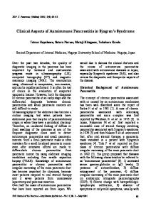
Acute Pancreatitis: Pathophysiology, Clinical Aspects, Diagnosis e Treatment Raffaele Pezzilli, Bahjat Barakat*, Dario Fabbri, Andrea Imbrogno*, Mario Cavazza** Pancreas Unit, Department of Digestive Diseases and Internal Medicine, *Emergency Department Sant’Orsola-Malpighi Hospital, Bologna
Acute pancreatitis is an acute inflammation of the pancreas. The clinical classification of the disease recognizes the mild acute pancreatitis, characterized by the absence of local and/or systemic complications, and the severe disease, characterized by the presence of local complications such as necrosis, abscess or pseudocysts and/ or distant organ failure. Gallstones constitute the predominant etiological factor. The severity assessment is essential for proper
initial treatment of the disease. Primary objectives to achieve in the treatment of acute pancreatitis essentially are: pain control, electrolyte support and energy intake, removal of the causal agent, attenuation of the inflammation, and prevention and eventual treatment of local and systemic complications of necrotizing forms. Keywords: Acute pancreatitis; Disease management; Severity assessment; Therapy.
Introduction
necrosis. Computed tomography with intravenous contrast bolus is currently the best diagnostic method (accuracy 80-90%). Pancreatic necrosis rarely involves the entire gland in its entire thickness; usually it remains confined to the periphery and spares the glandular core. Haemorrhagic foci are present in varying degrees. The necrosis may become infected (10-30% of cases) and the distinction between sterile and infected pancreatic necrosis is important because the therapeutic approach (mainly medical therapy in sterile pancreatic necrosis, surgical in the infected type) and prognosis (mortality rate about three times higher in infected pancreatic necrosis) differ considerably. The diagnostic gold-standard for suspected infection of pancreatic necrosis is represented by microbial cultures of material from percutaneous needle aspiration (Figure 2). Acute fluid collection is a localized effusion in or near the pancreas, without granulation fibrous wall. It tends to appear early and regresses spontaneously in most cases. It is not considered a sign of disease severity unless it becomes infected. Pseudocysts is a collection of pancreatic juice enclosed by a wall lacking epithelialization and appearing as a result of acute pancreatitis, chronic pancreatitis or pancreatic trauma. The maturation of a pseudocyst after acute pancreatitis requires at least 4 weeks after the onset of the disease. A post-acute pancreatitis pseudocyst is therefore an acute fluid collection persisting more than 4 weeks surrounded by a well-defined wall (Figure 3). Walled-off pancreatic necrosis is an intra-abdominal collection of pus (usually near the pancreas), appearing after an attack of acute pancreatitis or after pancreatic trauma. Pus predominates and there is only small amount of necrotic tissue, distinguishing it from non-infected pancreatic necrosis. A pseudocyst presenting pus within its walls is also correctly defined as a walled-off pancreatic necrosis [3].
Acute pancreatitis is an acute inflammation of the pancreas with variable involvement of peripancreatic tissues and/or distant organs. The inflammatory process may be limited to the pancreatic gland with edema or necrosis, or it may involve the surrounding tissues and/or distant organs, so the clinical manifestations range from mild abdominal pain to very serious presentations with high mortality rate [1]. Episodes of acute pancreatitis in patients who will subsequently develop anatomical, clinical and functional features compatible with chronic pancreatitis are classified as the former until the final diagnosis is established. The now widely accepted classification of the disease and its complications is a clinical classification prominently known as the Atlanta classification [2] and is shown below. Mild acute pancreatitis is characterized by a favorable clinical course in the absence of local and/or systemic complications. The predominant pathological expression is interstitial edema more or less associated with peripancreatic steatonecrosis (Figure 1). Severe acute pancreatitis is characterized by the presence of local complications such as necrosis, abscess or pseudocysts and / or organ failure. In most cases it is the clinical expression of the presence of pancreatic necrosis; in fact, patients with acute edematous-interstitial pancreatitis rarely present a clinically severe form of the disease. The organ failure was defined as shock (systolic blood pressure < 90 mmHg), pulmonary insufficiency (PaO2 < 60 mmHg), renal failure (serum creatinine > 2 mg/dl after rehydration) or gastrointestinal bleeding (> 500 cc/24h). Pancreatic necrosis is a focal or diffuse area of non-viable parenchyma, which typically is associated with peripancreatic steato-
emergency care journal - organizzazione, clinica, ricerca • Anno VII numero 2 • Giugno 2011 • www.ecj.it
ABSTRACT
5 Fig. 1 - Multidetector computer tomography: edematous pancreatitis.
Materiale protetto da copyright. Non fotocopiare o distribuire elettronicamente senza l’autorizzazione scritta dell’editore.
clinica e terapia
clinica e terapia
Fig. 2 - Multidetector computer tomography: Necrotizing pancreatitis.
emergency care journal - organizzazione, clinica, ricerca • Anno VII numero 2 • Giugno 2011 • www.ecj.it
Pathophysiology There are many recognized causes of acute pancreatitis, but surely gallstones constitute the predominant etiological factor in our geographical area [4]. Less frequently, acute pancreatitis is related to chronic use or abuse of alcohol and even more rarely is secondary to abdominal surgery, diagnostic and/or interventional endoscopical procedures on the papilla of Vater, abdominal trauma, dyslipidemia or the use of drugs with pancreatic toxicity [5-7]. The mechanisms by which the various etiological factors trigger pancreatic inflammation have not yet been fully identified but it seems proved with sufficient certainty that, whatever the initial pathogenic noxious stimuli, the earliest pathogenetic events are triggered inside the acinar cells [8] . Under normal conditions these cells produce digestive enzymes and lysosomal enzymes, the former segregated in lysosomal vacuoles, the latter in the vacuoles of zymogen. In acute pancreatitis this strict compartmentalization can be overridden by alteration of a complex biological process, calcium-dependent, defined as “stimulus-secretion coupling”. A colocalization of lysosomes and zymogen granules in a unique vacuole is thus determined: the lysosomal enzyme cathepsin B can activate trypsinogen at this point with consequent cascade activation of other proteases and phospholipases. It follows the rupture of vacuoles, cell
damage, necrosis and release of cellular activated enzymes in the interstitium. Local processes of vasoconstriction-dilatation determine infiltration of inflammatory cells and increased necrosis. In the most severe forms of acute pancreatitis it is present a complex biochemical cellular and humoral response not substantially different from what happens in other serious diseases such as septic shock, the poly-trauma and extensive burns. The magnitude and the continuation of such events, assignable to the so-called SIRS (systemic inflammatory response syndrome), affect the extent and severity of local damage and progression to systemic complications [9]. Implicated mediators are various cytokines such as interleukin-1 (IL-1), IL-6, IL-8, TNF (tumor necrosis factor), PAF (platelet activating factor) [9,10]. All these mediators are markedly elevated in the first 24 hours of illness, whereas the anti-inflammatory cytokines to (IL-2, IL-10) are reduced. The result is the activation of neutrophils, monocytes, lymphocytes, platelets and endothelial cells. The increased expression of cell adhesion molecules and integrins on neutrophils results in increased adhesion to the endothelium, diapedesis and invasion of distant organs (first of all the lungs) where hyperactive neutrophils call forth other polymorphonuclear leukocytes and result in extensive tissue destruction [11]. The presence of trypsin, chymotrypsin and elastase in the pancreatic interstitium, in serum and peritoneal fluid is responsible for activation
Fig. 3 - Ultrasonography: pancreatitis pseudocyst (Ps) The main pancreatic duct is dilated (W).
6
Materiale protetto da copyright. Non fotocopiare o distribuire elettronicamente senza l’autorizzazione scritta dell’editore.
of the coagulation-fibrinolysis systems, endothelial cells, PMN leukocytes and monocytes-macrophages with synthesis and release of cytokines, superoxide ions and PAF [8]. The latter is a key mediator capable of stimulating the release of other proinflammatory cytokines, increase vascular permeability, induce a negative inotropic effect, leukocyte chemotaxis, tissue edema and cellular damage. It is possible to clearly appreciate the possibility of a serious involvement of distant organs up to the development of the “fearsome” multi-organ failure syndrome.
outcome [15]. From these simple evaluations are derived three important corollaries: a) the majority of patients benefits from a conservative therapy, not surgery, b) the early identification of those patients at increased risk of developing complications is crucial for prognosis and therapy; c) surgical therapy is to be reserved for those patients who develop specific complications, primarily the infection of pancreatic necrosis and / or peripancreatic fluid collections.
Evaluation and stratification of severity of acute pancreatitis
Essentially, the severity of an acute attack leads to the development of a necrotizing form of the disease and pancreatic necrosis is not only responsible for the clinical severity, but also the onset of complications and, ultimately, mortality. At the present state of knowledge a severe form of acute pancreatitis can not adequately be dealt without the support of a CT scan available full-time and other multi-specialistic skills/human resources and equipment. The guidelines of the British Society of Gastroenterology [16] and the Italian of the Italian Association for the Study of the Pancreas [12] suggest that a specialized center for the treatment of severe acute pancreatitis should have the following characteristics: 1. allocation in a general hospital where the major medical and surgical specialties are present; 2. multidisciplinary team with specialists in Internal Medicine, Surgery, Endoscopy, Critical Care and Intensive Care and Pathology, 3. day and night availability of CT and ultrasound with staff expert in percutaneous treatments, the availability of magnetic resonance and angiography may be useful but not essential, 4. the presence of daytime endoscopists experienced in endoscopic retrograde cholangiopancreatography (ERCP) and related interventional procedures.
Severity assessment is essential for proper initial treatment in the management of acute pancreatitis and this constitutes a recommendation of grade A in the Italian guidelines on acute pancreatitis [12]. These guidelines also suggest that assessment of severity should be done by a scoring system such as Acute Physiology and Chronic Health Evaluation (APACHE) II [12]: an APACHE-II score greater than 8 is important for determining treatment policy and identifying the need for transfer to a referral unit. Serum C-reactive protein values grater than 150 mg/dL are useful for severity assessment, but they may not reflect severity within the first 48 h after onset. In addition contrast-enhanced CT scanning and contrast- enhanced MRI play an important role in severity assessment. The CT severity index, as proposed by Balthazar et al. [13], should be used. In fact, the gold-standard for the presence of pancreatic necrosis is the computed tomography (CT) with intravenous contrast medium, which should be done after 72 hours from pain onset and after rehydration of the patient; it may be possibly repeated according to the clinical situation. Management in, or referral to, high-volume units is necessary for patients with extensive necrotizing pancreatitis or other complications who may require care in the intensive therapy unit or interventional radiological, endoscopic or surgical procedures and this constitutes a recommendation of grade B [12].
Therapy Background One of the recurring features of acute pancreatitis is the frequent presence of a variably long period of time, sometimes days, between the onset of symptoms and hospitalization. This is a factor that affects the very effectiveness of therapeutic measures and ensures that treatment is more often aimed at controlling the progression of the disease rather than at interfering with initial pathogenetic phenomena. The delay in hospital makes it difficult to interpret results of therapeutic trials and impossible a homogeneous analysis of aggregate data from multiple studies, as the timing of treatment is often not specified, or the onset of symptoms is considered at the time of hospitalization. The time frame in which there is a reasonable chance of specifically antagonize the inflammatory mediators and activated pancreatic enzymes to mitigate or prevent the development of a partial or total impairment of distant organs is about 2-3 days after the onset of pain and this period is also called interventional window [14]. All this should lead to a “specific” treatment as early as possible and at the same time confirms that it is absolutely unnecessary and wasteful to use these same drugs in patients who come late to the observation, often in the second week of illness, at a stage where there are already signs of impairment of distant organs. In this clinical scenario, treatment should be more rationally targeted toward measures useful in supporting cardiovascular, respiratory and renal systems and preventing septic complications. It is possible to affirm that for every four patients with acute pancreatitis, three will respond favorably to conservative medical treatment, while the fourth will present complications with a one in three chance of suffering a fatal
Objectives and methods of conservative treatment Primary objectives to achieve in the treatment of acute pancreatitis essentially are: 1. pain control, 2. electrolyte support and energy intake, 3. removal of the causal agent, when possible, 4. attenuation of inflammatory and autolytic processes at the glandular level (“specific” therapy), 5. prevention and eventual treatment of local and systemic complications of necrotizing forms. For mild forms of disease, in most cases the first three steps are sufficient for clinical resolution. In severe forms, the therapeutic engagement is more complex and patients may, with reasonable frequency, require periods of hospitalization in intensive care units. The therapeutic approach to severe acute pancreatitis is reported in Figure 4. The control of pain must be swift and effective: for this purpose, meperidine is the drug of choice [15]. Supportive therapy is a measure of fundamental importance that counterbalances the seizure of the fluids and hypercatabolism particularly important in severe forms. The maintenance of cardiovascular parameters, renal and respiratory can in many cases prevent the onset of multisystem complications. The pancreatic hypoperfusion, secondary to inadequate maintenance of plasma volume is indeed able to trigger and increase the phenomena of pancreatic necrosis. Patients with mild forms, for which it is expected an oral refeeding within 4-6 days of hospitalization, do not need an aggressive nutritional approach [15]. In contrast, in severe forms total parenteral nutrition (TPN) must be used, which must take into account in its formulation of any metabolic imbalances (such as acidosis or alkalosis, hyperglycemia, hypocalcemia, and hypokalemia ipomagenesiemia) and cardiovascular complications [15]. Recently, enteral nutrition through naso-jejunal probe has been used with good results in patients with severe acute pancreatitis
Materiale protetto da copyright. Non fotocopiare o distribuire elettronicamente senza l’autorizzazione scritta dell’editore.
emergency care journal - organizzazione, clinica, ricerca • Anno VII numero 2 • Giugno 2011 • www.ecj.it
clinica e terapia
7
emergency care journal - organizzazione, clinica, ricerca • Anno VII numero 2 • Giugno 2011 • www.ecj.it
clinica e terapia instead of the NPT. The pathophysiological assumption is that the NPT does not provide all essential nutrients (eg glutamine) and does not fuction as intestinal barrier which can increase intestinal permeability to toxins and bacterial translocation. The early removal of the causative agent makes it paramount to achieve a sufficiently precise etiologic diagnosis and early intervention. In clinical practice, this translates, at least in our population, in the identification of a mechanism for biliary obstruction, transient or persistent, complete or partial, in about 2/3 of cases. The removal of biliary obstruction using endoscopic techniques has now entered into the routine treatment of these patients.[12, 16]. The “specific” therapy of acute pancreatitis relies on antisecretory and antiprotease drugs. The use of somatostatin and its synthetic analogue octreotide is much debated. The results of published studies, many uncontrolled and with small case series or not stratified by severity of illness, are controversial, although a metanalytical evaluation shows, in general, a therapeutic advantage. From the theoretical point of view, the negative effects of vasoconstriction of the splanchnic circulation and contraction of the sphincter of Oddi outstripped the hypothetical beneficial effects related to inhibition of exocrine pancreatic secretion. Among antiprotease drugs the gabexate mesylate showed a positive effect in patients with severe acute pancreatitis, with significant reduction of systemic complications and the need for surgery but not mortality compared with placebo. The dosage used in early studies was 3 g/day by continuous intravenous infusion, but subsequently it was found that a dose of 1.5 g/day in the same manner for a period of treatment of 7-8 days is also a viable option [17]. The best results are obtained when the administration starts earlier than the onset of symptoms. The use of systemic antibiotics for the prevention of pancreatic infections is one of the cornerstones of conservative treatment of severe forms of acute pancreatitis. Several studies have shown a significant reduction in the incidence of pancreatic and extrapancreatic infections but not mortality in patients treated with imipenem-cilastatin [18]. Quinolones, due to their pharmacokinetic characteristics and their range of action, should ensure an effective prophylactic action as well. However, in a recent randomized prospective trial, patients treated with pefloxacin showed an incidence of infected necrosis significantly higher than patients treated with imipenem (34% vs 10%) [19]. At present,
therefore, it is recommended for all patients with acute necrotizing pancreatitis an early administration of imipenem-cilastatin at a dose of 1.5-2 g/day, lasting for at least two weeks. Objectives and indications of surgical treatment The infection of pancreatic necrosis in the course of acute pancreatitis is a very serious medical condition and its presence is associated with a marked increase in risk of death; it developes in percentages varying from 15 to 70% of all patients with acute necrotizing pancreatitis and accounts for more than 80% of deaths from acute pancreatitis. The risk of infection increases with the extent of necrosis and the days after initiation of acute pancreatitis, reaching a peak incidence (70%) after three weeks [20]. In most cases the infection is caused by Gram-negative bacteria of enteric origin, and about two thirds of infections are caused by a single microbiological agent. Therefore, E. coli is the most frequent causative agent (26%), followed by Pseudomonas, Klebsiella and Proteus species. Frequently Gram positive infections are also detected, such as Staph.aureus (15%), Streptococcus faecalis and Enterococcus or other anaerobic bacteria, and in some cases by fungi. In clinical terms acute pancreatitis with sterile necrosis can be difficult to distinguish from a form with infected necrosis, because both can give fever, leukocytosis and abdominal pain. But this distinction is very important, since the mortality in patients with infected necrosis that did not underwent early surgery is high. TC or ultrasound-guided percutaneous suction of the necrotic material and/or peripancreatic fluid collections, with a fresh microscopic examination and bacterial culture, is safe and accurate (sensitivity and specificity exceeding 95%) and must be used, even repetitively, usually from the second week of illness, in patients whose clinical condition worsens or does not tend to improve, despite the removal of any causative agent and the implementation of a vigorous supportive treatment. Debridement is the surgical treatment of choice of infected necrosis and the only therapeutic doubt concerns the type of intervention to perform (classic necrosectomy with drainage-washing or open packing technique). Recently other treatment options, such as percutaneous, endoscopic or minimally invasive surgery have been proposed [21-23]. These methods require highly experienced operators, are not risk-free and should be for the moment limited to patients unfit for surgery because of a high anesthetic risk.
8 Fig. 4 - Therapeutic approach to severe acute p ancreatitis.
Materiale protetto da copyright. Non fotocopiare o distribuire elettronicamente senza l’autorizzazione scritta dell’editore.
The treatment of patients with sterile pancreatic necrosis remains controversial. Surgical treatment offers no demonstrable survival benefits compared with supportive treatment; in addition, because the surgery may cause postoperative infection of necrotic tissue in 25-50% of cases with a very high secondary mortality, a surgical indication in any case sterile necrosis should be considered with prudence and care. Even when the necrotic process determines rupture of the main pancreatic duct and necrosis remains sterile, there are good prospects for a resolution of the disease with conservative therapy. The precise role of surgery in the treatment of sterile necrosis is therefore limited and should be reserved for selected cases, such as those patients in whom repeated attempts at oral re-feeding after 5-6 weeks of therapy are associated with abdominal pain, nausea, vomiting or recurring pancreatitis. At this stage of the disease, however, generally necrosis is more demarcated and the surgical act is easier. In other cases, supportive care associated with prophylactic antibiotic treatment should be the primary treatment [24-28]. It is therefore very important to perform in due time (possibly during the same hospitalization for mild forms, usually at a distance of three to four weeks for severe) a cholecystectomy in case of gallstones in order to prevent recurrence of acute episodes [12].
clinica e terapia
13. 14. 15. 16. 17.
18. 19.
20.
References 1. Steinberg W, Tanner S. Acute pancreatitis. New Engl. J. Med 1994; 330: 1198-210. 2. Bradley EL. A clinically based classification system for acute pancreatitis. Arch Surg. 1993, 128: 586-90. 3. Stamatakos M, Stefanaki C, Kontzoglou K, Stergiopoulos S, G Giannopoulos, M. Safioleas Walled-off pancreatic necrosis. World J Gastroenterol 2010; 16:1707-12. 4. Pezzilli R, Uomo G, Gabbrielli A, Zerbi A, Frullone L, De Rai P, Castoldi L, Cavallini G, Di Carlo V; Proinfa-AISP Study Group. A prospective multi-center survey on the treatment of acute pancreatitis in Italy. Dig Liver Dis 2007; 39:838-46. 5. Pezzilli R, Cecilia R, Corinaldesi R, Barakat B. Acute pancreatitis two to simvastatin therapy: Increased severity after rechallenge. Dig Liver Dis 2004; 36:639-40. 6. Pezzilli R, Morselli-Labate AM, Corinaldesi R. NSAIDs and acute pancreatitis: A Systematic Review. Pharmaceuticals 2010; 3: 558-571. 7. Pezzilli R, Corinaldesi R, Morselli-Labate AM. Tyrosine kinase inhibitors and acute pancreatitis. JOP 2010; 11:291-3. 8. Pezzilli R, Romboli E, Campana D, Corinaldesi R. Mechanisms Involved in the onset of post-ERCP pancreatitis. JOP. 2002; 3:162-8. 9. Kusse AM Rongions AJ, Reber HA Cytokines and acute pancreatitis. Gastroenterology 1996; 110: 639-41. 10. Pezzilli R, L Bellacosa, Felicani C. Lung injury in acute pancreatitis. JOP 2009; 10:481-4. 11. Rinderknecht H. Genetic determinants of mortality in acute necrotizing pancreatitis. Int J. Pancreatol. 1994, 16: 11-8. 12. Pezzilli R, Zerbi A, Di Carlo V, Bassi C, Delle Fave GF and the Working Group of the Italian Association for the Study
21. 22.
23.
24. 25. 26. 27.
28.
of the Pancreas on Acute Pancreatitis. Practical guidelines for acute pancreatitis. Pancreatology 2010;10:523-35. Balthazar EJ, Freeny PC, van Sonnenberg E. Imaging and intervention in acute pancreatitis.Radiology 1994; 193: 297–306. Makhija R, Kingsnorth AN. Cytokine storm in acute pancreatitis. Pancreatitis J Hepatobiliary Surg 2002; 9:401-10. Pezzilli R. Pharmacotherapy for acute pancreatitis. Expert Opin Pharmacother. 2009, 10:2999-3014. UK guidelines for the management of acute pancreatitis. Gut 1998; 42 (s-2): 1-13. Pezzilli R, Miglioli M. Multicentre comparative study of two schedules of gabexate mesilate in the treatment of acute pancreatitis. Italian Acute Pancreatitis Study Group. Dig Liver Dis 2001, 33:49-57. Villatoro E, Bassi C, Larvin M. Against Antibiotic therapy for prophylaxis of pancreatic necrosis infection in acute pancreatitis. Cochrane Database Syst Rev 2006; 4: CD002941. Bassi C, Falconi M, Talamini G, Uomo G, G Papaccio, Dervenis C, Salvia R, Minelli EB, Pederzoli P. Controlled clinical trial of pefloxacin versus imipenem in severe acute pancreatitis. Gastroenterology 1998, 115:1513-7. Beger HG, Bittner R, Block S, Büchler M. Bacterial contamination of pancreatic necrosis. A prospective clinical study. Gastroenterology 1986; 91:433-8. Schoenberg MH, Rau B, Beger HG. New Approaches in surgical management of severe acute pancreatitis. Digestion 1999; 60 (s-1): 22-6. Bakker OJ, van Santvoort HC, Besselink MG, van der Harst E, Hofker HS, Gooszen HG; Dutch Pancreatitis Study Group. Prevention, detection, and management of infected necrosis in severe acute pancreatitis. Curr Gastroenterol Rep 2009; 11:104-10. Santvoort van HC, Besselink MG, Bakker OJ, Hofker HS, Boermeester MA, Dejong CH, van Goor H, Schaapherder AF, van Eijck CH, Bollen TL, van Ramshorst B, VB Nieuwenhuijs , R Timmer, Laméris JS, Kruyt PM, Manusama ER, van der Harst E, van der Schelling GP, Karsten T, Hesselink EJ, van Laarhoven CJ, Rosman C, Bossche K, de Wit RJ, Houdijk AP, van Leeuwen MS, Buskens E, Gooszen HG; Dutch Pancreatitis Study Group. A step-up approach or open necrosectomy for necrotizing pancreatitis. N Engl J Med 2010, 362:1491-502. Baron TH, Morgan DE. Acute necrotizing pancreatitis. N. Engl. J. Med 1999; 340: 1412-15. Steinberg WM, Bradley EL, Warshaw AL. Indications for debridement of necrotizing pancreatitis. Pancreas 1996; 13: 219-23. Bradley EL. Operative vs. nonoperative therapy in necrotizing pancreatitis. Digestion 1999; 60 (s-1): 19-21. Bassi C, Falconi M, Sartor N, Bonora A, Caldiron E, Butturini G et al. The role of surgery in the early Major Complications of severe acute pancreatitis. Eur.J.Gastroenterol. Hepatol. 1997, 9: 131-6. Banks PA. Practice guidelines in acute pancreatitis. Am J. Gastroenterol. 1997, 92: 377-86.
emergency care journal - organizzazione, clinica, ricerca • Anno VII numero 2 • Giugno 2011 • www.ecj.it
clinica e terapia
9
Materiale protetto da copyright. Non fotocopiare o distribuire elettronicamente senza l’autorizzazione scritta dell’editore.
clinica e terapia
Onset of angioedema following a viper bite: different options for diagnostic procedure in an emergency department Alessandro Riccardi, Pierangela Minuto, Anna Da Col, Viviana Panunzio*, Roberto Lerza SC Pronto Soccorso e Medicina e Chirurgia d’Accettazione e d’Urgenza, Ospedale San Paolo, Savona, Italy *SC Immunoematologia e Medicina Trasfusionale, ASL 2 Savonese, Savona, Italy
emergency care journal - organizzazione, clinica, ricerca • Anno VII numero 2 • Giugno 2011 • www.ecj.it
ABSTRACT
10
The unexpected onset of an angioedema in a patient observed in the emergency department after a viper bite, gave the opportunity to consider some different etiopathogenetic hypothesis and to discuss the most correct diagnostic procedure. The patient was a 60-year-old man who was treated with sartans for hypertension and reported a recent diag-
nosis of monoclonal gammopathy of uncertain significance. Based on the patient history and on the clinical aspects of the angioedema, the authors consider the possibility of an acquired non-allergic angioedema and its probable close relationship with a lymphoproliferative disorder. Keywords: angioedema, viper bite, emergency department.
Introduction
Among the first adopted measures antimicrobial prophylaxis with amoxicilline/clavulanate was started and antitetanus gammaglobulins were administered. The patient was then admitted to our ward for a short time of observation. After ten hours from his admission, during the night, the patient showed a sudden onset of a non-inflammatory oedema of the face, in particular of the palpebral region and lips (fig. 1a, b), and of the hands. Legs, foot and genitals were not involved. Breathing and vital parameters were not impaired, there was however only a slight involvement of the mucosa of tongue and throat. Then steroids, antihistamines and epinephrine were administered intravenously. All symptoms fully disappeared without complications within 24 hours. The patient told physicians that this episode was similar to those he had periodically complained in the last two years. A direct relationship between the snake venom and the onset of angioedema was excluded by the consultant of the Anti-poison Centre (Fondazione Maugeri-Pavia). The viper bite had only locally induced a slight haematoma confined to the upper part of the left foot (fig. 2). The patient was discharged about 48 hours after the admission, and he was addressed to a further haematological and allergologic evaluation with the aim to reconsider the diagnosis of MGUS and to perform specific immunological tests. The latter confirmed the presence of a serum monoclonal paraprotein IgMk and showed the positivity of the Bence Jones protein. The value of serum IgM was increased (1182 mg/dl normal range 47-230), IgA were 139 mg/dl (90-395) and IgG 696 mg/dl (840-1660). The level of the C1-inhibitor (C1-INH) resulted at the lowest level of the normal range. This is possible because there is a report of a lymphomaassociated angioedema with normal plasma concentration of C1-INH (4) and in lymphoproliferative disease it is hypothized that auto-antibodies to C1-INH make the protein functionally inactive or increase its catabolism (3). The M component might correspond to the anti-C1-INH auto-antibodies (5). To definitely confirm a diagnosis of acquired angioedema, reduced C1q molecule levels, normal level of C3 and a low C4 should be detected together with the demonstration of a decreased C1-INH plasma concentration or activity, but only few centers are able to give reliable results. Finally, the bone marrow biopsy showed the presence of a small monoclonal population of B lymphocytes. Phenotypically the cells expressed CD5, CD19, CD20, CD22 , were negative for CD10, CD 23 and restricted for k chains in a picture suggestive for a mature B cells lymphoproliferative disorder.
Angioedema is a life-threatening clinical condition because of the mucosal swelling of the upper airways which may impair breathing. Face, neck, limbs, genitals and the gastrointestinal mucosa can be involved by a recurrent unpredictable oedema. The pathophysiology of allergic angioedema recognizes a histamine-mediated mechanism, and consequently a pruritic oedema and urticaria are typically detected. On the contrary, non-allergic angioedema symptoms are generally mediated by a bradykinin release, and urticaria and/or inflammatory and pruritic swelling are classically absent. Five different types of non-allergic angioedema are defined in literature (1). They include hereditary, acquired, idiopathic, pseudo-allergic, and renin-angiotensin-aldosteron system blocker-induced angioedema. Among these, acquired angioedema is considered a rare condition (2, 3); however, its incidence is probably underestimated and frequently unrecognized. The pathophysiology of this condition reflects a non-genetic C1-esterase inhibitor (C1-INH) defective activity due to decreased plasma levels or impaired function (1). Here we present a case of a possibly acquired angioedema, which was an interesting opportunity for the emergency physicians to discuss the different hypothesis of the diagnostic procedure
Case report A 64-years-old man was seen for the first time in our emergency room in September 2010 because of a viper bite involving the fifth finger of the left foot. He had had a history of hypertension treated with angiotensin II receptor antagonist (candesartan) for approximately ten years. He reported a recent diagnosis of monoclonal gammopathy of uncertain significance (MGUS) IgMk made by a haematologist, and he had been submitted to many allergologic tests because in the last two years he had experienced sporadic and transient episodes, characterized by stuffy nose and swelling in the palpebral region. The checks results were negative and no specific agent had been found as precipitating factor. When admitted in the emergency room, he denied any known allergy. No physician had seen and visited the patient during the described episodes. At the time of admission he appeared in good general conditions, his physical examination was inexpressive, the vital parameters and the basic laboratory findings were normal. A slight haematoma was present at the fifth finger of the left foot together with small signs from the snake bites. The patient had killed the snake and taken it to the hospital.
Materiale protetto da copyright. Non fotocopiare o distribuire elettronicamente senza l’autorizzazione scritta dell’editore.
Fig. 1 - Non-inflammatory oedema of the face (a) and lips (b).
(7,8). According to this statement, a snake bite can be considered a minor trauma that certainly induces an emotional stress. In our patient two aspects were of importance in addressing the diagnosis toward a non-allergic angioedema. The first one was the absence of anamnestic data suggestive for a known allergy: allergologic cutaneous and blood tests previously carried out resulted negative. The second one is the typical non-inflammatory cutaneous swelling that wasn’t itchy or painful. The lack of signs of urticaria further support the hypothesis of a non-allergic angioedema (1). Among the different types of the latter condition a hereditary angioedema seems unlikely due to the absence of family history, the age of the patient and the relatively recent time of beginning of the disorder. It is well known that angioedema can be an important side effect not only of the angiotensin-converting enzyme inhibitors therapy, but also of a treatment with sartans (9-11). We think it was correct to advise the patient to stop the drug even if he had taken sartans for more than ten years, and then a correlation with the appearance of angioedema did not seem believable. On the basis of the considerations reported so far, we were more and more suspicious of a possible close relationship between the MGUS and the angioedema. In our patient both the first episode of angioedema and the diagnosis of MGUS were approximately of the same period. Moreover, monoclonal gammopathies, lymphoproliferative syndromes, autoimmune diseases, neoplasms and infections have been reported as diseases associated with acquired angioedema (3, 12). Although not frequently reported, lymphoproliferative disorders of mature B-lymphocytes expressing IgM kappa or lambda are the prominent disorders underlying an acquired angioedema (3, 13-15), and the latter is an important, potentially life-threatening complication of lymphoma. It is also important a prompt suspicion and recognition of these conditions, because an appropriate treatment of the associated disease could resolve angioedema in some patients (3). Obviously, in an emergency department there is no possibility to study these cases deeply from a haematological and immunological point of view, but it’s important to think of this possible association and to correctly address the patient to a diagnostic procedure. In an emergency setting it is not unusual to see repeatedly the same patient with episodic angioedema, but often the emergency physicians are not involved and informed of the final diagnosis, while their role is limited to the surveillance of the vital functions and the observation of patients who frequently have a self-limiting disturbance. We were extremely satisfied by the fact that, in the presented case, the emergency physician was the first one to hypothize the correct diagnosis and to put haematologist and allergologist in contact, in order to consider the close relationship between the two conditions they had separately evaluated.
Discussion Facing the onset of an angioedema physicians should ask themselves some questions. First of all, it is important to establish if the angioedema has an allergic or a non-allergic cause. The snake poison, the administration of antibiotics and gammaglobulin as anti-tetanus prophylaxis could theoretically be antigens inducing a histamine-mediated allergic angioedema. However, in our case this appears an unlikely etiopathogenetic hypothesis, because the patient complained recurrent episodes of angioedema in the last two years apart from the exposure to specific agents. Moreover, the Anti-poison Centre excluded a relationship between the viper venom and the angioedema, and literature reports only of some cases of angioedema following specific antivento treatment (6). We agree with the Anti-poison Centre, but trying to explain the coincidence between the viper bite and the episode of angioedema is suggestive to consider the snake bite as a precipitating stressing event. In fact, the old term “angioneurotic oedema” derived from the opinion that in non-allergic angioedema precipitating factors included minor trauma, estrogen, infection and emotional stress
emergency care journal - organizzazione, clinica, ricerca • Anno VII numero 2 • Giugno 2011 • www.ecj.it
clinica e terapia
11 Fig. 2 - Haematoma of the left foot induced by the viper bite.
Materiale protetto da copyright. Non fotocopiare o distribuire elettronicamente senza l’autorizzazione scritta dell’editore.
clinica e terapia
emergency care journal - organizzazione, clinica, ricerca • Anno VII numero 2 • Giugno 2011 • www.ecj.it
References 1. Bas M, Adams V, Suvorava T, Niehues T, Hoffmann TK, Kojda G. Non allergic angioedema: role of bradykinin. Allergy 2007;62:842-56. 2. Zingale LC, Castelli R, Zanichelli A, Cicardi M. Acquired deficiency of the inhibitor of the first complement component: presentation, diagnosis, corse and conventional management. Immunol Allergy Clin North Am 2006;26:669-90. 3. Cicardi M, Zanichelli A. Acquired angioedema. Allergy Asthma & Clin Immunol 2010;6:1492-96. 4. Gaur S, Cooley J, Aish L, Weinstein R. Lymphoma-associated paraneoplastic angioedema with normal C1-inhibitor activity : does danazol work? Am J Hematol 2004;77:296-98. 5. Cicardi M, Beretta A, Colombo M, Gioffre D, Cugno M, Agostoni A. Relevance of lymphoproliferative disorders and anti-C1 inhibitor autoantibodies in acquired angioedema. Clin Exp Immunol 1996;106:475-80. 6. Stahel E, Wellauer R, Freyvogel TA. Poisoning By domestic viper (vipera berus and vipera aspis). A retrospective study of 113 patients. Schweitz Med Wochenschr 1985;115:890-96. 7. Baxi S, Dinakkar C. Urticaria and angioedema. Immunol Allergy Clin North Am 2005;25:353-67. 8. Fay A, Abinum M. Current management of hereditary angioedema. J Clin Pathol 2002;55:266-70. 9. Roskiewicz F, Andriamanana I, Gras-Champel V, Andrejak
10.
11.
12.
13. 14.
15.
M, Massy ZA. Iatrogenic angioedema: the role of angiotensin converting enzyme inhibitor and angiotensin II receptor blockers. Nephrol Ther 2007;3:89-95. Malde B, Regalado J, Greenberger PA. Investigation of angioedema associated with the use of angiotensin-converting enzyme inhibitors and angiotensin receptor blokers. Ann Allergy Asthma Immunol 2007;98:57-63. Haymore BR, Yoon J, MikitaCP, Klote MM, DeZee KJ. Risk of angioedema with angiotensin receptor blockers in patients with prior angioedema associated with angiotensin-converting enzyme inhibitors: a meta-analysis. Ann Allergy Asthma Immunol 2008;101:495-9. Nettis E, Colanardi MC,Loria MP, Vacca A. Acquired C1 inhibitor deficiency in a patient with systemic lupus erythematosus: a case report and review of the literature. Eur J Clin Invest 2005;35:781-4. Bain BJ, Catovsky D, Ewan PW. Acquired angioedema as a presenting feature of lymphoproliferative disorders of mature B-Lymphocytes. Cancer 1993;72:3318-22. Bibi-Triki T, Eclache V, Frilay Y, Stirnemann J, FremeauxBacchi V, Fain O. Acquired C1 inhibitor deficiency associated with lymphoproliferative disorders: four cases. Rev Med Interne 2004;25:667-72. Wellwood J, Taylor K, Wright S, Bentley M, Eliadis P. Angioedema in the emergency department: a presentation of lymphoma. Emerg Med (Fremantle) 2001;13:465-8
12
Materiale protetto da copyright. Non fotocopiare o distribuire elettronicamente senza l’autorizzazione scritta dell’editore.
clinica e terapia
Victims of Violence in Accident & Emergency: Reporting Survey of Eleven Emergency Structures Out of Eighteen Maria Pia Ruggieri*, Anna Santa Guzzo**, Massimo Magnanti*, Giannantonio Cerqua* *UOC Medicina I per l’Urgenza, Azienda Ospedaliera San Giovanni Addolorata di Roma **UOC Medicina d’Urgenza e PS A.U. Policlinico Umberto 1° Sapienza Università di Roma
Sexual violence is a widespread and common phenomenon in our society, which unfortunately often goes unreported or undetected: a hidden “iceberg” of great dimension. Often the victims arrive to our emergency department where the sanitary workers are not prepared to help them professionally. By this work the authors want to evaluate health workers true understanding of the “problem of violence”; their human and professional capacity to manage it; their capacity to create
and promote sensitisation and training programmes for professionals working in the field; and the ability to design and implement protocols and procedures to be utilized when managing victims of violence by a questionnaire compiled and distributed to health professionals working in the critical areas (A&E and DEA) in the region Lazio. Keywords: sexual violence, victims of violence, critical areas, emergency department
Introduction
questionnaire was divided into six distinct sections, each containing questions designed to illicit data regarding particular aspects. The first section evaluated the subject population of the study by identifying: professional role - doctor or nurse, gender (male or female), age in years, number of years of experience working within the critical areas, the professional department they belonged to i.e. Accident & Emergency, Department of Emergency and Admissions or other. The second section evaluated health workers understanding of the problem of ‘violence’ by using the “triage colour code” system. This has been in use in all A&Es and DEAs in the Lazio Region since 2008 (6), implemented following a training programme delivered by experts and the Public Health Agency. Thanks to the single triage model and a unique regional database system (GIPSE) used in all A&Es and DEAs it is possible to evaluate the competency of health workers through their attribution of the triage colour code system. The third section evaluates the health workers level of understanding of the medical-legal aspects associated with the problem of ‘violence’ by utilising the indicator “referred to the judicial authorities (JA)”, taking into account that the law requires “… an obligatory medical report to be made to the judicial authorities to include contributions from all health professionals who have assessed or treated the referent”. The fourth section evaluates how the organization perceives and manages the problem “violence”, whether there is a management model in place for victims of violence and if so whether it is general knowledge to all the health workers in the structure. This evaluation is conducted using the indicator “presence/absence of management protocol for victims of abuse”. The fifth section evaluates the specific “violence” training for health workers through the indicator “attendance/non-attendance at specific related training”. The sixth and final section evaluates the sensibility of health workers towards dealing with this area through the indicator “proposal to improve/correct”. The questionnaire was distributed by colleagues doctors from Head Office SIMEU Lazio of different hospitals to 330 health professionals working in the critical areas, included doctors, nurses and midwifes.
Sexual violence is a widespread and common phenomenon in our society, which unfortunately often goes unreported or undetected: a hidden “iceberg” of great dimension. Different forms of rape, sexual abuse and blackmail exist, these can occur inside the family environment, externally or both (1). When one speaks of “types of violence” one refers to the different forms of violence: physical (ill-treatment), sexual (molestation, rape, exploitation), psychological (violation of oneself), economical (denying access to the economic resources of the family, even if earned by the victim) (2, 3). The 2006 ISTAT Survey, commissioned by the Ministry of Equal Rights and Opportunities (4, 5), has thrown light on some disturbing numerical data: from a sample of 25,000 women ranging in age from 16-70 years, 6,743,000 were victims of physical or sexual violence (31.9%); 5 million were victims of sexual violence (23.7%); 3.961.000 were victims of physical violence (18.8%); 6.092.000 were victims of psychological violence from their current partner (36.9%); 1.100.000 were victims of ‘stalking’, that is they had been constantly followed or harassed. In 2006 there were 74 thousand reported cases of rape or attempted rape, 69.7% of these offences had been committed by the victims partner or ex-partner. Abuse at home is very common place but often these acts are not perceived as abuse. Only 18.2% of victims are aware that the abuse they have endured are criminal offences, while 44% simply judge the abuse as ‘something wrong’ and 36% as ‘something that happened’. The proportion of episodes of violence which are reported is low (12.4%). The proportion increases when women have contact with Accident and Emergency (62.3%), Police, lawyers, and magistrates (47.6%) and doctors or nurses (35.9%). Based on this epidemiological data, it was decided to conduct an investigation in the Accident and Emergency units (A&E) and the Departments of Emergency and Admissions (DEA) in the Lazio Region. The aim of the investigation was to: evaluate health workers true understanding of the “problem of violence”; their human and professional capacity to manage it; their capacity to create and promote sensitisation and training programmes for professionals working in the field; and the ability to design and implement protocols and procedures to be utilized when managing victims of violence. A questionnaire was distributed to nurses and doctors working in the critical areas (A&Es and DEAs).
Materials and Methodology A questionnaire was compiled and distributed to health professionals working in the critical areas (A&E and DEA). The
Results Eleven structures out of a possible eighteen (61%) agreed to participate in the survey (2 A&E units, 7 DEAs level I and 2 DEAs level II) four of these were situated in the provinces of Rome and the remainder in the city itself. A total of 330 questionnaires were distributed, of these 293 (89%) were compiled and returned. The subject sample who participated in the survey included doctors,
Materiale protetto da copyright. Non fotocopiare o distribuire elettronicamente senza l’autorizzazione scritta dell’editore.
emergency care journal - organizzazione, clinica, ricerca • Anno VII numero 2 • Giugno 2011 • www.ecj.it
ABSTRACT
13
clinica e terapia •
Reception of victims of violence by health workers in A&E to maximise trust. The request for better information, also from hospitals where specific protocols have been put in place, where often the implementation of the protocol is inhibited by poorly trained health workers or the complete absence of dedicated space.
Discussion
emergency care journal - organizzazione, clinica, ricerca • Anno VII numero 2 • Giugno 2011 • www.ecj.it
Figure 1 - Results “Triage Colour Codes awarded for reported or overt violence”. % Triage “Reported or Overt Violence”
14
Fgure 2 - Results “Triage Colour Codes awarded for Suspected Violence”. % Triage “Suspected Violence”
nurses and midwifes, who had an average age of 40 and an average of 8.96 years work experience in the critical area under observation. Analysing the correct award of colour codes in accordance with the Lazio Triage Model (LTM), for a patient with “reported or overt violence”, 49.8% of the questionnaire respondents awarded code yellow, 25.9% code red and 6.1% code green (see figure 1) (7) this represents an evident underestimate of the importance of this category compared to the LTM where a patient with “reported or overt violence” corresponds to a priority code red (see figure 2) (7). The colour code awarded for a patient with “suspected violence” also resulted in errors compared to the requirements of the LTM where “suspected violence” corresponds to a priority yellow code (see appendix n°1) (6), however, 47.4% of the questionnaire respondents awarded code yellow, 5.4% code red, 25.2% code green and 1.3% code white (see figure 3) (7). Half of the health workers participating in the survey believed that referral to the Judicial Authorities was the sole responsibility of a doctor, 41% believed it was the sole responsibility of a nurse and 9% did not express an opinion. From the returned questionnaires it emerged that 7 of the 11 A&E/DEA structures did not have a protocol for the management of victims of violence; where a protocol was in place not all of the health workers knew of its existence; those who knew of its existence were unsure whether the protocol had been approved by the Hospital Mangers, by the Chief Physician or both. Only 21.8% of the health workers reported that they had received an adequate training in the field of “violence”, whilst 53.9% believed they had an adequate psychological attitude to receive victims; unfortunately, however, only 25.5% suggested or proposed alternatives or improvements to the way victims of violence are managed; those received included: • Training for the health workers with “Telephone Rosa”; • The creation of a direct route for victims from A&E to structures in the area; • Sensitisation and training to be constant and continuous for health workers in A&E;
From analysing the data gained from the questionnaire it emerged that medical and nursing staff who work in A&Es and DEAs in the Lazio Region, even though middle-aged and with good relevant work experience, lack the technical, professional and relationship training to successfully manage victims of violence. This scarcity in training together with a work context where too often protocols for the management of victims of violence are inexistent protocols which should be foreordained, shared and implemented by the senior managers and chief physicians of the structures. This situation is unfortunately widespread within A&Es and DEAs both within the city of Rome and its provinces. Furthermore, in spite of the methodology of the LTM, designed to protect possible and probable victims of violence, through the award of a code red or yellow to guarantee priority of treatment and cure, regardless if they are overt or suspected victims of violence and independent of whether their vital functions have been compromised, the triage nurses tend to underestimate these patients – proof of an enormous scarcity in sensibility and attention to duty from the health workers themselves and the health authorities more generally. As regards possible limitations of the study and source of errors the authors think that the possible bias could be searched in the number of questionnaires distributed which are not known; certainly they were much more that those collected. But this is not so important for the finality of the study because even if the number of collected questionnaires is fewer, they have demonstrated: 1) the absolutely absence of protocols for the management of victims of violence, 2) where there is a protocol is unknown from medical and nursing staff who work in A&Es and DEAs, 3) the absence of uniformity about the colour of triage, even if there is a regional sanitary educational and training project about triage from years.
Conclusions In the view of the authors, which is supported by national and international experience reported in research literature (8-10), this situation could change through the constitution of a group of experts, doctors and nurses, working in the field of emergency treatment in
Figure 3 - Algorithm Lazio Triage Model
Materiale protetto da copyright. Non fotocopiare o distribuire elettronicamente senza l’autorizzazione scritta dell’editore.
the Lazio Region, committed to create, in collaboration with the relevant institutions and in particular the Public Health Agency: a) a unique protocol for the management of victims of abuse to be implemented in all the Regions A&Es and DEAs (both level I and II); b) a training course, in collaboration with Voluntary Organisations and experts in the field, for all staff working in critical areas with periodical re-training; c) a register of victims (data collection for active monitoring and quality control); d) a network of assistance for victims after discharge from hospital to be organised in collaboration with anti-violence centres in Roma and its Provinces. In addition the authors hope that with this project it will be possible to develop hearth workers’ skills and ability to: identify, receive, listen to and initiate a helping relationship to respond to clinical and care needs of women who are victims of violence. In other words it is hoped that in the context of a dedicated diagnosis, care and treatment, violence becomes “a problem of Public Health, to be tackled with an interdisciplinary and scientific approach, that includes and integrates: medicine, epidemiology, sociology, psychology, criminology, education and economy” (11), a guarantee of ethics, humanity and professionalism.
Bibliography 1. Guidelines for medico-legal care for victims of sexual violence. (WHO) Geneva 2003.
2. Geneva Centre for the Democratic Control of Armed Forces (DCAF). Women in an Insecure World. Violence against Women. Facts, Figures and Analysis. Edited by Vlachovd and Lea Biason. 2005. 3. Muratore MG, Sabbadini LL. Italian survey on violence against woman. Statistical Journal of the United Nation ECE 22;2005:265-278. 4. Istat – Indagine Multiscopo, Sicurezza delle donne, 2006. 5. Istat. La violenza ed i maltrattamenti contro le donne dentro e fuori dalla famiglia 2006. Conferenza-evento a Palazzo Chigi, 2007. 6. Il Triage Ospedaliero, a cura del Gruppo “Triage Lazio” e Lazio Sanità Agenzia di sanità Pubblica. Edizione novembre 2007; pag. 148-150. 7. Ruggieri MP. La violenza sulla donna. IV Congresso Regionale SIMEU Nuove Prospettive Clinico-Organizzative; 20-21 novembre 2009 Roma. 8. Mincigrucci M. Gli strumenti di documentazione: il rapporto medico, i protocolli d’intesa. Servizio Consultoriale Azienda Sanitaria dell’Umbria USL n. 2. 9. Houry D, Cunningham RM, Hankin A, James T, Bernstein E, Hargarten S. Violence Prevention in the Emergency Department: Future Research Priorities. Academic Emergency Medicine 2009;16:1089-1095. 10. Linee Guida assistenza sanitaria, medico-legale, psico-sociale nelle situazioni di violenza alle donne e ai bambini, a cura del SVS “Soccorso Violenza Sessuale” Provincia di Milano. 2006. 11. World Report on Violence and Health, (WHO), Geneva 2002.
emergency care journal - organizzazione, clinica, ricerca • Anno VII numero 2 • Giugno 2011 • www.ecj.it
clinica e terapia
15
Materiale protetto da copyright. Non fotocopiare o distribuire elettronicamente senza l’autorizzazione scritta dell’editore.