Br J Sp Med 1993; 27(1)
Abdominal muscle training in sport C. M. Norris MSc MCSP Norris Associates, Chartered Physiotherapists, Altrincham, Cheshire This paper evaluates several abdominal exercises, and highlights factors which are important for their safe prescription and effective use. The function of the abdominal muscles and hip flexors is considered, and the importance of the infra-umbilical portion of the rectus abdominis is emphasized. The effects of flexion on the lumbar spine are outlined. The trunk curl, sit-up, and straight leg raise are analysed, together with modifications of these exercises. The effect of foot fixation and hip flexion during the performance of the sit-up is discussed. The sit-up performed with foot fixation, and the bilateral straight leg raise can compound hip muscle imbalance, and both hyperextend and hyperflex the lumbar spine and are therefore not recommended. The importance of muscular control of pelvic tilt is considered with reference to muscle imbalance around the pelvis. It is recommended that a musculoskeletal assessment should be performed before prescribing abdominal exercises. Exercise therapy to re-educate control of pelvic tilt is described. Intraabdominal pressure, and the effects of abdominal exercise on this mechanism, and lumbar stabilization are examined. The importance of training specificity is stressed. Keywords: Exercise, abdominal muscles, lumbar spine, muscle imbalance, injury
1 1K,\5
Transversus TA abdominis Rectus abdominis 'E'
J '1/
( 1) /
Serratus anterior
Latissimus dorsi
Internal
External oblique
Many adults begin an exercise programme with the aim of reducing their 'spare tyre', and athletes often
feel that a strong 'mid-section' is important. These two factors make abdominal strengthening exercises tremendously popular for fitness training and sport. Although adequate muscle tone in this area is important, abdominal exercises can be dangerous to the spine if performed incorrectly. Therefore, this paper will review several commonly used abdominal exercises, and highlight factors which are important for their safe prescription and effective use.
Psoas minor Psoas maior
/iacus
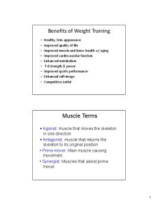
Abdominal muscles The anterior abdominal wall consists of four major muscles, the rectus abdominis, external oblique, internal oblique, and transversus abdominis. The iliopsoas must also be considered because of its important effect on the lumbar spine during trunk exercise. The muscles are illustrated in Figure 1.
\ Inguinal ligament
The rectus abdominis will flex the trunk by
approximating the pelvis and ribcage. The supraumbilical portion is emphasized by trunk flexion, Address for correspondence: Mr C. M. Norris, Norris Associates, 30 Navigation Road, Altrincham, Cheshire WA14 1NE, UK
(© 1993 Butterworth-Heinemann Ltd 0306-3674/93/010019-09
Figure 1. The anterior abdominal wall. Reproduced with the permission of the publishers from: Oliver J, Middleditch A. Functional Anatomy of the Spine. Oxford, UK: Butterworth-Heinemann, 1991I
Br J Sp Med 1993; 27(1) 19
Abdominal muscle training: C. M. Norris
while activity in the infraumbilical portion may be greater in posterior pelvic tilting1. The oblique abdominals work harder in twisting actions, the external oblique rotating the trunk to the opposite side, while the internal oblique rotates it to the same side. Thus, in right trunk rotation the right internal and left external obliques are active. The internal oblique and transversus have an important function in protecting the inguinal canal. They are continuously active in standing2, and this activity is increased during straining and expulsive actions. The transversus abdominis is thought not to participate greatly in trunk movements3. However, through its attachment to the thoracolumbar fascia it can exert an antiflexion effect on the lumbar spine4, a particularly important role in lifting. The function of the iliopsoas is mainly hip flexion, with slight adduction. It will tilt the pelvis anteriorly, holding it in this position if tight and increasing the lumbar lordosis. When the femur is fixed, the iliopsoas will pull the lumbar spine forwards, flexing it and rotating it to the opposite side, the rotation action being most noticeable at the L3 level5.
increases intradiscal pressure to only 150% 3, possibly because the lumbar spine itself does not flex. As well as intradiscal pressure variations, general lumbar compression and shear forces have been shown to alter considerably during the performance of different sit-up exercises. Reductions of 18% for compressive forces and 97% in shear forces were shown by Johnson and Reid14 using computer simulation during the performance of a sit-up exercise with the knees and hips flexed to 90° (bench curl-up).
Kinesiological analysis of abdominal exercises The term 'sit-up' is used to describe an action where the athlete moves from a supine lying to a sitting position by performing hip flexion without lumbar flexion (Figure 2). The term 'trunk-curl' (Figure 3) is used to describe flexion of the trunk, without hip flexion. Where an exercise involves both movements, the term 'curled trunk sit-up' will be used. too
Effects of flexion on the lumbar spine Lumbar flexion is limited by both contractile and inert structures. The spinal extensors will control flexion from the vertical body position by eccentric action. However, they will only limit movement if they are excessively tight, or if a flexion action is so rapid that a stretch reflex is stimulated. Of the inert structures on the posterior lumbar spine, the facet joints and spinal discs limit flexion more than the spinal ligaments6. The greatest resistance to flexion is provided by the apposing facet joints7. Facet joint movement in the lumbar spine on flexion is a combination of forward rotation and forward translation. The translation movement is limited by the facet joint, with the anterior component taking the horizontally imposed stress8. As these areas receive the highest pressures9 they are particularly vulnerable to degenerative changes3. Rapid, full change, flexion exercises on the lumbar spine are not recommended as they can stress the posterior structures excessively and may ultimately lead to hypermobility and facet degeneration. During flexion the intervertebral disc is compressed anteriorly causing the anterior annular fibres to bulge. The posterior annular fibres are stretched, placing them under tension. The discal nucleus is thought to move posteriorly with flexion10" 1. Intradiscal pressure within the lumbar spine varies tremendously with general alterations in posture, and variation is equally marked between the various sit-up procedures. If the pressure at the L3 disc for a 70-kg standing subject is said to be 100%, supine lying reduces this pressure to 25% 12. The pressure variations increase dramatically as soon as the lumbar spine is flexed, with the sitting posture increasing intradiscal pressure to 140%. The sit-up action from crook lying increases intradiscal pressure to 210%. Interestingly, the bilateral straight leg raise, although placing considerable stress on the lumbar spine, 20 Br J Sp Med 1993;
27(1)
Figure 2. The sit-up
C_ \ Figure 3. The trunk curl
Trunk curl At the beginning of this exercise, as soon as the head lifts from the ground, activity is seen in the rectus abdominis15, and as a consequence the rib cage is depressed anteriorly. This initial period of muscle activity emphasizes the supraumbilical portion of the rectus, the infraumbilical portion and the internal oblique contracting later1. As the internal oblique contracts, it pulls on the lower ribs, causing the ribs to flare out and so increase the infrasternal angle. Fixation of the pelvis is provided by the hip flexors, especially iliacus through its attachment to the pelvic rim. The strong pull of the hip flexors is partially counteracted by the pull of the lateral fibres of the external oblique which tend to tilt the pelvis posteriorly. Action of the external oblique, if powerful enough, will compress the ribs and reduce the infrasternal angle once more'. If during the execution of the trunk curl exercise, the trunk is rotated, by pulling the right shoulder towards the left leg for example (trunk curl with twist, Figure 4), extra stress is imposed on the oblique abdominals.
Abdominal muscle training: C. M. Norris
Figure 6. Bent knee sit-up Figure 4. The trunk curl with twist
Sit-up Often, the initial movement in a rapid sit-up is a momentary posterior tilting of the pelvis by action of the hip extensors. This will pre-stretch the hip flexors, giving them a mechanical advantage before hip flexion occurs. During this phase the abdominal muscles work eccentrically16. Later, the abdominals work isometrically to fix the pelvis and provide a stable base for the hip flexors to pull on. The trunk is lifted from the ground by concentric action of the hip flexors, working on the stationary femur. Only if the trunk is flexed (curled trunk sit-up) do the abdominal muscles act as primary or secondary movers. During the pure sit-up, the abdominal action felt by the athlete is mainly through action of these muscles as fixators. However, with subjects in poor physical condition, the abdominal muscles may be too weak to fix the pelvis. In this case, the iliopsoas can pull the lumbar spine into dangerous hyperextension.
Effects offoot fixation If the sit-up action is attempted from the supine lying position, there is a tendency for the legs to lift up from the supporting surface. This occurs because the legs constitute roughly one-third of the bodyweight and the trunk two-thirds, the lighter body part obviously lifting first. However, if the feet are fixed, the hip flexors can now pull powerfully without causing the legs to lift. In addition, the act of foot fixation itself may facilitate the iliopsoas17. Foot fixation requires the subject to pull against the fixation point by active dorsiflexion. This process stimulates the gait pattern at heel contact, increasing activity in the tibialis anterior, quadriceps and iliopsoas, a pattern known as flexor synergy18. As with the standard sit-up, when the feet are fixed the action of the iliopsoas is to hyperextend the lumbar spine (Figure 5). However, the increased activity seen in the iliopsoas through foot fixation will cause greater hyperextension, and therefore an increased likelihood of injury to the lumbar spine.
Effects of knee and hip flexion Bending the knees and hips to alter the starting position of a sit-up or trunk curl (Figure 6) will affect
0-0--
both the passive and active actions of the hip flexors, and the biomechanics of the lumbar spine. In the supine starting position, the iliopsoas is partially stretched and so able to exert its greatest active tension. As the muscle is shortened by flexing the hips and knees, tension produced by the iliopsoas on contraction will reduce. With 450-hip flexion tension development is 70-80% of its maximum, while with the hips and knees flexed to 900 this figure reduces to between 40 and 50% 14 However, passive tension developed by the iliopsoas due to elastic recoil must also be considered. If the hips are flexed, the iliopsoas will not be fully stretched, and will not be able to limit passively the posterior tilt of the pelvis. Instead, to fix the pelvis and provide a stable base for the abdominals to pull on, the hip flexors will contract earlier in the sit-up action. This contraction, although occurring earlier, will be of reduced intensity15 due to the length-tension relationship of the muscle19. When performing a sit-up in any starting position, contraction of the iliopsoas may place stress on the lumbar spine in severely deconditioned subjects. To reduce iliopsoas activity to a minimum, active hip flexion should be eliminated and the trunk curl exercise chosen.
Straight leg raising The bilateral straight leg raise (Figure 7) has been shown to create only slight activity in the upper rectus abdominis, although the lower rectus abdominis contributes a greater proportion of the total abdominal work than with the sit-up20. The rectus works isometrically to fix the pelvis against the strong pull of the iliopsoas21. The force of contraction of the iliopsoas is at a maximum when the lever arm of the leg is greatest, near the horizontal, and reduces as the leg is lifted towards the vertical. In subjects with weaker abdominals, the pelvis will tilt and the lumbar spine hyperextend (Figure 8). This forced hyperextension will dramatically increase stress on the facet joints, in the lumbar spine
Figure 7. Bilateral straight leg raise
I
Figure 5. At the beginning of a sit-up, the iliopsoas will hyperextend the lumbar spine if the abdominal muscles are weak
Figure 8. Anterior tilt of the pelvis and hyperextension of the lumbar spine during the bilateral straight leg raise in subjects with weak abdominal muscles Br J Sp Med 1993;
27(1) 21
Abdominal muscle training: C. M. Norris
particularly. The movement is likely to be limited by impaction of the inferior articular processes of the facet joints on the laminae of the vertebrae below, or in some cases by contact between the spinous processes . Where this action occurs rapidly, damage may result to the facet joint structures. Once contact has occurred between the facet and the lamina, further loading will cause axial rotation of the superior vertebra22. The superior vertebra pivots, causing the inferior articular process to move backwards, overstretching the joint capsule. In some cases the joint capsule may become trapped between the inferior articular process and the lamina, and eventual erosion of the laminal periosteum may 3 occur.
Modification of straight leg raising As none of the abdominal muscles actually crosses the hip, they are not prime movers of the straight leg raising movement. However, the action is an important one as it emphasizes the pelvic stabilizing function of the infraumbilical portion of the rectus
abdominis, a body area frequently neglected during training. It is common to find athletes who can perform sit-ups easily, but who are unable to demonstrate a bilateral straight leg raise while keeping the back flat on the ground. Two exercises serve as modifications of the bilateral straight leg raise to reduce the stress on the lumbar spine. The first movement is leg lowering (Figure 9). For this exercise the subject lies supine with the hips flexed to 900 but the knees extended. From this position, the legs are lowered by eccentric action of the hip flexors to a point where the pelvis begins to tilt. Immediately this occurs, the legs are brought up again to 900 hip flexion. The advantage of this exercise over the standard straight leg raise is one of changing leverage. With the standard leg raising action, the subject starts from a point of maximum leverage on the leg, forcing the hip flexors and abdominals to work maximally straight away. With leg lowering, the starting position is one of minimum leverage as the legs are held vertically. As the legs are lowered away from the vertical, leverage increases but the subject is able to control the descent of the legs and avoid the position of maximal leverage which would cause the spine to hyperextend. In cases where subjects find the leg lowering difficult to control, the knees should be bent, to reduce leverage on the leg. Alternatively the subject can perform the exercise close to a wall, so their legs cannot be lowered fully (Figure 10). The leg lowering movement is sometimes combined with a trunk curl (the V' sit-up). However, this action must be performed as a controlled movement,
I. .