in the clinic ®
in the clinic
Abdominal Aortic Aneurysm
Physician Writer Frank A. Lederle, MD Section Co-Editors Christine Laine, MD, MPH David R. Goldmann, MD Harold C. Sox, MD
Screening and Prevention
page ITC5-2
Diagnosis
page ITC5-4
Treatment
page ITC5-9
Practice Improvement
page ITC5-14
CME Questions
page ITC5-16
The content of In the Clinic is drawn from the clinical information and education resources of the American College of Physicians (ACP), including PIER (Physicians’ Information and Education Resource) and MKSAP (Medical Knowledge and Self-Assessment Program). Annals of Internal Medicine editors develop In the Clinic from these primary sources in collaboration with the ACP’s Medical Education and Publishing Division and with the assistance of science writers and physician writers. Editorial consultants from PIER and MKSAP provide expert review of the content. Readers who are interested in these primary resources for more detail can consult http://pier.acponline.org and other resources referenced in each issue of In the Clinic.
The information contained herein should never be used as a substitute for clinical judgment. CME objective: To review the screening and prevention, diagnosis, treatment, and practice improvement for abdominal aortic aneurysm. © 2009 American College of Physicians
Downloaded From: http://annals.org/ by a McGill University User on 07/15/2016
A
n aneurysm is a failure of the arterial wall that results in a balloonlike dilatation of a segment of the artery. Aortic aneurysms constitute the 14th leading cause of death in the United States and the 10th leading cause of death in older men, who are the principal victims (1). Diagnosis of abdominal aortic aneurysm (AAA) is important because the natural history is that of continued enlargement with potentially catastrophic consequences. Because the condition may be entirely asymptomatic, and when symptoms are present, they may be nonspecific, the internist is often the first to evaluate the patient. Thus, understanding the disease, optimal diagnostic methods, and management are crucial for primary care practitioners. Approximately 80% of aortic aneurysms occur between the renal arteries and the aortic bifurcation. Abdominal aortic aneurysm is present in more than 5% of older men who have smoked. Each year in the United States, AAA rupture causes 4500 deaths; another 1400 deaths result from the 45 000 repair procedures done to prevent rupture (2).
Screening and Prevention
1. Silverberg E, Boring CC, Squires TS. Cancer statistics, 1990. CA Cancer J Clin. 1990;40:9-26. [PMID: 2104569] 2. McPhee JT, Hill JS, Eslami MH. The impact of gender on presentation, therapy, and mortality of abdominal aortic aneurysm in the United States, 20012004. J Vasc Surg. 2007;45:891-9. [PMID: 17391899] 3. Lederle FA, Johnson GR, Wilson SE, et al. The aneurysm detection and management study screening program: validation cohort and final results. Aneurysm Detection and Management Veterans Affairs Cooperative Study Investigators. Arch Intern Med. 2000;160:1425-30. [PMID: 10826454] 4. The UK Small Aneurysm Trial Participants. Mortality results for randomised controlled trial of early elective surgery or ultrasonographic surveillance for small abdominal aortic aneurysms. Lancet. 1998;352:1649-55. [PMID: 9853436]
© 2009 American College of Physicians
What factors increase the risk for AAA? Smoking is the most important reversible risk factor for AAA. In the largest screening study (3), a history of cigarette smoking accounted for 75% of all AAAs 4.0 cm or greater in diameter. The association with AAA is stronger for current smokers than for former smokers, increases with number of years smoked, and decreases with the elapsed time after quitting. After adjustment for other risk factors, AAA is several times more common in men than in women. Other factors associated with AAA include age, white race, and family history of AAA (see Box). Abdominal aortic aneurysm is less common in patients with diabetes (3). In a cross-sectional screening study, 126 196 veterans age 50 to 79 years underwent ultrasonography to detect AAA. Aneurysms 3.0 cm or greater in diameter were present in 4.2% of participants. The principal positive associations with AAA were age, smoking, male sex, white race, family history of AAA, and atherosclerotic diseases. This study first described the surprising and still unexplained inverse association of AAA with diabetes (3).
ITC5-2
In the Clinic
Downloaded From: http://annals.org/ by a McGill University User on 07/15/2016
What is the natural history of AAA? The natural history of AAA is to enlarge slowly over a period of years. Abdominal aortic aneurysms of 4.0 to 5.5 cm enlarge at a mean rate of 0.3 cm per year, with less than 25% enlarging 0.5 cm or more per year (4, 5). Larger AAAs enlarge faster. The rate of enlargement for 3.0- to 4.0-cm AAAs is one half that of 4.0-to 5.5-cm AAAs (6), and one half again faster for AAAs greater than 5.5 cm (7). The enlargement rate differs among individuals and is faster with advancing age and continued smoking. Most AAAs never rupture, but when rupture occurs, the mortality rate is 80% (8). As a AAA enlarges, the risk for rupture increases. The rupture rate for AAAs of 4.0 to 5.5 cm in diameter is 0.7% to 1.0% per year (4, 5), higher than with AAAs less than 4.0 cm. The rupture rate for small AAA is higher in women than in men (9). Men and women have equivalent rupture rates for AAAs greater than 5.5 cm (10). Rupture rates of AAAs greater than 5.5 cm in otherwise healthy patients are unknown and unlikely to be known in the future because of
Annals of Internal Medicine
5 May 2009
general agreement that these patients need treatment. However, in patients with AAA greater than 5.5 cm who are unfit for surgery, the rupture rate is high (7). A 5-year prospective longitudinal study followed 198 patients with AAA 5.5 cm or greater who were not repaired electively because of high operative risk or patient refusal. More than one half of the patients died during the study, 30% within 1 year of enrollment. The rate of probable AAA rupture was 10% per year in the entire cohort, and more than 25% at 6 months for AAA 8.0 cm or greater. Whether these rates apply to healthier patients is unknown, but they are useful for decision making in high operative risk patients (7).
Who should clinicians screen for AAA and how should screening be done? The U.S. Preventive Services Task Force (USPSTF) now recommends one-time screening with ultrasonography to detect asymptomatic AAA in 65- to 75-year-old men who have ever smoked (11). The USPSTF does not make a recommendation for nonsmoking men and recommends against screening women. Medicare covers abdominal ultrasonography to screen for AAA for men who have ever smoked and anyone with a family history of AAA. Screening should take place at the Welcome to Medicare visit, which must occur within 6 months after beginning Medicare insurance coverage. The USPSTF based its recommendation to screen for AAA on a solid body of evidence. Four randomized trials of AAA screening have reported results. These trials randomly assigned one half of patients from a population list to receive an invitation for ultrasonography screening. Of these, 60% to 80% attended the screening. Of the men who were screened, 4% to 8%
5 May 2009
Annals of Internal Medicine
Risk Factors for Abdominal Aortic Aneurysm • Older age • Smoking • Male sex • White race • Family history of abdominal aortic aneurysm • Occlusive atherosclerotic disease
had AAAs 3.0 cm in diameter or larger, and the frequency of elective AAA repair was several-fold greater in the invited groups than in the uninvited control groups. In a meta-analysis of these 4 trials performed for the USPSTF, men invited to undergo screening had a lower rate of AAA-related mortality (odds ratio, 0.57 [95% CI, 0.45 to 0.74]) (12). In the Multicentre Aneurysm Screening Study (MASS) conducted in the United Kingdom, 67 770 men age 65 to 74 years were randomly assigned to ultrasonography screening for AAA or usual care. Patients with AAA 5.5 cm or larger were referred for elective repair. After a mean 7 years of follow-up, deaths related to AAA were significantly reduced from 196 in the control group to 105 in the invited group. All-cause mortality was lower in the screened group; the difference was of borderline statistical significance (13). A recent meta-analysis of longer-term outcomes from the screening trials reported a small but statistically significant reduction in all-cause mortality in the invited patients (odds ratio, 0.97 [95% CI, 0.94 to 0.99]) (14).
These trials, added to those of smaller studies, form the basis of the USPSTF recommendation for AAA screening (11). They were not limited to smokers and did not report their results by such subgroups as smoking status because the authors had little information about the control groups. The first trial included 9342 women, who did not benefit from screening. However, the number of
In the Clinic
Downloaded From: http://annals.org/ by a McGill University User on 07/15/2016
ITC5-3
5. Aneurysm Detection and Management Veterans Affairs Cooperative Study Group. Immediate repair compared with surveillance of small abdominal aortic aneurysms. N Engl J Med. 2002;346:143744. [PMID: 12000813] 6. Santilli SM, Littooy FN, Cambria RA, et al. Expansion rates and outcomes for the 3.0cm to the 3.9-cm infrarenal abdominal aortic aneurysm. J Vasc Surg. 2002;35:666-71. [PMID: 11932660] 7. Veterans Affairs Cooperative Study #417 Investigators. Rupture rate of large abdominal aortic aneurysms in patients refusing or unfit for elective repair. JAMA. 2002;287:2968-72. [PMID: 12052126] 8. Adam DJ, Mohan IV, Stuart WP, et al. Community and hospital outcome from ruptured abdominal aortic aneurysm within the catchment area of a regional vascular surgical service. J Vasc Surg. 1999;30:922-8. [PMID: 10550191]
© 2009 American College of Physicians
AAA-related events in women was too small (10 ruptures in the screened group vs. 9 in the control group after 10 years of follow-up) to draw firm conclusions about the effect of screening in women (15).
9. Brown LC, Powell JT. Risk factors for aneurysm rupture in patients kept under ultrasound surveillance. UK Small Aneurysm Trial Participants. Ann Surg. 1999;230:289-96; discussion 296-7. [PMID: 10493476] 10. Powell JT, Brown LC, Greenhalgh RM, et al. The rupture rate of large abdominal aortic aneurysms: is this modified by anatomical suitability for endovascular repair? Ann Surg. 2008;247:173-9. [PMID: 18156938] 11. U.S. Preventive Services Task Force. Screening for abdominal aortic aneurysm: recommendation statement. Ann Intern Med. 2005;142:198202. [PMID: 15684208] 12. Fleming C, Whitlock EP, Beil TL, et al. Screening for abdominal aortic aneurysm: a bestevidence systematic review for the U.S. Preventive Services Task Force. Ann Intern Med. 2005;142:203-11. [PMID: 15684209] 13. Multicentre Aneurysm Screening Study Group. A sustained mortality benefit from screening for abdominal aortic aneurysm. Ann Intern Med. 2007;146:699-706. [PMID: 17502630] 14. Lindholt JS, Norman P. Screening for abdominal aortic aneurysm reduces overall mortality in men. A meta-analysis of the mid- and long-term effects of screening for abdominal aortic aneurysms. Eur J Vasc Endovasc Surg. 2008;36:167-71, response to comment, 621-2. [PMID: 18485756]
Several screening programs have reported the findings of repeat screening (16, 17). Aneurysms were less frequent and smaller than on the initial screening, which is not surprising because the initial screening would have detected the long-standing and therefore larger AAAs that had developed over the preceding
years. Consequently, most authors and the USPSTF have concluded that one-time screening after age 65 is sufficient. What should clinicians tell patients to help them decrease their risk for AAA? Because the association with AAA is substantially stronger for current smokers than former smokers, it is reasonable to assume that smoking cessation will reduce the risk for AAA. Clinicians can cite increased risk for AAA as another reason for patients to stop smoking.
Screening and Prevention... Abdominal aortic aneurysm is the 10th most common cause of death in older men. Smoking is its most important modifiable risk factor, and former smokers have a progressively smaller risk for AAA than current smokers. Randomized trials show that screening substantially reduces the risk for AAArelated mortality. The USPSTF recommends one-time abdominal ultrasonography screening for male smokers age 65 to 75 years.
CLINICAL BOTTOM LINE
Diagnosis
© 2009 American College of Physicians
How does AAA present? Before rupture, AAAs are nearly always asymptomatic. Detection occurs after abdominal palpation, radiologic imaging for other purposes, or imaging tests done to screen for AAA. This section focuses on diagnosing ruptured AAA, because it happens infrequently in a physician’s career and is one of the true emergencies of medical practice. Ruptured AAA is a leading cause of sudden death (18). Its most frequent findings are pain and tenderness in the abdomen, flank, groin, or back. Other common symptoms include urinary retention, constipation, urge to defecate, hypotension, transient syncope, sudden collapse, and shock. Misdiagnosis of ruptured AAA remains disturbingly common,
ITC5-4
In the Clinic
Downloaded From: http://annals.org/ by a McGill University User on 07/15/2016
despite descriptions of its varied presentations dating back more than a century. Although ruptured AAA usually presents with catastrophic vascular collapse, it can present with normal blood pressure and hematocrit, although leukocytosis is usually present. Many physicians are unfamiliar with the syndromes of symptomatic unruptured AAA, in which abdominal, flank, or back pain precedes rupture, and contained AAA rupture, in which the hematoma is sealed off in the retroperitoneum. Either of these syndromes can persist for days or, rarely, weeks before uncontained rupture leads to death (19). The clinician must be suspicious of the possible presence of AAA when encountering such symptoms in the appropriate context. Intervention can be lifesaving.
Annals of Internal Medicine
5 May 2009
How should clinicians use the history and physical examination in diagnosing AAA? Unruptured AAA
Although unruptured AAAs are almost always asymptomatic, a history of heavy tobacco use or a family history of AAA should heighten awareness in older persons, particularly men. The only physical examination maneuver of demonstrated value is abdominal palpation to detect a widened aortic pulsation or “pulsatile mass”. Physicians who care for older patients should become proficient at measuring the width of the aorta between the 2 index fingers. The width of the aortic pulsation provides the key evidence of AAA, not its prominence or force, the presence of bruits, or other findings. Palpating an AAA seems to be safe: No one has reported rupture immediately following palpation. However, abdominal palpation misses many AAAs 5.0 cm or larger in diameter and is even less sensitive for smaller AAAs or in obese patients. A systematic review pooled 15 studies involving 2955 participants not previously known to have AAA. Each was screened with both abdominal palpation and ultrasonography, which served as the diagnostic reference standard. The sensitivity of abdominal palpation ranged from 29% for AAA 3.0 to 3.9 cm, to 50% for AAA 4.0 to 4.9 cm, and 76% for AAA 5.0 cm or greater. The positive predictive value of palpation for detecting AAA 3.0 cm or greater was 43%. Abdominal obesity seemed to decrease the sensitivity of palpation. The authors concluded that abdominal palpation had moderate sensitivity for detecting AAAs large enough to be referred for surgery, but could not be used to exclude AAA (20).
On the basis of the estimates from this systematic review, a negative physical examination reduces the odds that the patient has an AAA 5.0 cm or larger by approximately 25%. On the other hand, if the
5 May 2009
Annals of Internal Medicine
Signs and Symptoms of Ruptured AAA • Pain in the flank, abdomen, groin, or back • Abdominal, flank, or back tenderness • Gastrointestinal dysfunction (constipation, distention, bleeding, urge to defecate) • Urinary dysfunction (retention, difficulty voiding, renal colic) • Syncope • Hypotension and shock
examiner feels a pulsatile mass, imaging confirmation, usually ultrasonography, is mandatory. Ruptured AAA
The diagnosis of ruptured AAA can be obvious or difficult. The most important prerequisite is a high index of suspicion, especially in an older man who has smoked. Physicians must know the various presentations and be particularly suspicious when the patient’s condition seems more serious than would be typical with an alternative explanation for flank or abdominal pain, such as pyelonephritis, diverticulitis, or lumbar disk herniation. The Box lists signs and symptoms of ruptured AAA. A pulsatile mass may be present but is often absent and should not be relied on to confirm rupture. Although ruptured AAAs tend to be large, palpating one can be difficult because of abdominal tenderness, guarding, and intestinal distention due to ileus. Studies of ruptured AAA that include postmortem diagnoses of rupture have reported that a pulsatile mass is palpable only about 50% of the time (20), which means that a negative examination is very unreliable. Table 1 describes the differential diagnosis of pulsatile mass and ruptured AAA, and Table 2 lists the history and physical examination elements for ruptured and unruptured AAA.
In the Clinic
Downloaded From: http://annals.org/ by a McGill University User on 07/15/2016
ITC5-5
15. Scott RA, Bridgewater SG, Ashton HA. Randomized clinical trial of screening for abdominal aortic aneurysm in women. Br J Surg. 2002;89:283-5. [PMID: 11872050] 16. Lederle FA, Johnson GR, Wilson SE, et al. Yield of repeated screening for abdominal aortic aneurysm after a 4year interval. Aneurysm Detection and Management Veterans Affairs Cooperative Study Investigators. Arch Intern Med. 2000;160:1117-21. [PMID: 10789604] 17. Hafez H, Druce PS, Ashton HA. Abdominal aortic aneurysm development in men following a “normal” aortic ultrasound scan. Eur J Vasc Endovasc Surg. 2008;36:553-8. [PMID: 18718773] 18. Thomas AC, Knapman PA, Krikler DM, et al. Community study of the causes of “natural” sudden death. BMJ. 1988;297:1453-6. [PMID: 3147014] 19. Lederle FA, Parenti CM, Chute EP. Ruptured abdominal aortic aneurysm: the internist as diagnostician. Am J Med. 1994;96:163-7. [PMID: 8109601] 20. Lederle FA, Simel DL. The rational clinical examination. Does this patient have abdominal aortic aneurysm? JAMA. 1999;281:77-82. [PMID: 9892455]
© 2009 American College of Physicians
Table 1. Differential Diagnosis of Pulsatile Mass and Ruptured Abdominal Aortic Aneurysm Disease
Characteristics
Pulsatile mass Normal aorta
Notes
Examiner may be misled by prominent Clinical observation* pulsation or subcutaneous fat Mass adherent to normal aorta that can transmit the aortic pulsation
Para-aortic nodes Ruptured abdominal aortic aneurysm† Gastrointestinal disease (such as diverticulitis, bowel ischemia, pancreatitis, penetrating duodenal ulcer, malignant conditions)
Abdominal pain, constipation, leukocytosis
The expanding hematoma can cause gastrointestinal symptoms by compressing the bowel. Reduced blood pressure and obstruction of aortic branch arteries can compromise blood supply to the gastrointestinal and urinary tracts. The expanding hematoma can compress the ureters. Reduced blood pressure and obstruction of aortic branch arteries can compromise blood supply to the urinary tract.
Urinary tract disease (such as stones, infection)
Flank pain, urinary retention, leukocytosis
Spinal diseases (such as herniated disk, bony metastases, lower extremity neuropathy) Incarcerated hernia
Severe back pain, nerve impingement syndromes Inguinal mass with pain and tenderness
AAA often diagnosed at surgery for incarcerated hernia‡
AAA = abdominal aortic aneurysm. * Nusbaum JW, Freimanis AK, Thomford NR. Echography in the diagnosis of abdominal aortic aneurysm. Arch Surg. 1971;102:385-8. [PMID: 5553311] † Lederle FA, Parenti CM, Chute EP. Ruptured abdominal aortic aneurysm: the internist as diagnostician. Am J Med. 1994;96:163-7. [PMID: 8109601] ‡ Khaw H, Sottiurai VS, Craighead CC, Batson RC. Ruptured abdominal aortic aneurysm presenting as symptomatic inguinal mass: report of six cases. J Vasc Surg. 1986;4:384-9. [PMID: 3761483]
Table 2. History and Physical Examination Elements for Abdominal Aortic Aneurysm Category
Element
Unruptured abdominal aortic aneurysm History Physical examination
Ruptured abdominal aortic aneurysm History 21. Lederle FA, Wilson SE, Johnson GR, et al. Variability in measurement of abdominal aortic aneurysms. Abdominal Aortic Aneurysm Detection and Management Veterans Administration Cooperative Study Group. J Vasc Surg. 1995;21:945-52. [PMID: 7776474]
Physical examination
Sensitivity, %
Usually asymptomatic Abdominal palpation AAA ≥3.0 cm AAA ≥4.0 cm AAA ≥5.0 cm All AAA
Pain in abdomen, flank, or back Constipation Urinary retention Syncope Abdominal palpation for pulsatile abdominal mass Hypotension Abdominal tenderness Ecchymosis anywhere between diaphragm and knees
39 60 76 ≤81
Specificity, %
Likelihood Ratio Positive
Likelihood Ratio Negative
97
12.0 15.6
0.72 0.51
99.2
80–100 22 22 26 40–60 50–70 70–90
AAA = abdominal aortic aneurysm.
© 2009 American College of Physicians
ITC5-6
In the Clinic
Downloaded From: http://annals.org/ by a McGill University User on 07/15/2016
Annals of Internal Medicine
5 May 2009
What imaging studies and other laboratory tests should clinicians order to evaluate patients with suspected AAA? Unruptured Aneurysm
Ultrasonography is the preferred test for diagnosis and surveillance of AAA in asymptomatic patients. It is safe, accurate, and relatively inexpensive. When the examination is adequate, ultrasonography has a sensitivity and specificity of nearly 100%. Bowel gas may cause an inadequate study, but a repeated examination after fasting is usually successful. Lack of precision (usually 11 x 109 cells/L Hemoglobin 5.5 cm; ask about symptoms Drug therapy Treatment of Antihypertensives Ongoing concurrent medical problems, such as hypertension Patient education
Symptom recognition in patients with AAA under observation
Ask patients to report abdominal, flank, or back pain
Notes
Optional, does not replace ultrasonography
Treatment of hypertension of undetermined value for preventing rupture
Each visit
AAA = abdominal aortic aneurysm.
ITC5-12
In the Clinic
Downloaded From: http://annals.org/ by a McGill University User on 07/15/2016
Annals of Internal Medicine
5 May 2009
imaging surveillance to assess aneurysm diameter is recommended every 6 to 12 months if the AAA diameter is 4.0 to 5.4 cm and every 2 to 3 years if it is less than 4.0 cm (49). As noted previously, a large randomized trial (5) showed that imaging at intervals of 6 months was equivalent to a strategy of immediate repair of AAAs measuring 4.0 to 5.4 cm. Observational studies that monitored enlargement rates suggest that surveillance intervals of up to 3 years are safe for smaller AAAs (6, 50, 51). In deciding when to recommend repair, physicians should remember that errors of up to 0.5 cm in measuring the diameter of an AAA are common with abdominal ultrasonography or CT (21). How should clinicians monitor patients after aortic aneurysm repair? The monitoring strategy depends on the method of AAA repair. After open repair, the graft failure
rate is very low (about 0.3% per year), and no specific follow-up is recommended (5, 52, 53). Endovascular repair is associated with more late graft problems (for example, endoleaks, graft migration or kinking, continued AAA enlargement) and resulting reinterventions. In the EVAR-1 trial (40), 20% of patients randomly assigned to endovascular repair required reintervention within 4 years compared with 6% of those randomly assigned to open repair. Current practice, which is derived from the device approval trials but not based on comparative trials, is to perform CT at 1, 6, and 12 months after endovascular repair and then annually for the rest of the patient’s life. This strategy is a considerable burden for both patients and vascular surgeons; is expensive; and results in substantial radiation exposure, especially with the commonly used triphasic CT protocol. Alternative followup methods are under active investigation.
Treatment... Elective repair of AAA is indicated when the external diameter
reaches 5.5 cm. Below that size, randomized trials show that repair does not improve outcomes. Smoking cessation treatment and control of high blood pressure are indicated. Surveillance abdominal ultrasonography is indicated for aneurysms smaller than 5.5 cm, with more frequent surveillance as the diameter approaches 5.5 cm. The choice between open elective repair and endovascular repair is difficult. Endovascular repair has less mortality and morbidity in the perioperative period but all-cause mortality and other outcomes are equivalent by 2 years, and the very long-term outcomes of endovascular repair are unknown. The treatment for ruptured AAA is immediate repair, either open or endovascular.
CLINICAL BOTTOM LINE
5 May 2009
Annals of Internal Medicine
In the Clinic
Downloaded From: http://annals.org/ by a McGill University User on 07/15/2016
ITC5-13
49. American Association for Vascular Surgery. ACC/AHA 2005 guidelines for the management of patients with peripheral arterial disease (lower extremity, renal, mesenteric, and abdominal aortic): executive summary a collaborative report from the American Association for Vascular Surgery/Society for Vascular Surgery, Society for Cardiovascular Angiography and Interventions, Society for Vascular Medicine and Biology, Society of Interventional Radiology, and the ACC/AHA Task Force on Practice Guidelines (Writing Committee to Develop Guidelines for the Management of Patients With Peripheral Arterial Disease) endorsed by the American Association of Cardiovascular and Pulmonary Rehabilitation; National Heart, Lung, and Blood Institute; Society for Vascular Nursing; TransAtlantic Inter-Society Consensus; and Vascular Disease Foundation. J Am Coll Cardiol. 2006;47:1239-312. [PMID: 16545667] 50. Grimshaw GM, Thompson JM, Hamer JD. A statistical analysis of the growth of small abdominal aortic aneurysms. Eur J Vasc Surg. 1994;8:741-6. [PMID: 7828753] 51. McCarthy RJ, Shaw E, Whyman MR, et al. Recommendations for screening intervals for small aortic aneurysms. Br J Surg. 2003;90:821-6. [PMID: 12854107] 52. Hallett JW Jr, Marshall DM, Petterson TM, et al. Graftrelated complications after abdominal aortic aneurysm repair: reassurance from a 36-year population-based experience. J Vasc Surg. 1997;25:277-84; discussion 285-6. [PMID: 9052562]
© 2009 American College of Physicians
53. Johnston KW. Nonruptured abdominal aortic aneurysm: sixyear follow-up results from the multicenter prospective Canadian aneurysm study. Canadian Society for Vascular Surgery Aneurysm Study Group. J Vasc Surg. 1994;20:16370. [PMID: 8040938] 54. Joint Council of the American Association for Vascular Surgery and Society for Vascular Surgery. Guidelines for the treatment of abdominal aortic aneurysms. Report of a subcommittee of the Joint Council of the American Association for Vascular Surgery and Society for Vascular Surgery. J Vasc Surg. 2003;37:1106-17. [PMID: 12756363] 55. Lederle FA, Powell JT, Greenhalgh RM. Repair of small abdominal aortic aneurysms [Letter]. N Engl J Med. 2006;354:1537-8. [PMID: 16598058]
in the clinic
Tool Kit Abdominal Aortic Aneurysm
© 2009 American College of Physicians
What do professional organizations recommend about the care of patients with AAA? The U.S. Preventive Services Task Force (USPSTF) recommends onetime screening for AAA with ultrasonography in men age 65 to 75 years who have ever smoked (11). It does not recommend for or against screening in 65- to 75-year-old men who have never smoked. The USPSTF recommends against routine screening in women. The Joint Council (54) recommended 5.5-cm diameter as the appropriate threshold for elective repair in most patients, on the basis of the finding of 2 randomized trials (4, 5, 39) that patients did not benefit from repairing AAA less than 5.5 cm in diameter. However, the Council also recommended repair of smaller AAA in various patient groups, including women, younger patients, and patients of surgeons whose operative mortality rate is low. The last recommendation is based on weak evidence (observational studies) and personal opinion and conflicts with randomized trial evidence that repair of small aneurysms confers no benefit. The American College of Cardiology and the American Heart Association’s peripheral arterial disease guideline (49) contains a number of well-supported recommendations about AAA, including one-time
ultrasonography screening in men age 65 to 75 years old who have ever smoked or have a first-degree relative with AAA; advice to stop smoking; open or endovascular repair of AAA 5.5 cm or larger in diameter; imaging surveillance of AAA 4.0 to 5.4 cm at 6- to 12-month intervals and 2- to 3-year intervals for AAA less than 4.0 cm; and immediate evaluation and repair of ruptured AAA. However, the guideline contains several recommendations that conflict with randomized trial findings, including long-term b-blockade to slow the rate of AAA enlargement. The guideline also states that “Repair can be beneficial in patients with infrarenal or juxtarenal AAAs 5.0 to 5.4 cm in diameter,” an opinion that prompted a letter of dissent from the directors of the 2 randomized trials (55). How should clinicians educate patients with AAA? Patients with AAA should know about the course, management, and prognosis of AAA and important self-management issues, including the importance of smoking cessation, the need to report symptoms immediately, the scheduling of regular follow-up visits for surveillance before repair and after endovascular repair, and the available surgical options. Physicians have a responsibility to provide them with this information.
PIER Modules www.pier.acponline.org Access the following PIER module: Abdominal aortic aneurysm. PIER modules provide evidence-based, updated information on current diagnosis, treatment, and management, in an electronic format designed for rapid access at the point of care. Patient Education Resources www.annals.org/intheclinic/AAA.html Access the Patient Information material on the following page for duplication and distribution to patients. www.medicinenet.com/abdominal_aortic_aneurysm Abdominal Aortic Aneurysm index from Medicinenet.com Clinical Guidelines www.annals.org/cgi/reprint/142/3/198.pdf U.S. Preventive Services Task Force on screening for AAA Quality Measures for AAA qualityindicators.ahrq.gov/downloads/iqi/iqi_guide_v31.pdf Guide to Inpatient Quality Indicators, from the Agency for Healthcare Research and Quality (AAA is discussed on pp. 25, 26, 39, and 40) www.qualitymeasures.ahrq.gov/summary/summary.aspx?doc_id=12739 Abdominal aortic aneurysm repair, from the National Quality Measures Clearinghouse
ITC5-14
In the Clinic
Downloaded From: http://annals.org/ by a McGill University User on 07/15/2016
Annals of Internal Medicine
in the clinic
Practice Improvement
5 May 2009
WHAT YOU SHOULD KNOW ABOUT ABDOMINAL AORTIC ANEURYSM
In the Clinic Annals of Internal Medicine annals.org
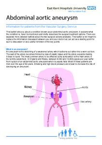
What is an abdominal aortic aneurysm? Blood moves from the heart to other parts of the body through arteries. The abdominal aorta is the main artery in the belly, which is also called the abdomen. An abdominal aortic aneurysm (AAA) occurs when the wall of the aorta gets too big. This is caused by a weakening of the wall of the aorta. If the enlarged aorta breaks, blood can then leak into the body.
Is AAA a big problem? Yes. It is the 14th most common cause of death in the United States. In older men, it is the 10th most common cause of death.
What are the symptoms of AAA?
How is AAA found? The doctor may feel the AAA when pressing on the belly. The best way to find AAA is by doing a test called an ultrasonography of the abdomen.
Who should have a screening test for AAA? A screening test is done to look for a disease when there are no symptoms. Experts recommend that older men who smoke should have a screening test for AAA. Experts do not recommend a screening test for other people.
Who is most likely to have an AAA? Cigarette smoking makes the chances of having an AAA greater than anything else. Being older, of white race, male, and a smoker all add to the risk for having an AAA.
If screening finds AAA, what should be done? The greatest chance that the AAA could break is if it is larger than 2 in (5.5 cm). If it is smaller than 2 in, the doctor will do an ultrasonography test, tell the patient to stop smoking, and treat the patient’s high blood pressure. If the AAA is 2 in or more, doctors usually do surgery to fix it.
For More Information Web Sites With Information on Abdominal Aortic Aneurysm: www.medem.com/medlib/article/ZZZVTBUUBZE JAMA Patient Page: Aortic Aneurysms
www.medicinenet.com/abdominal_aortic_aneurysm Abdominal Aortic Aneurysm index from Medicinenet.com
Downloaded From: http://annals.org/ by a McGill University User on 07/15/2016
Patient Information
There are no symptoms of AAA until it breaks. If blood does leak out, there may be pain in the back or belly, weakness, or fainting.
CME Questions 1. A 75-year-old asymptomatic man undergoes his annual examination. He is obese and has a long history of smoking, hypertension, and chronic kidney disease (creatinine level, 2.7 mg/dL [238.73 µmol/L]). His medications are furosemide, ramipril, atorvastatin, and aspirin.
pulsatile mass. A follow-up ultrasonography shows a 5.1-cm aneurysm with thrombus. The patient is again encouraged to stop smoking. What is the most appropriate next step in this patient’s management?
A. Repeat ultrasonography in 6 On physical examination, the blood months pressure is 170/78 mm Hg bilaterally, B. Increase atenolol, repeat and pulse rate is 70/min and regular. ultrasonography in 6 months There is a 2/6 holosystolic murmur heard C. Elective aneurysm repair at the apex with radiation to the axilla. D. Start warfarin (international There is a soft systolic abdominal bruit normalized ration, 2–3), repeat and a mildly tender midline pulsatile ultrasonography in 6 months mass. There are bilateral femoral bruits with absent distal pulses. Laboratory 3. A 67-year-old woman comes to your studies show a total cholesterol level of office because of intermittent lower back 200 mg/dL (5.17 mmol/L), HDL cholespain of 4 days’ duration. The pain is dull, terol level of 32 mg/dL (0.83 mmol/L), nonradiating, and nonpositional, and is and LDL cholesterol level of 132 mg/dL unrelated to meals or exertion. She has (3.41 mmol/L). The patient is encouraged no dyspnea, chest pain, or dysuria, and no to stop smoking. history of trauma. She has a history of hypercholesterolemia and underwent a In addition to increasing the antihypercholecystectomy at age 45 years. She also tensive medications, what is the most has a history of recurrent urinary tract appropriate next step in this patient’s infections. She stopped smoking 10 years management? ago. Her only medication is simvastatin, A. Abdominal radiograph 20 mg every night orally. B. Abdominal computed tomography On physical examination, blood pressure scan with contrast is 145/85 mm Hg and heart rate is 86 C. Abdominal ultrasound beats/min and regular. The patient is D. Contrast aortography afebrile. Jugular venous pressure is nor2. A 74-year-old woman undergoes a roumal. Carotid pulses are 2+ bilaterally, tine evaluation. She is a smoker and has without bruits. Cardiac examination hypertension, hypercholesterolemia, and shows S4; the findings are otherwise type 2 diabetes mellitus. Last year, she normal. The lungs are clear to auscultahad an asymptomatic 4.4-cm infrarenal tion. Abdominal examination shows a abdominal aortic aneurysm diagnosed midline pulsatile mass. No spinal or cosduring an ultrasonography for suspected tovertebral angle tenderness is noted. gallstones, at which time she was Distal pulses are normal. encouraged to stop smoking. She is Laboratory findings include hematocrit petite, active, asymptomatic, and comof 36%, leukocyte count of 9.5 × 10⁹ pliant with her medications, which cells/L, platelet count of 290 × 109 include atenolol, glyburide, metformin, cells/L, and serum creatinine level of 1.5 lisinopril, and aspirin. mg/dL (114.38 µmol/L). Results of uriOn physical examination, the blood pressure is 125/78 mm Hg, and the pulse rate is 70/min and regular. The lungs are clear; cardiac examination shows an S4, and abdominal examination shows a nontender abdomen with a
nalysis are normal. The LDL cholesterol level is 115 mg/dL (2.98 mmol/L). Abdominal ultrasound shows a 4.8-cm abdominal aortic aneurysm that originates from the celiac trunk and extends below the renal arteries.
Which of the following is the most appropriate next step?
A. Initiate treatment with metoprolol, 25 mg twice a day orally, and schedule a follow-up visit with ultrasonography in 6 months. B. Increase the dose of simvastatin to 40 mg/d and schedule a follow-up visit with ultrasonography in 3 months. C. Hospitalize the patient, order a high-resolution computed tomography scan, and obtain a vascular surgery consultation. D. Hospitalize the patient; initiate treatment with metoprolol, 25 mg twice a day; and refer the patient to a vascular surgeon. E. Initiate treatment with metoprolol, 25 mg twice a day orally, and order magnetic resonance imaging of the spine. 4. A 72-year-old man is evaluated during a routine examination. He has a 45pack-year history of smoking but quit smoking 10 years ago. He is fit and exercises aggressively. He has no known coronary artery disease and no medical problems. On physical examination, BMI is 26.4 kg/m2. Pulse rate is 62/min, and blood pressure is 118/64 mm Hg. Serum total cholesterol level is 175 mg/dL (4.53 mmol/L), serum high-density lipoprotein cholesterol level is 52 mg/dL (1.34 mmol/L), serum low-density lipoprotein cholesterol level is 102 mg/dL (2.64 mmol/L), serum triglyceride level is 105 mg/dL (1.19 mmol/L). Which of the following is most appropriate next step in the management of this patient?
A. Electron-beam computed tomography for calcium score B. Carotid artery ultrasonography C. Ultrasound to evaluate for abdominal aortic aneurysm D. Statin therapy
Questions are largely from the ACP’s Medical Knowledge Self-Assessment Program (MKSAP). Go to www.annals.org/intheclinic/ to obtain up to 1.5 CME credits, to view explanations for correct answers, or to purchase the complete MKSAP program.
© 2009 American College of Physicians
ITC5-16
In the Clinic
Downloaded From: http://annals.org/ by a McGill University User on 07/15/2016
Annals of Internal Medicine
5 May 2009