Biota Vol. 16 (2): 342−347, Juni 2011 ISSN 0853-8670
A Sensitive Method for Identification of Porcine Contaminant in Unprocessed Food by PCR Amplification Technique Metode Sensitif untuk Identifikasi Pencemaran Babi pada Makanan Tanpa Diolah dengan Teknik Amplifikasi PCR Endang Tri Margawati*, Muhamad Ridwan, and Indriawati Research Center for Biotechnology, The Indonesian Institute of Sciences (LIPI) Jln. Raya Bogor KM. 46, Cibinong E-mail:
[email protected] *Corresponding author
Abstract In the era of globalization currently, it is almost impossible to avoid from either processed or non-processed food from overseas. This research was aimed to examine the sensitivity of PCR technique in the identification of small contents (%) of porcine contaminant in the fresh beef meat. Five-tiny porcine contents of 0.05; 0.10; 0.15; 0.20 and 0.25% in a total weight of 5 gram mixed meat were examined in this study. Positive control (100% porcine meat) and negative control (100% beef meat) were also involved. Five mixed meat, positive and negative control were collected their DNA by using commercial kit (QIAGEN). A pair of specific primer for porcine Leptin was used for DNA amplification. PCR optimization was conducted in prior to PCR work to get approximately annealing temperature of the Leptin primer. Two PCR cycles of 25 and 35 were applied in the PCR works. PCR products were visualized in the 1020% PAGE Gradient. Results showed that Lepten fragments were identified at all 5 mixed meat samples and positive control (100% pork) with size of 152bp, however it was none Leptin in the negative control (100& beef). Amplification of DNA with 35 cycles of PCR showed more clear band compared to that 25 cycles of PCR in all samples. This study suggests that a quick method of PCR amplification with 35 cycles is suitable as a sensitive method for identification of up to 0.05% porcine contaminant in fresh beef meat. Key words: Leptin, sensitivity, PCR method, Contamination, porcine
Abstrak Pada era globalisasi akhir-akhir ini, tidak mungkin menghindar dari masuknya bahan makanan olahan atau tanpa diolah dari luar negeri. Tujuan penelitian ini yaitu menguji sensitivitas (kerentanan) teknik PCR untuk deteksi kandungan rendah kontaminan daging babi pada daging sapi mentah. Sebanyak 5 tingkat kontaminan daging babi (0,05; 0,10; 0,15; 0,20 dan 0,25%) dalam 5 gram berat total campuran daging telah diuji. Ke lima campuran daging dan 100% daging sapi serta 100% daging babi, dikoleksi DNA nya menggunakan kit DNA (QIAGEN). Satu pasang primer spesifik untuk porcine Leptin digunakan dalam amplifikasi DNA dengan kit PCR. Suhu annealing ditentukan dengan optimasi PCR terlebih dahulu. Dua siklus PCR (25 dan 35) diaplikasikan dalam amplifikasi. Produk PCR divisualisasi pada 1020% gradient PAGE untuk spesifik porcine Leptin. Hasil menunjukkan bahwa ukuran potongan Leptin (152pb) telah teridentifikasi pada ke lima sampel sampuran maupun pada control positif (pork), namun tidak terindikasi pada kontrol negatif (daging sapi). PCR dengan 35 siklus menghasilkan tampilan pita lebih baik dari 25 siklus. Studi ini menyarankan bahwa PCR dengan 35 siklus dapat digunakan sebagai metode cepat untuk identifikasi pencepamaran daging babi dengan dengan tingkat sensitivitas pencemaran daging babi sampai 0,05%. Kata kunci: Leptin, sensitiviitas, PCR method, pencemaran, daging babi
Diterima: 31 Januari 2011, disetujui: 16 Mei 2011
Margawati et al.,
Introduction Entering the era of market globalization, people in the world could not avoid from imported foods. There has recently been tremendous demand in quality product consumption and changing attitudes with certain respect. Consumers demand quality product with well-labeled even though fraudulent or unintentional mislabeling still exist and may not be detected (Calvo et al., 2001). It would be resulting in a poor-quality product. In respect to some population in the world such as vegetarian those of Jewish and Arabic or Arabic descent, all Moslem are not permissible or prohibited to eat pork. Demand of quality product consumption needs detection methods for pork or other species contaminant. Martin et al., (2007) developed a speciesspecific PCR method based on the 12S ribosomal RNA mitochondrial gene for detection and identification of cat, dog, rat or mouse tissues in food and foodstuffs. Regional issue of porcine contaminant in beef meat sold at the traditional markets of several places in Indonesia was also exposed in some electronic and printed mass media several times ago. It is of course interferes certain religious in the community. Similar issue of imported foods positive porcine contaminated or with other species that different from the original meat (beef) was also happening in other countries (Farouk et al., 2006 and Al Araid, 2008). Mixture of meat with pork often occurs compared with other species meat due to more economic reason (Lees and Popping, 2003; Girisha et al., 2005). The adulterated meat products can not be easily detected by naked eyes. This kind of fraudulent often occurs in food industry where beef meat is used as the main component. Detection or identification of certain animal species in food product consumption is to be crucial to protect consumers from illegal or unneeded material in the products. Therefore, it is important to detect the ingredients or compounds of beef meat products with more sensitive, accurate with specific manners. There were several analytical methods of detecting meat contaminated with different material from the original main material based
Biota Vol. 16 (2), Juni 2011
on protein analysis that has been developed for porcine identification. Those methods are such as fatty acid, electrophoresis techniques, liquid chromatography and immunoassay (Less and Popping, 2003). Detection of species in meat and meat products can also be analyzed by using enzyme-linked immunosorbent assay (Ayaz et al., 2006). However, analytical methods based on protein have limitations on protein lose their biological activity after animal death which the presence and characteristic pork depends on cell types (Calvo et al., 2001). Nowadays, polymerase chain reaction (PCR) is the accurate technique of choices for species identification (Céspedes et al., 1999) or meat species (Ilhak and Arslan, 2007). Some PCR approaches can be implemented by RAPD-PCR, DNA mitochondrial D-loop analysis, PCRRFLP mitochondrial 12-S rRNA gene (Girisha et al., 2005) and by PCR assay (Matsunaga et al., 1999). Previous study in detection of porcine contaminant was also conducted by PCR technique in processed beef meat balls (Margawati and Ridwan, 2010). An obese gene (ob/ob) encoding porcine Leptin is often used for certain animal species detection due to has a specific fragment size of 152bp (Meyer et al., 1995; Farouk et al., 2006; Al Araid, 2008). With regard to the technique development, this study was therefore designed to determine sensitivity and specificity detection of porcine contaminant in several designed pork contents in beef meat by using PCR technique. This study was also involved a hundred percentage of fresh beef meat sample as a negative control to guarantee that specific Leptin fragment only comes from the pork.
Materials and Methods Sample Preparation Fresh beef meat was collected from supermarket and fresh pork was collected from illegal traditional market. Both beef and pork meat were considered as a negative and a positive control, respectively. The samples were mixed up according to the research design at various percentages of pork content in beef meat as presented in Table 1.
343
Identification of Porcine Contaminant in Unprocessed Food
DNA Collection DNA extraction of all samples was performed according to the manufacturer’s instruction of the DNeasy Blood and Tissue kit (QIAGEN). Each mixture sample at total of 5gram was overlaid with liquid Nitrogen and grinded separately as designed porcine contents. The samples were incubated in lyses buffer ATL and proteinase K (20 mg/ ml) at 56o C overnight then homogenized by vortexing. Buffer of AL and ethanol 96% were added into the meat mixture and homogenized by vortexing. The mixture solution was then applied into the DNA spin column for DNA purification. The DNA bound in the column was washed in twice centrifugation steps using two different wash buffers (AW1 and AW2) to improve purity of the eluted DNA. The purified DNA was eluted from the column using 200 ul Elution Buffer (QIAGEN). This purified DNA is ready to be used for PCR amplification. PCR Amplification The PCR amplification followed a procedure of HotStartTaq DNA Polymerase (QIAGEN) in a final volume of 20μl. The PCR reaction consisted of 10x Buffer, 2 mM each of dNTPs (NEB), 25 mM MgCl2, 50μM of each Leptin Primers, 1μl of 30ng/μl DNA template, Q solution and 5U/μl Taq DNA Polymerase. A pair of the used specific primer for Leptin was forward: 5’-TGCAGTCTCTCCTCCAAA-3’ and reverse: 5’-CGATAATTGGATCACATT CTG-3’ (Meyer et al., 1995; Farouk et al., 2006, Alaraidh, 2008). Optimization of PCR was conducted prior to PCR work at several annealing temperatures of 50.6oC, 51.9oC, 53.3oC, 54.8oC, 55.0oC and 56.2o in one DNA sample. Amplification was performed by PCR program with initial denaturation at 94oC for 3 min followed by step-cycle profile: strand denaturation at 94oC for 1 min, primer annealing at 55oC for 1min and primer extension 72oC for 1 min and extended for last extension step at 72oC for 7 min and standby step of PCR products at 4oC. The PCR work was set up for 25 and 35 cycles. Visualization of PCR Products PCR products were determined by visualizing their products in the 10 to 20%
344
PAGE (Poly-Acrylamide Gel Electrophoresis) gradient. The gradient gel preparation was a modified method for protein visualization (Coligan et al., 1997) that was used in previous research (Margawati and Ridwan, 2010). The electrophoresis was run in 1xTBE buffer for 4 hours with power supply of 10 mA then stained in the ethidium bromide (10 ng/ml) for 20 min.
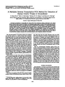
Results and Discussion Isolation of DNA from single meat (either positive or negative control) and mixed meat samples was initially performed manually. The collected DNA was not pure enough for subsequent PCR work. Therefore, DNA collection was then conducted by using a commercial kit of DNeasy Blood and Tissue (QAGEN). As the designed research study, all 5 mixed meat samples and two samples of positive and negative controls were amplified in two PCR program cycles which were set up at 25 and 35 cycles. This present study was a development of the previous technique of PCR for detection of porcine contaminant in several meat ball products that collected from supermarkets and a traditional market in Bogor (Margawati and Ridwan, 2010). Development of the PCR technique was conducted to examine the sensitivity method that applied in the percentage of various porcine contents of 0.05; 0.10; 0.15; 0.20 and 0.25 in beef meat. Besides the sensitivity examination, applying two step cycles of PCR was intended to examine time consuming that needed in the process of porcine contaminant detection. The result of amplification showed an extremely significant of emerging the Leptin bands. The PCR program that set up at 25 cycles did not show any band on those samples and even on the positive control (Figure 1) while the PCR program at 35 cycles emerged clearly band of all five samples and in positive control and none in negative control (Figure 2). This result showed that more tiny porcine contents in this study needed a bit longer cycle step profiles for detection of contaminant. It was proved when the PCR cycle was increased to be 35 cycles, all samples and positive control showed the Leptin bands
Biota Vol. 16 (2), Juni 2011
Margawati et al.,
(Figure 2: No. 2 to No. 7). With regard to the PCR cycle, this recent finding proved a bit different especially in 25 cycles and porcine contents from the previous report (Calvo et al., 2001). They reported that applying PCR at 20 cycles could detect a more small porcine content up to 0.001 while the larger porcine content up to 1% also needed 20 cycles. However this present result showed similar report to Al Araid (2008) which amplified the Leptin fragment of 152bp at 35 cycles while the same cycles in detection of 0.1% pork content was agreed with Ilhak and Arslan (2007). It might be understood that DNA extraction was used in the present study using the same kit of ASL buffer (QIAGEN), (Al Araid, 2008) and was different from other researchers (Calvo et al., 2001; Ilhak and Arslan, 2007; Al Araid, 2008). The common differences in PCR results might be due to differences in setting up the time and temperature of each PCR step among researchers and might be differences in the each concentration of reaction compounds but needs to be further detail evaluated. There are some factors influencing PCR work, one of them is DNA template concentration and its purity (Altshuler, 2006). The important is, each amplification work needs to adjust the
annealing temperature in advanced in order to find the approximately exact temperature of annealing. As presented in the Figure 2, the porcine Leptin fragment was identified at size of 152bp for all mixed meat samples (No. 2 to No. 6) and at positive control of pork (No. 7) while it was not detected in No. 1 as a negative control (beef meat). This 152bp Leptin size was exactly the same as two other previous researchers (Farouk et al., 2001; Al Araid, 2008) which used Leptin-specific primer for porcine (F/TGCA GTCTCTCCTCCAAA and R/CGATAATTGG ATCACATTCTG). The fragment size of 152bp for porcine Leptin was also detected in chocolates and chicken nuggets by previous investigation (Farouk et al., 2006). They used porcine-specific primer to amplify a 152bp fragment from the porcine Leptin gene which is homologues of murine obese. The latest report also detected positive porcine contaminant with 152 bp Leptin fragment in two foods (beef steak and beef sausage) with “Halal” label out of thirty three imported food samples (Al Araid, 2008). Both investigations used a PCRbased methodology in their research. The length of Leptin size was confirmed as alignment in the frame of the Leptin size of the porcine sequence that reported by Ramsey et al., (1998).
Table 1. The Percentage of Pork Contents in Beef Meat. Meat Pork (g) Beef (g)
% Pork Contents in Beef Meat of 5g Total Weight 0.05 0.10 0.15 0.20 0.25 A B C D E 0.0025 0.0050 0.0075 0.0100 0.0125 4.9975 4.9950 4.9925 4.9900 4.9875
Positive Control
Negative Control
100 %
100 %
5.0000 -
5.0000
No found any DNA fragment of 152bp Leptin
100bp
M 1
2
3
4
5
6
7
Figure 1. PCR products derived from 25 cycles (M= 100bp DNA ladder, No.1= negative control or beef meat, No. 2 to No. 6 = 0.05; 0.10; 0.15; 0.20 and 0.25% pork in beef meat, respectively; No. 7= positive control or pork).
Biota Vol. 16 (2), Juni 2011
345
Identification of Porcine Contaminant in Unprocessed Food
152bp
100bp M
1
2
3
4
5
6
7
Figure 2. PCR products derived from 35 cycles (M= 100bp DNA ladder, No.1= negative control or beef meat, No. 2 to No. 6= 0.05; 0.10; 0.15; 0.20 and 0.25% pork in beef meat, respectively; no. 7= positive control or pork).
Conclusion This study reported that a PCR technique could be used as a tool for detection of porcine contaminant in unprocessed food of meat. Furthermore, this technique is a quick method with sensitivity of contaminant detection as small as 0.05% porcine contaminant in beef meat at 35 cycles of PCR amplification. This technique could be recommended as a sensitive method based on molecular technique to examine either processed or unprocessed foods (meat) whether or not bearing “Halal” (religious permissible) tags.
References Al Araidh, I.A. 2008. Improved DNA extraction method for porcine contaminants, detection in imported meat to the Saudi market. Saudi J. of Biolog. Sci., 15 (2): 225229. Altshuler, M.L. 2006. PCR Trouble Shooting: The Essential Guide. Academic Press, NY. 80 pp. Ayaz, Y., Ayaz, N.D. and Erol, I. 2006. A Research Note: Detection of species in meat and meat products using enzyme-linked immunosorbent assay. J. of Muscle Foods, 17 (2): 214220. Calvo, J.H., Zaragona, P. and Osta, R. 2001. Technical note: A quick and more sensitive method to identify pork in processed and unprocessed food by PCR amplification of a new specific DNA fragment. J. of Animal Sci., 79: 21082112.
346
Céspedes, A., Garcia, T., Cerrera, E., Gonzáles, I., Fernández, A., Hernández, P.E. and Martin, R. 1999. Identification of sole (Solea solca) and Greenland halibut (Reinhardtius hippoglossoides) by PCR amplification of the 5S rDNA gene. J. of Agric. Food Chem., 47: 10461050. Coligan, J.E., Dunn, B.M., Ploegh, H.L., Speicher, D.W. and Wingfield, P.T. 1997. Current Protocols in Protein Science Volume 1. New York, USA: John Wiley & Sons, Inc. Farouk, A., Batcha, M.F., Griner, R., Salleh, H.M., Salleh, M.R. and Sirajudin, A.R. 2006. The use of a molecular technique for the detection of porcine ingredients in the Malaysian food market. Saudi Medicine J., 27: 447450. Girisha, P.S., Anjaneyulua, A.S.R., Viswasb, K.N., Shivakumarc, B.M., Anandd, M., Patele, M. and Sharmad, B. 2005. Meat species identification by polymerase chain reactionrestriction fragment length polymorphism (PCR-RFLP) of mitochondrial 12S rRNA gene. Meat Sci., 70 (1): 107112. Ilhak, O.I. and Arslan, A. 2007. Identification of meat species by polymerase chain reaction (PCR) technique. Turk J. of Vet. Animal Sci., 31 (3): 159−163. Lees, M. and Popping, B. 2003. Meat and meat products. In: M. Lees. (Ed.) Food Authenticity and Traceability. Pp. 347352. CRC Press. France. Margawati, E.T. and Ridwan, M. 2010. Pengujian pencemaran daging babi pada beberapa produk bakso dengan teknologi PCR: Pencarian system pengujian efektif (Analysis of Porcine Contamination by Using PCR Technology in Several Meat Ball Products: To Find an Effective Assessment System). J. Berita Biologi, 10 (1): 9398.
Biota Vol. 16 (2), Juni 2011
Margawati et al.,
Martin, I., Garcia, T., Fajardo, V., Rojas, M., Hernández, P.E., Gonzáles, I. and Martin, R. 2007. Technical Note: Detection of cat, dog and rat or mouse tissues in food and animal feed using species-specific polymerase chain reaction. J. of Animal Sci., 85: 27342739.
Meyer, R., Hofelein, C., Luthy, J. and Candrian, U. 1995. Polymerase chain reaction-restriction fragment length polymorphism analysis: A simple method for specific identification of in food. J. of Association of Analytical Chem., 78: 15421551.
Matsunaga, T., Chikuni, K., Tanabe, R., Muroya, S., Shibata, K., Yamada, J. and Shinmura, Y. 1999. A quick and simple method for the identification of meat species and meat products by PCR assay. Meat Sci., 5 (2): 143148.
Ramsey, T.G., Yan, X. and Morrison, C. 1998. The obesity gene in swine: Sequence and expression of porcine leptin. J. of Animal Sci., 76: 484490.
Biota Vol. 16 (2), Juni 2011
347