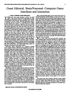
Opinion
TRENDS in Cognitive Sciences
Vol.9 No.10 October 2005
A mechanism for cognitive dynamics: neuronal communication through neuronal coherence Pascal Fries1,2 1 2
F.C. Donders Centre for Cognitive Neuroimaging, Radboud University Nijmegen, 6525 EN Nijmegen, The Netherlands Department of Biophysics, Radboud University Nijmegen, 6525 EZ Nijmegen, The Netherlands
At any one moment, many neuronal groups in our brain are active. Microelectrode recordings have characterized the activation of single neurons and fMRI has unveiled brain-wide activation patterns. Now it is time to understand how the many active neuronal groups interact with each other and how their communication is flexibly modulated to bring about our cognitive dynamics. I hypothesize that neuronal communication is mechanistically subserved by neuronal coherence. Activated neuronal groups oscillate and thereby undergo rhythmic excitability fluctuations that produce temporal windows for communication. Only coherently oscillating neuronal groups can interact effectively, because their communication windows for input and for output are open at the same times. Thus, a flexible pattern of coherence defines a flexible communication structure, which subserves our cognitive flexibility. Introduction Because we are equipped with mechanisms of selective attention, we can do tasks such as the following: we can fixate on a central cross and press a button only when a green dot is flashed to the right while ignoring the same dot anywhere else in the visual field. And we can switch attention to do this task at any other spatial position, now ignoring the formerly relevant position. Although in both conditions, the same physical stimuli are given and the same behavioral responses are issued, there is obviously a strong cognitive control over the routing of information from sensory to motor areas. Conceptually, the effect of cognitive top-down control is a modification in the communication structure between brain areas. But how do groups of neurons communicate? And how do top-down influences modify the communication structure within a few hundred milliseconds when anatomical connections stay unchanged on that timescale? Although we still know very little about neuronal communication mechanisms, we often have an implicit concept or model about it. In very general terms, the dominant model of neuronal communication is that a neuron sends its message (encoded in e.g. action potential rate or in the degree of action potential synchronization) down its axons Corresponding author: Fries, P. (
[email protected]).
to all neurons to which it is anatomically connected. Those receiving neurons combine (e.g. sum and threshold) all the different inputs that they receive from all neurons to which they have connections. An important aspect of this model is that both the distribution and the reception of neuronal signals is governed solely by the structure of the anatomical connections, that is, there is no further communication structure beyond the one imposed by anatomical connectedness. However, cognitive functions require flexibility in the routing of signals through the brain. They require a flexible effective communication structure on top of the anatomical communication structure that is fixed, at least on the timescale at which cognitive demands change. In this article, I hypothesize that this effective communication structure is mechanistically implemented by the pattern of coherence among neuronal groups, that is, the pattern of phase-locking among oscillations in the communicating neuronal groups. Specifically, I hypothesize that neuronal communication between two neuronal groups mechanistically depends on coherence between them and the absence of neuronal coherence prevents communication. I will address this hypothesis as the ‘communication-through-coherence’ (CTC) hypothesis. It is based on two realizations: first, activated neuronal groups have the intrinsic property to oscillate [1,2]. Second, those oscillations constitute rhythmic modulations in neuronal excitability that affect both the likelihood of spike output and the sensitivity to synaptic input. Thus, rhythmic excitability peaks constitute rhythmically reoccurring temporal windows for communication. Only coherently oscillating (or phase-locked) neuronal groups can communicate effectively, because their communication windows for input and for output are open at the same times. Previous work has hypothesized that neuronal coherence (or phase-locking or synchronization) could provide a tag that binds those neurons that represent the same perceptual object [3–7]. This binding tag would be a flexible code for linking neurons into assemblies and thereby greatly enlarging the representational capacity of a given pool of neurons. This hypothesis is known as the bindingby-synchronization (BBS) hypothesis. The CTC and the BBS hypotheses are fully compatible with each other, but they are also clearly distinct. Whereas the BBS hypothesis
www.sciencedirect.com 1364-6613/$ - see front matter Q 2005 Elsevier Ltd. All rights reserved. doi:10.1016/j.tics.2005.08.011
Opinion
TRENDS in Cognitive Sciences
is primarily suggesting a representational code, the CTC hypothesis considers the mechanistic consequences of neuronal oscillations for neuronal communication. It suggests that at the heart of our cognitive dynamic is a dynamic communication structure and that the neuronal substrate is the flexible neuronal coherence pattern. In the following, I will first review neurophysiological data that suggest an important role of synchronous neuronal oscillations for neuronal communication. I will then present some evidence that directly suggests that neuronal coherence can serve neuronal communication and can be dynamically modulated by cognitive demands. Finally, I will review neurophysiological data about the attentional modulation of synchronous neuronal oscillations and speculate about the detailed implementation of a flexible communication structure through a flexible coherence pattern.
Feedforward models of neuronal communication and their neurophysiological tests Neuronal communication through firing rate modulation The predominant, but often only implicit, model of neuronal communication is that a neuronal group sends a message through enhancing its firing rate and the receiving group of neurons integrates this input over some time window and modulates its firing rate accordingly [8] (Figure 1a). Probably the most important reason why this model is dominant is that many experiments have demonstrated modulations in firing rate that correlate in a meaningful way with either stimulus parameters or cognitive parameters. However, in the search for mechanisms of neuronal communication, the crucial question is whether the receiving group of neurons is actually affected by modulations in input rate. Thus, experiments must be done that record from receiving neurons at the same time as recording or manipulating the input to those receiving neurons. Recent experiments that have achieved this cast some doubt on the model of neuronal communication through firing rate modulations. Tsodyks and Markram found that the size of the excitatory postsynaptic potential decreases with increasing input rate such that under some conditions, there is no postsynaptic net effect of increased input rate [9] (Figure 1b). Azouz and Gray recorded
Vol.9 No.10 October 2005
475
intracellularly in neurons of primary visual cortex of anesthetized cats [10]. They stimulated the cells visually and investigated variations in spike threshold (the membrane potential at which a spike is elicited) as a function of the average subthreshold membrane potential during the 250 ms preceding each spike. This average pre-spike membrane potential reflects the input rate and the spike threshold increased linearly with the input rate, thus reducing the effect of changes in input rate (Figure 1c). Neuronal communication through modulations in the degree of spike synchronization Although in the study by Azouz and Gray, the spike threshold increased with increasing average subthreshold membrane potential, it decreased with increasing slope of the membrane potential immediately preceding the spike [11] (Figure 2b), indicating that neurons are particularly sensitive to high temporal densities of spike input. Thus, for a group of neurons sending a message to be maximally effective, each neuron in the group should bundle its spikes into bursts and all neurons of the group should synchronize those bursts with each other [12,13] (Figure 2a). This is exactly what happens when neurons engage in synchronous gamma-frequency oscillations which have been found to be modulated by stimulus parameters [14–24] as well as cognitive parameters [22,25,26]. Neuronal communication through neuronal coherence In this article, I explore the potential that neuronal oscillations offer as mechanisms for neuronal communication and propose that neuronal communication is not only subserved by oscillatory synchronization within the group of neurons sending a message, but also by coherence (or phase-locking) between the oscillations in the sending group and the receiving group (Figure 3a). The central argument is that activated neuronal groups have the intrinsic property to oscillate [1,2]. Those oscillations constitute excitability fluctuations that do not only affect the output of the neuronal group, but also its sensitivity to input [27,28]. Thus, oscillations of a neuronal group rhythmically open and close the group’s windows for communication. It is obvious that different groups of neurons can only communicate effectively with each other
Figure 1. ‘Neuronal communication through firing rate modulation’ and some neurophysiological evidence against this model (see text for discussion). (a) Red and green filled circles illustrate a sending and a receiving neuronal group, respectively. The small vertical lines illustrate action potentials of neurons in the two groups and the arrows illustrate those action potentials traveling along the connecting axons. (b) Excitatory postsynaptic potentials (EPSP) as a function of input rate (from [9]). (c) Spike threshold as a function of the average membrane potential in a 250 ms period before the spike (Vm,psZpre-spike membrane potential). (Adapted from [10].) www.sciencedirect.com
476
Opinion
TRENDS in Cognitive Sciences
Vol.9 No.10 October 2005
Figure 2. ‘Neuronal communication through modulation of synchronization’ and some supporting evidence. (a) Same general format as in Figure 1a. (b) Spike thresholds as a function of the membrane potential slope immediately preceding the spike. (Adapted from [11].)
if the rhythmic opening of their communication windows is coordinated between the groups. Specifically, for a sending group to communicate a message effectively to a receiving group, the sending group’s output has to be timed such that it arrives at the receiving group when that group is excitable. For this prospective timing to be possible, several requirements have to be met: first, the excitability fluctuations in the receiving neuronal group must be predictive. This requirement is met by the physiologically occurring neuronal oscillations that are sufficiently regular to allow prediction of the next excitability peak [17,20,23,29,30]. Second, the spike traveling time from the sending to the receiving group must be reliable. This requirement seems to be met in general. In fact, conduction velocities appear to be regulated in the different parts of an axonal tree such that they ensure synchronous arrival of a spike at all receiving neurons, irrespective of physical distance [31,32]. Third, the sending group has to time its output to arrive, after the
spike traveling time, when the receiving neuronal group is excitable. If the excitability of the receiving group is indeed predictable because it oscillates, then this last requirement is obviously met if the sending group also oscillates and if the oscillations in the two groups are phase-locked or coherent to each other. Thus, communication will be achieved through coherence. The putative role of different frequency bands One intricate issue in this scheme of communication through coherence is the precise timing of events. In essence, the frequency of the coherent oscillations, the relative phase between them and the conduction delay need to match. For unidirectional communication, an oscillation in a sending group might entrain an oscillation that is intrinsically generated in the receiving group or it might even simply drive an oscillation in the receiving group. In this case, the conduction delay would, for a given frequency, directly translate into a relative phase, as is the
Figure 3. ‘Neuronal communication through neuronal coherence’ and some supporting evidence. (a) Same general format as in Figure 1a. Spikes that arrive at excitability peaks of the receiving neuronal group have pointed arrowheads. Spikes that miss excitability peaks have blunt arrowheads. The red and green neuronal groups undergo coherent excitability fluctuations and their communication is therefore effective. The black neuronal group however undergoes excitability fluctuations that are not coherent with the green neuronal group and therefore communication between the green and the black neuronal group is prohibited. (b) Membrane potentials during combined injection of sinusoidal current and electrical stimulation of one afferent axon. The timing of the axon stimulation was varied such that the synaptic input arrived at the recorded neuron either precisely at its excitability peak or shortly before or after it. (Adapted from [28].) www.sciencedirect.com
Opinion
TRENDS in Cognitive Sciences
case in the unidirectional coherence between motor cortex and the spinal cord [33]. The situation is more complicated for bi-directional communication, which is likely to be a frequent form of communication between cortical areas. If conduction delays were on the same order as the cycle length of the oscillation, then two communicating groups could be coherent at zero phase (i.e. synchronized) and spike output generated in one cycle would always arrive at the respective receiving group at the peak of the next cycle. However, if one considers the physiologically occurring frequencies and conduction delays, another scenario appears more plausible: for a given communication ‘link’, conduction delays are typically an order of magnitude shorter than the cycle length of the oscillation. Thus, sending and receiving probably happen within one and the same excitability peak (Figure 3a, red and green neuronal groups). For neuronal groups within one cortical area, coherent oscillations have mostly been found in the gamma-frequency range, between 30 and 100 Hz [17,20,23,29,30]. Although this corresponds to cycle lengths as short as 10 to 30 ms, the local conduction delays are around 1–3 ms, an order of magnitude shorter. For neuronal groups in different cortical areas, coherent oscillations have been found in the b-frequency range, between 15 and 25 Hz [34–37]. Although in this case, conduction delays might amount to 5 ms, this is again an order of magnitude faster than the relevant cycle length of around 50 ms. Apart from gamma- and b-band oscillations, also slower rhythms can be found in the brain. Notably, a-frequency rhythms around 10 Hz, thetafrequency rhythms between 4 and 8 Hz and deltafrequency rhythms between 1 and 3 Hz. In principle, those rhythms might also subserve communication according to the CTC scheme. It will be interesting for future research to investigate whether each of those rhythms contributes to CTC and if so, what the differential roles are and whether interactions between different rhythms might also contribute to CTC [38,39]. Selective coherence for selective communication At least as important as the facilitation of communication is the preclusion of communication. It is the very nature of a communication structure that communication is facilitated in a structured or selective way and not globally. Global phase-locking is found during states of epilepsy and is obviously incompatible with normal cognitive functioning. The fact that the brain normally manages to avoid epileptic states, but rather generates an intricate communication structure, suggests that it is equipped with robust regulation mechanisms. Neuronal oscillations could prevent communication in at least two ways: as is clear from the above, two neuronal groups that exhibit coherent oscillations might nevertheless fail to communicate if the frequency of the coherent oscillations, the relative phase between them and the conduction delay do not match. In that case, input from a sending group would repeatedly miss the excitable phase of the receiving group’s oscillation. This might look like a powerful way to prohibit communication, but the available physiological data suggest another mechanism, namely non-communication through non-coherence. In this case, communication is www.sciencedirect.com
Vol.9 No.10 October 2005
477
prevented by the absence of a reliable phase relationship between the oscillations in the sending and the receiving group. In order for this to be possible, the oscillations need to be sufficiently irregular (or broad-band in the frequency domain) to restrict spurious coherence to short time periods. This requirement is met by physiologically observed oscillations, which are typically predictable for few cycles but not more [17,20,23,29,30]. Note that for a given short time segment, non-coherence can ‘look like’ out-of-phase coherence (Figure 3a, green and black groups), whereas over longer time periods, the variable phase relationship becomes apparent. Cortico-spinal coherence subserves cortico-spinal communication There is also experimental evidence that the coherence between neuronal groups subserves their communication. Several studies have demonstrated coherence between different areas involved in visuo-motor transformations, starting from early visual areas and reaching through parietal cortex and motor cortex to the spinal cord [34–37,40–49]. A recent study tested directly one prediction of the CTC hypothesis, namely that neuronal coherence has a functional role in human cognition and that this can be demonstrated through a behavioral correlate of coherence between distant groups of neurons [33] (Figure 4). Human subjects performed a simple reaction-time task in which they extended their right wrist and had to keep this extension until a visual gosignal occurred. The subjects’ readiness to respond was manipulated by training them on different hazard rates for the go-signal, that is, different conditional probabilities that a go-signal would occur at a certain time in a trial if it had not occurred before. When the hazard rate was high, subjects were particularly ready to respond and, when probed with an actual go-signal, did so with shortened reaction times (Figure 4a and e). Before the go-signal, motor cortex and spinal cord produce a constant motor output, but prepare for a reaction according to the hazard rate. If coherence were to subserve the cortico-spinal interaction, it should be modulated with the hazard rate. Cortico-spinal coherence was assessed between the EMG of the involved muscles and the MEG recorded over contralateral motor cortex. This coherence was indeed highly correlated with the hazard rate and this correlation was selective for the gamma-frequency range (Figure 4). Coherence, competition and binding Although the study on cortico-spinal coherence demonstrates that neuronal communication is rendered more effective through neuronal gamma-band coherence, it did not yet address another prediction of the CTC hypothesis, namely that neuronal coherence renders neuronal communication also selective. I am not aware of experimental evidence that tests this prediction directly, but let us speculate about a scenario in which such a potential mechanism might have profound importance. It has been shown that an attended visual stimulus induces stronger local gamma-band synchronization in monkey extrastriate cortex than an unattended stimulus [22,26] (Figure 5a and b). If the two stimuli are
478
Opinion
TRENDS in Cognitive Sciences
Vol.9 No.10 October 2005
Figure 4. Cortico-spinal communication through cortico-spinal coherence. (a)–(d) show the effect of increasing hazard rate on reaction times and cortico-spinal coherence. (a) Hazard rate (gray line) with the resulting reaction times (pink line). Please note the inverted reaction time axis on the right of the plot. (b) Time-frequency representation of cortico-spinal coherence. (c) Time course of cortico-spinal coherence in the gamma-band (40 – 70 Hz). The gray line is a scaled copy of the hazard rate for comparison. (d) Pearson correlation coefficient between hazard rate and cortico-spinal coherence as a function of frequency of the latter. Bars indicate significant frequency bands (p!0.05, non-parametric randomization test, corrected for multiple comparisons). (e)–(h) show the effect of decreasing hazard rate on reaction times and cortico-spinal coherence in the same format as (a)–(d). (Adapted from [33].)
neighboring in the visual field, then the respective two activated groups of neurons will provide converging and competing input to a receiving neuronal group at the next stage of visual processing (Figure 5a and c). This receiving neuronal group now receives two inputs that both consist of rhythmic, synchronized bursts of spikes, but that are not synchronous with each other, because they are driven by two different visual stimuli [15,18]. The spikes from the sending neurons driven by the attended stimulus are more precisely gamma-band synchronized than the spikes
driven by the unattended stimulus [22,26] (Figure 5b), and the receiving group will therefore tend to phase-lock to this ‘attended input’ rather than to the ‘unattended input’. If this reasoning holds, the resulting pattern of selective coherence is bound to have consequences for neuronal communication. The coherence between the receiving group and the attended sending group is likely to render this interaction highly effective. As a result, the receiving group should respond as if it received only the
Figure 5. Coherence and competition. (a) Stimulus configuration used in a selective visual attention experiment [22]. The lower patch of grating falls into the receptive field of a neuronal group in V4 indicated in red (and black for the upper patch). Both grating patches fall into the receptive field of a neuronal group in IT cortex (green). The purple ‘spotlight’ indicates that spatial selective attention is directed to the grating patch contained in the red receptive field. (b) Although the firing rates of the attended V4 neurons are only slightly enhanced, they show a strong enhancement of gamma-band coherence. (Data from [22]; new analysis of spike-field coherence, z-transformed and pooled across pairs of recording sites). (c) The different neuronal groups in V4 and IT that are activated by the stimuli shown in (a). Experimental evidence suggests that the attended V4 neurons communicate effectively with the IT neurons but the unattended V4 neurons fail to do so. This is indicated with pointed and blunt arrowheads, respectively. This might be the result of modulatory input from parietal cortex that gives a competitive bias towards the attended V4 neurons. www.sciencedirect.com
Opinion
TRENDS in Cognitive Sciences
attended input and this is indeed found experimentally when two stimuli are placed within the receptive field of neurons in higher visual areas and attention is directed to one of them [50]. Equally important, the absence of coherence between the receiving group and the unattended sending group will leave their interaction highly ineffective. In addition, feedback from the receiving group is likely to be more effective at the coherent sending group (the attended one) than at the non-coherent sending group (the non-attended one), even if it is anatomically directed to both. Thus, a flexible pattern of coherence would be able to dynamically modulate the effective gain of neuronal connections despite the fact that anatomical connections are constant at that timescale. This might be a general mechanism for the preferential routing of selected signals [23]: if the rhythm of the selected sending group is passed onto other groups of neurons, then the subthreshold membrane potential fluctuations of those neurons can become entrained to the ‘selected rhythm’. All those entrained neuronal groups would thereby be made sensitive for selected input and they would at the same time be rendered deaf for deselected input. Neuronal ‘broadcasting centers’ like some thalamic nuclei that have widespread reciprocal connections with neocortex would be in the ideal position to distribute the selected rhythm. Their anatomical connections might be fairly unselective, but their influence on other neuronal groups would acquire functional selectivity through the specific rhythm that they broadcast. In this concept, so called top-down mechanisms that reflect the attentional selection of behaviorally relevant stimuli, are transformed from a spatial to a temporal code. The topdown mechanism first provides some yet unknown modulatory input to the selected neuronal group in early sensory areas. This group is defined by its spatial position in the respective sensory map. Because the selected group’s oscillatory synchronization is strengthened, it is distributed more successfully and sets the ‘selected rhythm’, that is, the top-down signal then resides in the temporal information. In summary, I propose that the CTC hypothesis presents a fascinating link from simple neuronal oscillations to a flexible effective communication structure that might represent the neuronal substrate of our cognitive flexibility. Future experiments will be needed to test all the concrete predictions that flow from it. Acknowledgements I would like to thank Nancy Kopell and Wolf Singer for helpful discussions and comments. Supported by The Netherlands Organization for Scientific Research, grants 452–03–344 and The Human Frontier Science Program Organization, grant RGP0070/2003.
References 1 Kopell, N. et al. (2000) Gamma rhythms and beta rhythms have different synchronization properties. Proc. Natl. Acad. Sci. U. S. A. 97, 1867–1872 2 Tiesinga, P.H. et al. (2001) Computational model of carbachol-induced delta, theta, and gamma oscillations in the hippocampus. Hippocampus 11, 251–274 3 von der Malsburg, C. and Schneider, W. (1986) A neural cocktail-party processor. Biol. Cybern. 54, 29–40 www.sciencedirect.com
Vol.9 No.10 October 2005
479
4 Singer, W. and Gray, C.M. (1995) Visual feature integration and the temporal correlation hypothesis. Annu. Rev. Neurosci. 18, 555–586 5 Singer, W. (1999) Neuronal synchrony: a versatile code for the definition of relations? Neuron 24, 49–65 6 Engel, A.K. et al. (1997) Role of the temporal domain for response selection and perceptual binding. Cereb. Cortex 7, 571–582 7 Engel, A.K. et al. (2001) Dynamic predictions: oscillations and synchrony in top-down processing. Nat. Rev. Neurosci. 2, 704–716 8 van Rossum, M.C. et al. (2002) Fast propagation of firing rates through layered networks of noisy neurons. J. Neurosci. 22, 1956–1966 9 Tsodyks, M.V. and Markram, H. (1997) The neural code between neocortical pyramidal neurons depends on neurotransmitter release probability. Proc. Natl. Acad. Sci. U. S. A. 94, 719–723 10 Azouz, R. and Gray, C.M. (2003) Adaptive coincidence detection and dynamic gain control in visual cortical neurons in vivo. Neuron 37, 513–523 11 Azouz, R. and Gray, C.M. (2000) Dynamic spike threshold reveals a mechanism for synaptic coincidence detection in cortical neurons in vivo. Proc. Natl. Acad. Sci. U. S. A. 97, 8110–8115 12 Salinas, E. and Sejnowski, T.J. (2000) Impact of correlated synaptic input on output firing rate and variability in simple neuronal models. J. Neurosci. 20, 6193–6209 13 Salinas, E. and Sejnowski, T.J. (2001) Correlated neuronal activity and the flow of neural information. Nat. Rev. Neurosci. 2, 539–550 14 Gray, C.M. and Singer, W. (1989) Stimulus-specific neuronal oscillations in orientation columns of cat visual cortex. Proc. Natl. Acad. Sci. U. S. A. 86, 1698–1702 15 Eckhorn, R. et al. (1988) Coherent oscillations: a mechanism of feature linking in the visual cortex? Multiple electrode and correlation analyses in the cat. Biol. Cybern. 60, 121–130 16 Engel, A.K. et al. (1991) Direct physiological evidence for scene segmentation by temporal coding. Proc. Natl. Acad. Sci. U. S. A. 88, 9136–9140 17 Gray, C.M. et al. (1992) Synchronization of oscillatory neuronal responses in cat striate cortex: temporal properties. Vis. Neurosci. 8, 337–347 18 Gray, C.M. et al. (1989) Oscillatory responses in cat visual cortex exhibit inter-columnar synchronization which reflects global stimulus properties. Nature 338, 334–337 19 Kreiter, A.K. and Singer, W. (1996) Stimulus-dependent synchronization of neuronal responses in the visual cortex of the awake macaque monkey. J. Neurosci. 16, 2381–2396 20 Fries, P. et al. (1997) Synchronization of oscillatory responses in visual cortex correlates with perception in interocular rivalry. Proc. Natl. Acad. Sci. U. S. A. 94, 12699–12704 21 Fries, P. et al. (2001) Rapid feature selective neuronal synchronization through correlated latency shifting. Nat. Neurosci. 4, 194–200 22 Fries, P. et al. (2001) Modulation of oscillatory neuronal synchronization by selective visual attention. Science 291, 1560–1563 23 Fries, P. et al. (2002) Oscillatory neuronal synchronization in primary visual cortex as a correlate of stimulus selection. J. Neurosci. 22, 3739–3754 24 Logothetis, N.K. et al. (2001) Neurophysiological investigation of the basis of the fMRI signal. Nature 412, 150–157 25 Gruber, T. et al. (1999) Selective visual-spatial attention alters induced gamma band responses in the human EEG. Clin. Neurophysiol. 110, 2074–2085 26 Taylor, K. et al. Coherent oscillatory activity in monkey area V4 predicts successful allocation of attention. Cereb. Cortex (in press) 27 Burchell, T.R. et al. (1998) Gamma frequency oscillations gate temporally coded afferent inputs in the rat hippocampal slice. Neurosci. Lett. 255, 151–154 28 Volgushev, M. et al. (1998) Modification of discharge patterns of neocortical neurons by induced oscillations of the membrane potential. Neuroscience 83, 15–25 29 Friedman-Hill, S. et al. (2000) Dynamics of striate cortical activity in the alert macaque: I. Incidence and stimulus-dependence of gammaband neuronal oscillations. Cereb. Cortex 10, 1105–1116 30 Maldonado, P.E. et al. (2000) Dynamics of striate cortical activity in the alert macaque: II. Fast time scale synchronization. Cereb. Cortex 10, 1117–1131 31 Innocenti, G.M. et al. (1994) Computational structure of visual callosal axons. Eur. J. Neurosci. 6, 918–935
480
Opinion
TRENDS in Cognitive Sciences
32 Salami, M. et al. (2003) Change of conduction velocity by regional myelination yields constant latency irrespective of distance between thalamus and cortex. Proc. Natl. Acad. Sci. U. S. A. 100, 6174–6179 33 Schoffelen, J.M. et al. (2005) Neuronal coherence as a mechanism of effective corticospinal interaction. Science 308, 111–113 34 Lachaux, J.P. et al. (2005) The many faces of the gamma band response to complex visual stimuli. Neuroimage 25, 491–501 35 Brovelli, A. et al. (2004) Beta oscillations in a large-scale sensorimotor cortical network: directional influences revealed by Granger causality. Proc. Natl. Acad. Sci. U. S. A. 101, 9849–9854 36 Tallon-Baudry, C. et al. (2001) Oscillatory synchrony between human extrastriate areas during visual short-term memory maintenance. J. Neurosci. 21, RC177 37 Tallon-Baudry, C. et al. (2004) Oscillatory synchrony in the monkey temporal lobe correlates with performance in a visual short-term memory task. Cereb. Cortex 14, 713–720 38 Palva, J.M. et al. (2005) Phase synchrony among neuronal oscillations in the human cortex. J. Neurosci. 25, 3962–3972 39 Bragin, A. et al. (1995) Gamma (40-100 Hz) oscillation in the hippocampus of the behaving rat. J. Neurosci. 15, 47–60 40 Kilner, J.M. et al. (2000) Human cortical muscle coherence is directly related to specific motor parameters. J. Neurosci. 20, 8838–8845 41 Roelfsema, P.R. et al. (1997) Visuomotor integration is associated with zero time-lag synchronization among cortical areas. Nature 385, 157–161
Vol.9 No.10 October 2005
42 Bressler, S.L. et al. (1993) Episodic multiregional cortical coherence at multiple frequencies during visual task performance. Nature 366, 153–156 43 Frien, A. et al. (1994) Stimulus-specific fast oscillations at zero phase between visual areas V1 and V2 of awake monkey. Neuroreport 5, 2273–2277 44 Engel, A.K. et al. (1991) Synchronization of oscillatory neuronal responses between striate and extrastriate visual cortical areas of the cat. Proc. Natl. Acad. Sci. U. S. A. 88, 6048–6052 45 Bernasconi, C. et al. (2000) Bi-directional interactions between visual areas in the awake behaving cat. Neuroreport 11, 689–692 46 Salazar, R.F. et al. (2004) Directed interactions between visual areas and their role in processing image structure and expectancy. Eur. J. Neurosci. 20, 1391–1401 47 Salazar, R.F. et al. (2004) Effects of training on neuronal activity and interactions in primary and higher visual cortices in the alert cat. J. Neurosci. 24, 1627–1636 48 Gross, J. et al. (2004) Modulation of long-range neural synchrony reflects temporal limitations of visual attention in humans. Proc. Natl. Acad. Sci. U. S. A. 101, 13050–13055 49 Liang, H. et al. (2002) Synchronized activity in prefrontal cortex during anticipation of visuomotor processing. Neuroreport 13, 2011–2015 50 Reynolds, J.H. et al. (1999) Competitive mechanisms subserve attention in macaque areas V2 and V4. J. Neurosci. 19, 1736–1753
Five things you might not know about Elsevier 1. Elsevier is a founder member of the WHO’s HINARI and AGORA initiatives, which enable the world’s poorest countries to gain free access to scientific literature. More than 1000 journals, including the Trends and Current Opinion collections, will be available for free or at significantly reduced prices. 2. The online archive of Elsevier’s premier Cell Press journal collection will become freely available from January 2005. Free access to the recent archive, including Cell, Neuron, Immunity and Current Biology, will be available on both ScienceDirect and the Cell Press journal sites 12 months after articles are first published. 3. Have you contributed to an Elsevier journal, book or series? Did you know that all our authors are entitled to a 30% discount on books and stand-alone CDs when ordered directly from us? For more information, call our sales offices: +1 800 782 4927 (US) or +1 800 460 3110 (Canada, South & Central America) or +44 1865 474 010 (rest of the world) 4. Elsevier has a long tradition of liberal copyright policies and for many years has permitted both the posting of preprints on public servers and the posting of final papers on internal servers. Now, Elsevier has extended its author posting policy to allow authors to freely post the final text version of their papers on both their personal websites and institutional repositories or websites. 5. The Elsevier Foundation is a knowledge-centered foundation making grants and contributions throughout the world. A reflection of our culturally rich global organization, the Foundation has funded, for example, the setting up of a video library to educate for children in Philadelphia, provided storybooks to children in Cape Town, sponsored the creation of the Stanley L. Robbins Visiting Professorship at Brigham and Women’s Hospital and given funding to the 3rd International Conference on Children’s Health and the Environment. www.sciencedirect.com