2. LITERATURE REVIEW Chilli (Capsicum annuum L.) one of the most important commercial crop of India belongs to the Solanaceae family which represents a diverse plant group. The genus name Capsicum derived from the latin word ‘capsa’ meaning chest or box because of the shape of fruit which encloses seeds very neatly, as in the box (Berke and Shieh, 2000). Chillies are cultivated mainly in tropical and sub-tropical countries like India, Japan, Mexico, Turkey, United States of America and African countries (Panda, 2010). Genus Capsicum represents a diverse plant group which contains approximately 30 species, 5 of which C. baccatum, C. annuum, C. chinense, C. frutescens and C. pubescens are domesticated and cultivated in different parts of the world. Among the five species of Capsicum cultivated, C. annuum is one of the most common cultivated crop worldwide (Tong and Bosland, 1999) followed by C. frutescens (Ince et al., 2010; Wang and Bosland, 2006). Chillies are known from pre-historic times in Peru and believed to have originated in the tropical America. It is also said that chillies have originated in the Latin American regions of the New Mexico and Guatemala as a wild crop around 7500 BC, as per the remains of the pre-historic Peru. Columbus carried chilli seeds to Spain in 1493. The cultivation of chilli spread rapidly from Spain to Europe. The Portuguese brought capsicum from Brazil to India during the year 1584. Chillies became popular in the whole of Asia rapidly and native Asians started cultivating this crop as well (Raju and Luckose, 1991). The south Asian climate suited this crop, and since its introduction in the 16 th century chilli has been increasingly cultivated in south Asia. Chillies are the cheapest spices available in India and are eaten across all groups (Shinoj and Mathur, 2006).
2.1 International scenario The world area and production of chilli is around 1.5 million ha and 7 million tonnes respectively. In Asia, India, China, Pakistan, Indonesia, Korea, Turkey and Sri Lanka; Nigeria, Ghana, Tunisia and Egypt in Africa; Mexico, United States of America in North and Central America; Yugoslavia, Spain, Romania, Bulgaria, Italy and Hungary in Europe and Argentina, Peru and Brazil in South America are the major chilling growing countries. India is the world leader in chilli production followed by China and Pakistan (Hussain and Abid, 2011). The bulk share of chilli production is held by Asian countries. India, China, Mexico, Thailand, United States of America, United Kingdom, Germany and Sweden (Hanamashetti et al., 2009) are the major chilli consumers in the world. The major chilli exporting countries with their percentage share in world total exports are India (25 %), China (24 %), Spain (17 %), Mexico (8 %), Pakistan (7.2 %), Morocco (7 %) and Turkey (4.5 %). The world trade in chilli account for 16 % of the total spice trade in the world. United Arab Emirates, European Union, Sri Lanka, Malaysia, Japan and Korea (Thampi, 2004) are major chilli importing countries. 2.2 National scenario India is not only the largest producer but also the largest consumer of chilli in the world. Chilli is the most common spice cultivated in all States and Union Territories of India contributing about 36% to the world total production. Andhra Pradesh is the largest chilli producer in India contributing about 26% to the total area, followed by Maharashtra (15%), Karnataka (11%), Orissa (11%), Madhya Pradesh (7%) and other states contributing nearly 22% to the total area under chilli cultivation (Jagtap et al., 2012). “Naga Jolokia” the world’s hottest chilli is cultivated in the hilly terrain of Assam in a
small town, Tezpur in India (Goudappa et al., 2012). The crop is a significant source of income making India the world’s single largest producer and exporter to the USA, Canada, UK, Saudi Arabia, Singapore, Malaysia, Germany and many more countries across the world (Chandra Nayaka et al., 2009). India is not only largest producer but also exporter of chilli in the world, after China. In India, it is grown practically all over the country. In India, chilli is cultivated over an area of 0.81 million ha during 2010-11 with an annual production of 1.22 million tonnes green chillies (Anonymous, 2011). Chilli occupies number one position in export of spices with 2,09,000 metric tonnes volume worth Rs. 1097 crores (Anonymous, 2009). Another important export component of the Indian spice export is value added production like oils and oleoresin with Rs. 563 crores for which the major share of the raw material used is chilli and hence in the recent year, chilli is gaining greater importance in global market (Saideswara Rao, 2008). India is the largest exporter of chilli and about 2.5 to 3.0 % of country's total production is exported. Andhra Pradesh and Karnataka account for 75% of the country's net area under chilli and its production. At present, Karnataka ranks second in area (0.1322 million ha) and production of chilli (0.148 million tonnes) and is being extensively cultivated in Dharwad, Haveri, Belgaum, Gadag, Bellary, Gulbarga, Chikkamagalur and Raichur (Goudra et al., 2011). Karnataka state currently possesses 10 to 15 varieties and produces about 10 varieties of chillies which contain different ranges of colour, pungency, size and shape. The main varieties grown in Karnataka are namely Byadagi Kaddi, Byadgi Dabbi, Guntur and NP-46A (Jwala) (Rajur and Patil, 2013).
2.3 Cultivation Chilli is an annual herbaceous crop that reaches a height of one meter and has glabrous or pubescent lanceolate leaves with white flowers and fruits with varied, colour and pungency. Chilli plant requires a warm and humid climate for its best growth and dry weather during the maturation of fruits. Chilli grows best at 20 -30°C. It can be grown in higher altitude up to 2000 metres above the sea level. It can be grown successfully as a rain-fed crop in areas receiving an annual rainfall of 850-1200 mm and a soil pH of 4.3-8.7. Capsicum species are cold sensitive and generally grow best in welldrained, sandy/ silt-loam soil. Plantings are established by seeding or transplanting. Usually flowering usually occurs three months after planting. High temperature associated with low relative humidity at flowering increases the transpiration resulting in shedding of buds, flowers and small fruits (Rammohan et al., 2001). 2.4 Growth phases in chilli The crop duration of chilli is about 150-180 days depending on variety, climate, fertility and water management. The growth of chilli consists of vegetative and reproductive phases. Vegetative phase in chilli extends to 75-85 days followed by 75-95 days of reproductive phase. The vegetative phase is characterized by increase in plant height with branching. Flowering starts from 80-85 days of the crop or 40-45 days after transplanting. Chilli plant is an often cross pollinated crop with 50% of natural crossing. For fruit development and maturity about 40 days time is required after anthesis and pollination (Rajput and Paraluke, 1998).
2.5 Economic importance of chilli Chilli has been used since ancient times, traditionally in the form of spice. It is also used as a natural flavour and colorant in food industry (Vinaya et al., 2009) as well as raw material for the pharmaceutical industry. Chilli is nutritious crop, every 100 gm of green and dry chilli yield about 229 and 297 calories of energy (Table 2.1). It is mainly cultivated for three constituents of fruits viz., capsaicin, capsanthin and oleoresin (Amusa et al., 2004). It is grown for its pungent fruits which are used both as green and ripe to impart pungency and flavour to the food. Pungency, one of the important attributes of Capsicum species is due to the presence of alkaloid ‘capsaicin’ in the fruit. It is used primarily in the flavouring of pickles, meats, barbecue sauces, ketchup, cheese, snack food, dips, chilli cake, salads, and sausages (Pugalendhi et al., 2010). As a medicinal plant, the Capsicum species has been used as a carminative, stomachic, stimulant, rubefacient and tonic. It prevents heart diseases by dilating blood vessels. Chilli stimulates saliva and gastric juices and aids in digestion. Oleoresin of capsicum is used in pain balms and vapour rubs. Chilli extracts are used in wide range of medicines against tonsillitis, loss of appetite, flatulence, intermittent fever, sore throat, swellings and hardened tumours (Bosland and Votava, 2003). Chilli leaves are used as a dressing for wounds and sores and the leaf sap is squeezed into eyes against headache (Molnar
et
al.,
2004).
(Medvedeva et al., 2003).
Chilli
leaves
are
also
used
to
treat
toothache
Table 2.1 Nutritive value of chilli
Dry chillies (per 100 gm)
Green chillies (per 100 gm)
Carbohydrates Proteins
31.60 gm 15.00 gm
3.00 gm 2.90 gm
Fats Minerals Fibre Calcium Phosphorous
6.20 gm 6.10 gm 30.20 gm 160.00 mg 370.00 mg
0.60 gm 1.00 gm 6.80 gm 30.00 mg 80.00 mg
Iron Moisture
2.30 mg 10.00 gm
4.40 mg 85.70 gm
Carotene Thiamine
345.00 µg 0.93 mg
175.00 µg 0.19 mg
Riboflavin Niacin Vitamin C
0.43 mg 9.50 mg 50.00 mg
0.39 mg 0.90 mg 111.00 mg
Parameters
Vitamins
Minerals & trace elements Sodium Potassium Phytin phosphorous Magnesium
14.00 mg 530.00 mg
-
71.00 mg
7.00 mg
-
272.00 mg
Copper Manganese Molybdenum Zinc Chromium
-
1.40 mg 1.38 mg 0.07 mg 1.78 mg 0.04 mg
Oxalic acid Caloric values
229
67.00 mg 297
(Source: The National Institute of Nutrition, Hyderabad, Gopalan et al., 2004)
2.6 Diseases of chilli Plant diseases are an ongoing limiting factor in crop production. Diseases of crops lead to yield losses and are gaining importance with an increase in world population. Several abiotic and biotic stresses affect the productivity of chilli crop worldwide. Fifty one different pathogens have been reported to cause diseases on various parts of chilli (Saha and Singh, 1988) (Table 2.2). Out of them, thirty nine belong to the fungi of classes Mastigomycotina, Ascomycotina and Deuteromycotina. Fungal diseases are still an obstacle to the economic production of chilli. The most serious disease for agriculturist cultivating chilli are anthracnose and root rot (Vudhivanich, 2003). Anthracnose disease caused by Colletotrichum species, root rot caused by Rhizoctonia solani are the most serious destructive diseases of chilli (Isaac, 1992). 2.6.1 Chilli anthracnose Anthracnose, derived from a Greek word meaning ‘coal’, is the common name for plant diseases characterized by very dark, sunken lesions, containing spores (Isaac, 1992). Anthracnose of chilli was first reported from New Jersey, USA, by Halsted in 1890 who described the causal agents as Gloeosporium piperatum and Colletotrichum nigrum (Halsted,
1891).
These
taxa
were
then
considered
as
synonyms
of
Colletotrichum gloeosporioides by von Arx (1957). Anthracnose of chilli is one of the most economically important disease reducing marketable yield from 10% to 80% (Poonpolgul and Kumphai, 2007; Than et al., 2008b). Colletotrichum is capable of causing disease on virtually all parts of the chilli plant during any stage of plant growth. However, fruit lesions are the most economically important
aspect of anthracnose disease (Phoulivong et al., 2011). Under favourable conditions of disease development, up to 50% of the fruits can be damaged. Fruit rot takes place at 28 ° C and 95.7% relative humidity (Hyde et al., 2009). Table 2.2. Some of the important diseases of chilli in India S.No
Name of the disease
Causal organism
Fungal diseases Colletotrichum gloeosporioides Colletotrichum capsici
1.
Anthracnose/ Die back and fruit rot
2.
Root rot
3.
Cercospora leaf spot
Phytophthora capsici Rhizoctonia solani Cercospora capsici
4.
Damping off
Pythium aphanidermatum
5.
Powdery mildew
Leveillula taurica/ Oidiopsis taurica
6.
Fusarium wilt
Fusarium oxysporum
7.
Southern blight, collar rot
Sclerotium rolfsii
8.
Gray leaf spot
Stemphylium solani
9.
Gray mold
Botrytis cinerea
10.
Phytophthora Blight
Phytophthora capsici
11.
Verticillium wilt
Verticillium alboatrum Verticillium dahlia
Bacterial diseases 12.
Bacterial wilt
Ralstonia solanacearum
13.
Bacterial leaf spot
Xanthomonas vesicatoria
14.
Bacterial Soft Rot
Erwinia carotovora
Viral diseases 15.
Leaf curl
ChiLCV (chilli leaf curl virus)
16.
Chilli mosaic Nematode disease
Cucumber mosaic virus, Potato virus Y
17.
Root-Knot Nematode Meloidogyne incognita (Source: Pepper disease: a field guide, Black et al.,1991)
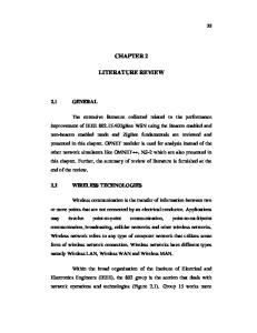
Typical anthracnose symptoms on chilli fruit include water soaked, sunken necrotic tissues, with concentric rings of acervuli. In some cases, the lesions are brown and then turn black, due to the formation of setae and sclerotia. The lesion may increase to 2-3 cm in diameter on larger fruits (Fig. 2.1). The attack commences from the growing point of the flower bud, the tops of the affected branches wither and turn brown. The infected plants bear fewer fruits of low quality (Voorrips et al., 2004). Fruit rot reduces dry weight, capsaicin and oleoresin content of affected fruits (Mistry et al., 2008), leading to reduction in the medicinal properties of chilli.
Figure 2.1. Anthracnose of chilli Anthracnose is mainly a problem on mature fruits, causing severe loss due to both pre and postharvest fruit decay exhibiting the phenomenon of quiescence in which symptoms do not develop until the fruit ripens (Bosland and Votava, 2003). Appressoria formed on immature fruits may remain quiescent until the fruits mature or ripen. The biotrophic life strategies adopted by Colletotrichum sp. may also contribute to their prominence as symptomless endophytes of living plant tissues (Yuan et al., 2011; Rojas et al., 2010).
2.6.1.1 Causal agent of chilli anthracnose – Colletotrichum gloeosporioides Anthracnose disease caused by C. gloeosporioides belongs to the Kingdom Fungi, Phylum Ascomycota, Class Sordariomycetes; Order Glomerellsles and Family Glomerellaceae (Agrios, 2005). Sixty-six species of Colletotrichum has been recently described by Hyde et al. (2009) to cause plant diseases. C. gloeosporioides Penz is so far the most predominant Colletotrichum sp. and can attack about 470 different host genera (Cannon et al., 2008). Many plant pathologists had recorded anthracnose disease throughout the world. C. acutatum, C. gloeosporioides and C. capsici were considered to be major causal agents of chilli anthracnose. Anthracnose of chilli has been shown to be caused by C. capsici and C. gloeosporioides in India, Indonesia, Korea, Thailand (Sharma et al., 2005; Pakdeevaraporn et al., 2005; Voorrips et al., 2004); C. acutatum in Australia and Indonesia (Nirenberg et al., 2002). Yield loss up to 50% in Thailand, 21- 47% in Sri Lanka, 15% in Korea and 50% in Malaysia has been reported (Kumaran et al., 2013; Than et al., 2008b). In India, the disease was first reported by Sydow in 1913 from Coimbatore of Madras Presidency (Sydow, 1928). Bansal and Grover (1969) during their studies on C. frutescens Linn. reported 10-35% fruit loss due to anthracnose disease in 1966 and 20 to 60 % fruit loss during 1967 in six districts of Punjab and Haryana. Thind and Jhooty (1985) reported fruit loss of 66-84% in Northern Karnataka. Kim et al. (2004) reported that different species Colletotrichum cause diseases of different organs of the chilli plant, C. acutatum and C. gloeosporioides infect chilli fruits at
all developmental stages, but not the leaves or stems, which are damaged by C. coccodes and C. dematium. The primary source of inoculum is seed and can survive well in soil by growing saprobically on dead plant fragments, and spreads via water-splash dispersal of conidia and air transmission of ascospores from the sexual morphology. Anthracnose usually appears after a rain or prolonged dew period. The initial infection processes of Colletotrichum spp. involves the attachment of conidia to plant surfaces, conidial germination, adhesive appressoria production and penetration of plant epidermis, plant tissue colonization and production of acervuli and sporulation (Prusky et al., 2000). Colletotrichum species can survive in and on seeds as acervuli and micro-sclerotia. Colletotrichum may also be introduced into fields by infected transplants or it may survive between seasons in plant debris or on weed hosts. Microsclerotia are naturally produced by Colletotrichum sp. to allow the fungus to lie dormant in the soil during winter or under stressed conditions. Microsclerotia can survive for many years even after 2 or 3 years of crop rotation. Colletotrichum sp. may also persist on alternative hosts such as other Solanaceous or legume crops (Pernezny et al., 2003). The fungi produces 17-18 x 3-4 µm, colourless, one- celled, ovoid, cylindrical and sometimes curved or dumb bell shaped conidia in acervuli (Agrios, 2005). The acervulus is saucer-shaped and surrounded by stiff, black, unbranched hairs, which are typically referred to as setae. The conidiogenous cells form a closely packed palisade of phialides. The curved elongated phialoconidia are produced in slimy droplet which is held in place by stiff dark setae surrounding the acervulus. The phialides are very small in size, producing phialoconidia at their apex. The conidia are aseptate, fusoid, and somewhat
curved or sickle-shaped (Webster and Weber, 2007). They are often hyaline or slightly pink-coloured with rounded ends. The conidia germinates by producing 1 to 4 (typically 2) germ tubes to give rise to new mycelium. The mycelium has richly-branched, septate hyphae, which are hyaline in the beginning, later turning to dark at maturity (Sharma, 2005). Asalmol et al. (2001) reported both seed borne and air borne transmission of the diseases. Kumudkumar et al. (2004) in a study on infected chilli seeds showed C. dematium presence in the seed coat of 31.25 % infected seeds and reported that the pathogen was transmitted to young seedling through infected seeds by local contact giving a ratio of 8:1 in seed infection and seed transmission. Chilli seed samples collected from different chilli growing districts of Northern Karnataka revealed C. capsici to be most predominant fungi (Vinaya et al., 2009). Sharma et al. (2005) reported the existence of 15 pathotypes of Colletotrichum capsici from the Himachal Pradesh area of northern India based on quantitative differences in lesion development on inoculated fruit of C. annuum genotypes. Than et al. (2008b) showed pathotype differences within C. acutatum isolates from infected strawberry and chilli fruit. 2.6.2 Rhizoctonia root rot Rhizoctonia solani Kuhn is a common soil borne pathogen with worldwide distribution and great diversity of host plants (Thomton et al., 2004). Species of Rhizoctonia infect over 500 plants, mainly in the family’s Compositae, Gramineae, Leguminosae, Solanaceae and Cruziferae (Ogoshi, 1996). The most widely recognized
species of Rhizoctonia was originally described by Julius Kuhn on potato in 1858 (Ceresini, 1999). The genus R. solani belongs to Form Class Deuteromycetes that does not make vegetative spores and can be present as mycelium, sclerotia or basidiospores. It produces shade of brown, thread-like growth called hypha. It is characterized by the diameter of vegetative hyphae (8-12 µm), constriction at the point of branching, and right angle branching of matured hyphae (Parmeter and Whitney, 1970). R. solani is found in agricultural soils and survives on plant residues as microsclerotia (Laemmlen, 2004). Once R. solani is in the soil or seed it moves quickly through the seedlings, the plant tissues become water- soaked and mushy. It attacks seeds (turning brown) and plants on the lower stem near the soil. As the fungus moves up and down the stem, the tap root rots and the developing lesions turn reddish-brown. These reddish-brown lesions are a diagnostic characteristic for this disease (Dorrance, 2003). Diseased plants often produce an abundance of secondary roots above the rotted tap root, but wilting, stunted growth and death of plants scattered throughout the field are the most noticeable symptoms of Rhizoctonia root rot. Once a plant is infected, vigour is greatly reduced and production is poor and the result is a poor stand that is mistakenly ascribed to poor seed quality or seed maggots rather than to the presence of a diseases (Harikrishnan and Yang, 2004). R. solani is the imperfect state of the basidiomycete fungus that does not produce any asexual spores (called conidia) and only occasionally produce sexual spores (basidiospores). In nature, R. solani exists primarily as vegetative mycelium and/or sclerotia. Depending on active stage of this pathogen, it may cause different symptoms
and diseases. The anamorph stage causes damping off, root rot, crown blights and fruit rots. However, it causes leaf blights when it is in its teleomorph stage and when it has the right environmental conditions (Carroll, 2004). 2.6.2.1 Lifecycle of Rhizoctonia solani The disease cycle of this pathogen consists of two stages; a rarely observed basidiomycetous perfect stage where the teleomorph is known as Thanatephorus cucumeris and an imperfect stage known as Rhizoctonia solani, where the fungus survives in the soil as a sterile mycelium (Parmeter and Whitney, 1970). 2.6.2.1.1 The perfect state - Thanatephorus cucumeris When exposed to certain environmental conditions, some isolates of R. solani will produce the teleomorph, T. cucumeris, which is characterised by the production of basidiospores. The hymenium consists of basidia produced on mats of interwoven branched hyphal cells, most often located on the lower aerial parts of the host plant near the soil surface (Sneh et al., 1991). Sterigmata are produced from the basidia, with numbers ranging from 1 to 7 observed, and basidiospores produced at the tips of the sterigmata (Sneh et al., 1991). 2.6.2.1.2 The imperfect state - Rhizoctonia solani In general, R. solani hyphae can be identified by pale/dark brown hyphal pigmentation, branching near the distal septum of young hyphal cells and the constriction of branch hyphae at the origin, where the branch attaches to the main hyphae. Septum formation occurs near the origin of hyphal branches, with the presence of dolipore septa and multinucleate cells in young, actively growing hyphae characteristic of R. solani
(Laroche et al., 1992). Branch hyphae form at 45° and 90° angles to the main hyphae. Other features often present in isolates of R. solani, but not universal, include a rapid growth rate, the presence of monolloid cells and sclerotia, with many strains exhibiting varying levels of host specificity and pathogenicity. Monolloid cells, also called barrel-shaped cells, are formed from buds or at the ends of pre-existing cells, and can form into infection cushions and sclerotia. Characteristics never present in a fungus belonging to the genus Rhizoctonia include clamp connections, conidia, rhizomorphs and sclerotia that are differentiated into a rind and medulla (Sneh et al., 1991). 2.7 Conventional control of C. gloeosporioides and R. solani Although controlling plant diseases with chemicals is common method, some plant diseases such as anthracnose and root rot lack effective chemicals to manage the disease. The fungicide traditionally recommended for anthracnose management in chilli is manganese ethylenebisdithiocarbamate (Maneb) (Smith, 2000), although it does not consistently control the severe form of anthracnose on chilli fruit. The strobilurin fungicides azoxystrobin (Quadris), trifloxystrobin (Flint), and pyraclostrobin (Cabrio) have recently been used for the control of anthracnose of chilli (Lewis and Miller, 2003). Hegde (1998) reported that among the non-systemic fungicides mancozeb (DM-45) was found to be highly effective in inhibiting growth and germination of conidia of C. capsici at 3000 ppm, whereas among systemic fungicides carbendazim was found effective at 1500 ppm. The efficacy of Bavistin against the fruit rot pathogen was reported by several workers (Voorrips et al., 2004). Kumudkumar et al. (2004) reported that among 8 fungicides tested as seed treatment for the management of dieback and anthracnose of chilli, Companion, JKstein
and bavistin + thiram were found significantly superior in eliminating the infection from the seed. Sitara and Hasan (2011) showed that out of 8 fungicides tested, chilli seed treated with fungicide ridomyl gold at 0.15 and 0.25% inhibited the growth of all fungi Aspergillus flavus, A. niger, A. fumigatus, A. alternata, Drechslera hawaiiensis, Fusarium moniliforme, F. oxysporum and F. solani. Study on efficacy of nine fungicides against C. gloeosporioides showed that Bavistin (0.05 and 0.1%), thiophanate methyl (0.05 and 0.1%), emisan (0.15 and 0.25%), propiconazole (0.05 and 0.1%) and hexaconazole (0.2%) completely inhibited mycelial growth (Nandoskar, 2001). Mehendale (1994) studied efficacy of eight fungicides against C. gloeosporioides causing anthracnose of ‘Bakul’ and reported that mancozeb (0.20% and 0.25%), bordeaux mixture (1%), benlate, carbendazim and thiophanate at 0.1 and 0.15 % concentrations were very effective. The application of the azoxystrobin and prothioconazole completely contained fungal growth when applied at the full recommended rate under the conditions tested. Fungicide fludioxonil was found to be effective in controlling Rhizoctonia stem canker and black scurf in potatoes (Bains et al., 2002). Foliar application of azoxystrobin effectively reduced 64% sheath blight disease incidence and increased 60% rice yield (Sundravadana et al., 2007). A new combination fungicide having azoxystrobin (1.25ml/l) and difenoconazole (1.0ml/l) was found effective against sheath blight recording least disease incidence of 9.36% and 16.43% respectively (Bhuvaneswari and Krishnam Raju, 2012). Although chemical compounds have been used to diminish crop and yield loss caused by plant pathogens and pests, there are numerous reports of negative effects of
using chemicals which include decrease in biodiversity of the soil-inhabiting microorganisms, hazardous effects of pesticides/ fungicides runoff on the aquatic systems (Johnston, 1986); contamination of environment and water resources, reduction or elimination of beneficial organisms, development of fungicidal resistant pathogen, and contamination of non target vegetation and acute health problems resulting from exposure of farmers to chemical pesticides (Arcury and Quandt, 2003). Furthermore, the increasing cost of pesticides, particularly in low-income countries of the world (Gerhardson and Wright, 2002) is also a limitation. Health concerns and environmental hazards associated with the use of chemical fungicides have resulted in an increasing interest in the use of microbes to control plant diseases which is an environment-friendly approach. With an objective to reduce residual levels of these chemicals on food, soil and water resources, restrictions were imposed on a number of registered chemical fungicides and efforts have been put towards research for alternative or complementary control methods. Alternative control methods include stimulation of plant defences, cultural practices and biological control. 2.8 Biological control Biocontrol organisms offer environmentally friendly alternatives to chemical control methods to manage plant diseases or pests. Biological control agents could be used where chemical pesticides are banned (organ chlorines) or being phased out (methyl bromide) or where pests or pathogens have developed resistance to conventional pesticides or to grow organic food to satisfy consumer perception (Butt et al., 2001). According to Cook and Baker (1983) ‘‘Biological control is the reduction of the amount of inoculum or disease producing activity of a pathogen accomplished by or
through one or more organisms other than man.’’ Biological control method has potential to control crop diseases while causing no or minimal detrimental environmental impact. Controlling plant disease with biocontrol microorganisms will lead to reduction of environmental pollution and resistance development as compared to chemical methods. This is because they produce degradable chemical in low amounts at targeted locations. This approach fits well in the worldwide strategy to grow healthy plants in a sustainable way and, therefore produce high quality food (Haggag et al., 2007). Baker and Paulitz (1996) outlined three strategies for biological control 1) protection of infection courts, 2) reduction of inoculum potential in sites not necessarily associated with the infection court, and 3) induction of host resistance. Over the years, many bacterial isolates have been evaluated as potential biocontrol agents against soil borne fungal phytopathogens. However, few of them are ultimately successful after evaluation in field trials. 2.9 Rhizosphere and PGPR The rhizosphere can be defined as any volume of soil specifically influenced by plant roots and/or in association with roots and hairs and plant-produced material. This space includes soil bound by plant roots, often extending few mm from the root surface and includes the plant root epidermal layer. The root rhizosphere is the place of an intense microbial life with a high microbial activity (Mohamed, 2009). During plant growth, various substances are deposited by roots into the rhizosphere is referred to as rhizodeposition, which are divided into two main groups. First group comprises a wide variety of water-soluble compounds including sugars, amino acids, organic acids, fatty acids, vitamins and enzymes (Brimecombe et al., 2001) while second
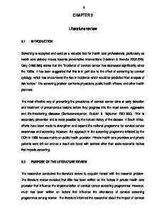
group comprises sloughed-off root cap cells and other debris and mucilage (polysaccharide) originating from the root cap or from lysates released during autolysis. Most of the bacterial colonization occurs in the areas of maximum root exudation viz. the elongation zone junctions between epidermal cells, on root hairs, and at lateral root emergence sites (Fig. 2.2).
Figure 2.2 Rhizodeposition root zones in the rhizosphere (Gobat et al., 2004) Rhizosphere is relatively rich in nutrients due to the loss of as much as 20-40% of plant photosynthesis from the roots (Lugtenberg et al, 2001). Thus, root exudates viz. carbohydrates, amino acids, organic acids and mucilage-derived nutrients attract deleterious rhizobacteria as well as beneficial and neutral bacteria allowing them to colonize and multiply in the rhizosphere (Walker et al., 2003). PGPR have to be highly competitive to colonize the root zone successfully (Compant et al., 2010).
Most rhizosphere organisms occur within 50 mm of root surface and populations within 10 mm of root surface may reach 109-1012 microbial cells/ g of soil. Despite large numbers of bacteria in the rhizosphere, only 7-15% of the total root surface is generally occupied by microbial cells (Gray and Smith, 2005). Root colonization is influenced by many biotic factors (genetic traits of the host plant and the colonizing organism) and abiotic factors (growth substrate, soil humidity, soil and rhizosphere pH, and temperature). To colonize the rhizosphere during an extended period characterized by strong microbial competition and to exert plant growth promoting traits, soil bacteria need to be rhizosphere competent (Whipps, 2001). Root colonization and rhizosphere competence are heavily influenced by the plant changes in the physical and chemical composition of the rhizosphere soil compared to the bulk soil. These differences are manifested by changes in water potential, partial pressure of O 2, and other physical and chemical characteristics due to plant exudations (Vessey, 2003). 2.10 Plant Growth Promoting Rhizobacteria Microorganisms that colonize the rhizosphere are classified as beneficial, deleterious
and
neutral
groups
on
the
basis
of
their
effects
on
plant
(Dobbelaere et al., 2003). Beneficial microorganisms that can grow in the rhizosphere are ideal for use as biocontrol agents. Kloepper and Schroth (1981) termed these beneficial rhizobacteria as Plant Growth Promoting Rhizobacteria (PGPR). PGPR are defined by three intrinsic characteristics: (i) they must be able to colonize the root (ii) they must survive and multiply in micro-habits associated with the root surface, in competition with other micro-biota at least for the time needed to express their plant growth promotion /protection activities (iii) they must promote plant growth. Plant growth promoting
rhizobacteria are thus free- living, soil-borne bacteria which when applied to seeds/soils or crops, enhance the growth of the plant directly by providing nutrients to plants or indirectly by reducing the damage from soil borne plant pathogens (Klopper et al.,1994). Plant growth promoting rhizobacteria have first been used for agricultural purposes in the former Soviet Union and India in the early 20 th century and are now being tested worldwide. In general, PGPR can be divided into two categories (i) Extracellular PGPR (ePGPR, free living) existing in the rhizosphere on the rhizoplane or in the spaces between cells on the root cortex and (ii) intracellular PGPR (iPGPR, symbiotics) which exist inside root cells (Gray and Smith, 2005). Generally, iPGPR include the members of the family Rhizobiaceae, capable of forming nodules on the root systems of leguminous plants (Figueiredo et al., 2011).
Among ePGPR Pseudomonas and Bacillus are the most
commonly described genera (Bhattacharyya and Jha, 2012). Based on their mechanism of action, PGPRs can be classified into three general forms viz. biofertilizer, phytostimulator and biopesticide (Table 2.3). Recent investigations on PGPR revealed that they promote plant growth by [1] producing ACC deaminase to reduce the level of ethylene in the roots of developing plants (Dey et al., 2004) [2] producing plant growth regulators like indole acetic acid (IAA), gibberellic acid, cytokinins (Castro et al., 2008) and ethylene (Saleem et al., 2007) [3] asymbiotic nitrogen fixation (Ardakani et al., 2010) [4] exhibition of antagonistic activity against chitinases,
phytopathogenic microorganisms by producing
antibiotics,
siderophores
and
cyanide
(Pathma
et
β-1,3-glucanase, al.,
2011)
and
[5] solubilization of mineral phosphates and other nutrients (Hayat et al., 2010). PGPR may use more than one of above mechanisms to enhance plant growth as experimental evidence suggests that the plant growth stimulation is the net result of multiple
mechanisms that may be activated simultaneously (Martinez- Viveros et al., 2010) (Fig. 2.3). Table 2.3. PGPR forms and their mechanisms of action stimulating plant growth PGPR form
Biofertilizer
Phytostimulator
Biopesticide
Definition
Mechanism of action
A substance which contains - Biological N2 fixation live microorganisms which when applied on the seed, - Utilization of insoluble plant surface or the soil, forms of phosphorous colonizes the rhizosphere or the interior of the plant and promotes growth through increased supply of primary nutrients of the host plant
Microorganisms with the ability to produce growth regulators such as indole acetic acid, gibberellic acid and cytokinins and ethylene
- Production of phytohormones
Reference FuentesRamirez, and CaballeroMellado, 2006 Vessey, 2003
Lugtenberg et al., 2002 Somers et al., 2004
Microorganisms that - Production of promote plant growth by antibiotics controlling phytopathogenic (siderophores, HCN, agents antifungal metabolites) - Production of enzymes that degrade the cell wall of the fungi - Competitive exclusion - Acquired and induced systemic resistance
Somers et al., 2004
Chandler et al., 2008
Figure 2.3 Functional diversity among plant growth promoting rhizobacteria (Ahemad and Kibret, 2014) In addition, PGPR have great adaptation to harsh environments including drought stress, salt stress, high temperatures, dryness or heavy rainfall in tropical countries, and contaminated environments (Arzanesh et al., 2011; Dell Amico et al., 2008; Mayak et al., 2004), indicating that they could contribute to ameliorate plant crops in areas with poor agricultural potential. The first reports of PGPR on potato noted that growth promotion was associated with a reduction of total fungal propagules on the rhizoplane (Kloepper and Schroth, 1981). This suggested that the select PGPR strains could also be used to reduce pathogen populations in the root zone. 2.11 Pseudomonas as PGPR Members of the genus Pseudomonas (γ-Proteobacteria subclass, Pseudomonadales order, Pseudomonadaceae family) are non-sporulating rods with Gram-negative reaction, motile (one or several polar flagella), and a high genomic G+C content 58–69% (Palleroni, 2008). They are catalase and oxidase positive and chemo organotrophic, with a strictly respiratory metabolism (using oxygen and in some cases nitrate as terminal electron
acceptor). The fluorescent Pseudomonads include all Pseudomonas species with the ability to produce fluorescent pyoverdine siderophore(s), noticeably Pseudomonas aeruginosa, P. syringae, P. putida and P. fluorescens
(Bossis et al., 2000). The generation time of
Pseudomonas spp. in the rhizosphere was found to be 5- 14 h, whereas it was found to be 77 h in the bulk soil (Bowen and Rovira, 1973). There are many species such as P. fluorescens, P. putida, P. aeaureofasciens and P. chloraphis, which may act as plant beneficial bacteria by antagonizing plant pathogens and through the production of traits that directly influence plant disease resistance and growth (Ortiz-Castro et al., 2009). Pseudomonads have been mostly studied for protection of
crop
plants
from
phytopathogenic
oomycetes
(Pythium spp.)
and
fungi
(F. oxysporum, Gaeumannomyces graminis, R. solani, etc.), and to a lesser extent bacteria (Pectobacterium carotovorum) and nematodes (Meloidogyne spp.) (Table 2.4). Disease suppression by these bacteria often entails inhibition of phytopathogens in soil or on roots, by competition and/or antagonism (Haas and Defago, 2005). Plant protection may also result from direct interactions with the host plants, especially in the case of Induced Systemic Resistance (ISR) (Bakker et al., 2007). Several lines of evidence indicate that siderophore production when iron is limited is responsible for the antagonism of some strains of P. aeruginosa against Pythium spp., the causal agents of damping-off and root rot of many crops (Charest et al., 2005, Mavrodi et al., 2012). Pseudomonas spp. produces wide varieties of antibiotics, which confer a competitive advantage and microbial fitness to survive in most environments (Paulsen et al., 2005). Due to their ability to produce variable metabolites and to utilize several organic compounds most biocontrol pseudomonads are not specific for one
pathogen or plant species only, but have a wide host range and can suppress several pathogens (Fig. 2.4). Table 2.4. Disease suppressed by Pseudomonas biocontrol species Strain
Host
P. fluorescens Pf29A
wheat
P. fluorescens SBW25
pea
P. fluorescens DR54
sugarbeet
P. fluorescens 2P24
Pathogen
Reference
G. graminis var. tritici
Barret et al., 2009
Pythium ultimatum
Sanguin et al., 2008
R. solani P. ultimum
Sanguin et al., 2008
wheat
R. solanacearum, F. oxysporum, R. solani,
Sanguin et al., 2008
P. chlororaphis PA-23
canola
S. sclerotiorum
Fernando et al., 2007
P. fluorescens Pf-5
cotton
R. solani Pythium ultimum
Loper et al., 2007
R. solani
Jung et al., 2007
Wheat
G. graminis var. tritici
Weller et al., 2007
P. aeruginosa FP10
Banana
F.oxysporum f.sp cubense
Ayyadurai et al., 2006
P. aeruginosa 7NSK2
Rice
P. aureofaciens P. fluorescens Q2-87
soybean
Magnaportha grisea
De Vleesschauwer et al., 2006
Figure 2.4 Overview of plant-protection mechanisms in closely-related species of fluorescent Pseudomonas (Couillerot et al., 2009)
2.12 PGPR traits of fluorescent Pseudomonads 2.12.1 Phosphate solubilization Phosphorus is second important mineral nutrient after nitrogen required for plant development and growth making up about 0.2% of plant dry weight (Vikram and Hamzehzarghani, 2008). It is also involved in photosynthesis, signal transduction, macromolecular biosynthesis, and respiration (Fernandez et al., 2007). Although Phosphate (P) is abundant in soils in both inorganic (originating mainly from applied P fertilizer) and organic forms (derived from microorganisms, animals and plants), it is still one of the major plant growth limiting nutrients. About 10-25% of P fertilizer is required by the plants (Saha and Biswas, 2009) for promoting their functions. The low availability of P to plants is because majority of soil P is found in insoluble forms. Most of the P is
found in form of iron, calcium or aluminum phosphates (Fe-P, Ca-P, or Al-P). Plant roots can only absorb P in two soluble forms, the monobasic (H 2PO4-) and the diabasic (HPO42-) ions (Vessey, 2003). To circumvent the problem, chemical phosphate fertilizers are used. However, due to its high reactivity, almost 75 to 90% of added P fertilizer is precipitated by Al, Fe, and Ca complexes present in the soils, creating a demand for suitable alternatives to mobilize this fixed fraction of the important bio- element (Glick, 2012). Organisms with phosphate solubilizing activity are called phosphate solubilising microorganism (PSM), may provide the available forms of P to the plants and are viable substitute to chemical phosphatic fertilizer. Members of the genus Azotobacter, Bacillus, Enterobacter, Erwinia, Pseudomonas, Rhizobium and Serratia are reported as the most significant phosphate solubilising bacteria (Bhattacharyya and Jha, 2012). Several species of fluorescent pseudomonads such as P. aeruginosa (Ahemad and Khan,
2011a),
P.
putida
(Ahemad
and
Khan,
2012a),
P.
fluorescens
(Shaharoona et al., 2008), P. chlororaphis (Liu et al., 2007), P. jessenii (Rajkumar and Freitas, 2008) were reported as phosphate solubilizers. Solubilization of inorganic phosphorus occurs as a consequence of the action of low molecular weight organic acids which are synthesized by various soil bacteria (Zaidi et al., 2009). Conversely, the mineralization of organic phosphorus occurs through the synthesis of a variety of different phosphatises by catalyzing the hydrolysis of phosphoric esters (Glick, 2012). Importantly, phosphate solubilization and mineralization can coexist in the same bacterial strain (Tao et al., 2008). Several mechanisms have been proposed to explain the microbial solubilization of P compounds. The mechanisms include: (1) release of organic acids produced during
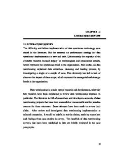
organic residue decomposition (Hameeda et al., 2006), (2) excretion of protons due to NH4+ assimilation by microorganisms (Whitelaw, 2000), (3) formation of complexes between organic acids/ anions with cations [Al3+, Fe3+, Ca2+] (Welch et al., 2002) and (4) desorption of P sorbed onto soil clay and/or oxides (Osorio, 2008). Nitric and sulfuric acid produced by Nitrosomonas and Thiobacillus species, respectively, have also been reported to dissolve P compounds (Azam and Memon, 1996). Equally, P compounds may be solubilized by carbonic acid formed as a result of organic matter decomposition (Memon 1996) (Fig. 2.5).
Figure 2. 5. Microbial contribution to plant phosphorous nutrition (Richardson, 2009).
Kumar et al. (2008) isolated metal tolerant, plant growth-promoting bacteria (Enterobacter sp.) which decreased the pH of the growth medium from 7 to 2, thereby achieving the maximum solubilization of P (229 mg/l). Chen et al. (2006) also showed that
the P solubilizing activity of isolated strains was related to the release of organic acids and the subsequent pH reduction in the medium. Hameeda et al. (2006) reported that phosphate solubilizing bacteria with cellulolytic activity enhanced the mineralisation and decomposition of crop residues. Pratibha and Arvind (2009) found that phosphate-solubilising fluorescent Pseudomonas strains producing 2-ketogluconic acid, gluconic acid, oxalic acid, succinic acid, lactic acid, formic acid, citric acid and malic acid in the culture filtrates during the solubilisation of tricalcium phosphate. De Werra et al. (2009) concluded that the ability of fluorescent Pseudomonas strain CHAO to acidify its environment and to solubilise mineral phosphate is strongly dependent on its ability to produce gluconic acid. Phosphate-solubilizing microbes are reported to reduce the toxicity of metals and protect the plants against the toxic effects of these metals and consequently enhance the growth and yield of plants in contaminated soils (Wani et al., 2007). Attia et al. (2009) reported that inoculation of phosphate-solubilizing bacteria with mineral phosphorus increased the efficiency of P fertilizer and decreased the required P rate to plants with enhanced vegetative growth and fruit quality. 2.12.2 Siderophore production Iron is a vital nutrient for almost all forms of life. In the aerobic environment at physiological pH, the reduced ferrous (Fe2+) form is unstable and is readily oxidized to oxidized ferric (Fe3+) which normally occurs as poorly soluble iron hydroxides and oxyhydroxides, basically unavailable to biological systems (Rajkumar et al., 2010). Siderophores are low molecular weight molecules (400-1000 daltons) which have high affinity for iron and thus bind ferric ions available in the soil. Many PGPR strains
like Pseudomonas, Bacillus, Acinetobacter, Serratia known to produce siderophores, thus improves the availability of iron to plants. Indirectly they control the pathogens by scavenging the limited amount of ferric ions available in the rhizosphere and thus inhibiting the pathogens in their immediate vicinity (Yu et al., 2011; Sarode et al., 2009). In both Gram positive and Gram negative rhizobacteria, Fe 3+ - siderophore complex on bacterial cell membrane is reduced to Fe2+ which is released into the cell from the siderophore via a gating mechanism linking the inner and outer membranes (Rajkumar et al., 2010). Based on the structural features, functional groups and types of ligands, bacterial siderophores are classified into four main classes namely, carboxylate, hydroxamates, phenol catecholates and pyoverdines (Crowley, 2006). Most of the bacterial siderophores are catecholates, and few are hydroxamates and carboxylates, whereas most fungal siderophores are hydroxamates (Schalk et al., 2011). Siderophores are reported to form stable complexes with other heavy metals such as Al, Cd, Cu, Pb, Zn (Chamongkolpradit et al., 2008). Siderophore production by various bacteria in response to iron deficiency normally occurs in neutral to alkaline pH soils, due to low iron solubility at elevated pH have been reported (Sharma and Johri, 2003). Of the several mechanisms used to facilitate plant growth, siderophore synthesized by microbes including rhizobia and species of Bacillus, Pseudomonas and Azotobacter (Ahmad et al., 2008) is well documented due to their iron sequestration ability from the soil. Rane et al. (2008) showed that biocontrol ability of P. aeruginosa ID 4365 against groundnut (Arachis hypogaea) phytopathogens was due to production of pyoverdin and pyochelin siderophores. Application of cadmium- resistant plant growth-promoting P. aeruginosa exhibiting siderophore production, when used as inoculant for blackgram
(Vigna mungo L.) plants grown in soil treated with a gradient of CdCl2 concentration, reduced the toxicity of metal to plants (Ganesan, 2008). Wani et al. (2008b) and Tripathi et al. (2005) suggested that plant chlorosis due to heavy metals can be prevented by providing them with siderophore producing bacterium that supplements sufficient amounts of iron to the plant. Vansuyt et al. (2007) have reported increased plant growth in Arabidopsis thaliana due to intake of Fe-pyoverdine complex synthesized by P. fluorescens C7. Charest et al. (2005) have demonstrated the contribution of siderophore towards inhibitory potential of P. aeruginosa towards Pythium spp. in an iron-depleted medium. Siderophore mediated suppression of rice fungal pathogens R. solani and Pyricularia oryze in an in-vitro assay on Kings-B medium has been reported by Battu and Reddy (2009). 2.12.3 Indole Acetic Acid Many important plant-microbial interactions center on the production of auxins. It is reported that 80% of microorganisms isolated from the rhizosphere of various crops possess the ability to synthesize and release auxins as secondary metabolites. IAA is produced by PGPR by using the rich supplies of substrates exuded from the roots and release of auxin in the rhizosphere as secondary metabolites. Several PGPR are reported to produce IAA which stimulates cell division, seed and tuber germination, increase the rate of xylem and root development (lateral and adventitious root formation) and thereby play a significant role in increasing the root surface area and number of root tips in many plants (Bhattacharyya and Jha, 2012). A greater root surface area and length enables the plant to access more nutrients from soil and thus contribute to plant growth promotion (Tsavkelova et al., 2007).
Tryptophan has been identified as a main precursor for IAA biosynthesis pathways in bacteria (Zaidi et al., 2009). Tryptophan inhibits anthranilate (reduces IAA synthesis) formation by a negative feed-back regulation on anthranilate synthase, resulting in indirect induction of IAA production. The identification of intermediates led to the identification of five different pathways for IAA synthesis using tryptophan as a precursor for IAA (1) IAA formation via indole-3-pyruvic acid and indole-3-acetic aldehyde is found in a majority of bacteria
like
Erwinia
herbicola;
saprophytic
species
of
the
genera
Agrobacterium and Pseudomonas; certain representatives of Bradyrhizobium, Rhizobium, Azospirillum, Enterobacter and Klebsiella (2) Conversion of tryptophan into indole-3acetic aldehyde may involve an alternative pathway in which tryptamine is formed (pseudomonads and azospirilla) and (3) IAA biosynthesis via indole-3-acetamide formation is reported for phytopathogenic bacteria P. syringae, and
Agrobacterium tumefaciens,
E. herbicola; saprophytic pseudomonads like P. putida and
P. fluorescens (4) IAA biosynthesis that involves tryptophan conversion into indole-3acetonitrile is found in the cyanobacterium (Synechocystis sp.) and (5) the tryptophanindependent pathway, which is common in plants and also found in azospirilla and cyanobacteria (Fig. 2.6). A positive correlation between auxin production and growth-promoting activity of diverse PGPR has been also reported in Brassica juncea and wheat (Khalid et al., 2004). Species of Bradyrhizobium and Rhizobium produced a substantial amount of IAA under in vitro conditions (Wani et al., 2008a; Ahmad et al., 2008). Among other PGPR strains, Pseudomonas, Bacillus, Agrobacterium sp., Alcaligenes piechaudii and two strains of Comamonas acidovorans secreted IAA (Rajkumar et al., 2006). In another study, numerous bacterial isolates recovered from wheat (Triticum aestivum) rhizosphere
demonstrated the production of auxins (ranging from 1.1 to 12.1 mg/ l) under in vitro conditions. However, when the medium was supplemented exogenously with tryptophan, it significantly enhanced the auxin biosynthesis which was confirmed by high performance liquid chromatography (HPLC) analysis (Khalid et al., 2004).
Figure 2.6. Overview of different pathway to synthesize IAA in bacteria (Spaepen et al., 2009) Tryptophan increased production of IAA in B. amyloliquefaciens FZB42 (Idris et al., 2007). IAA production, even in the culture without tryptophan supplementation has also been reported (Wahyudi et al., 2011). It has also been reported by Patten and Glick (2002) that the enzyme indolepyruvic decarboxylase (IPDC) is the principal enzyme which determines IAA biosynthesis and stimulates the development of the root system of the host plant. It has been reported that IAA production by PGPR can vary among different species and strains and is also influenced by growth stage, culture condition and substrate availability (Ashrafuzzaman et al., 2009).
Production of IAA has been shown in species of Bacillus, Pseudomonas, Azotobacter, Azospirillium and Glucanoacetobacter. It functions as an important signal molecule in the regulation of plant development and indirectly by influencing bacterial amino cyclopropane-1-carboxylate (ACC) deaminase activity (Wahyudi et al., 2011; Saharan and Nehra 2011). 2.12.4 Volatile organic compounds (VOC) Volatile organic compounds (VOCs) are defined as compounds that have high enough vapour pressures under normal conditions to significantly vaporize and enter the atmosphere. This class of chemicals includes compounds of low molecular weight (