11th International Conference on Tooth Morphogenesis and Differentiation Organized by Laurent VIRIOT (Chair), Françoise BLEICHER, and Vincent LAUDET
Plenary poster sessions abstract program
May 26-31, 2013 - La Londe Les Maures (France) ______________________________________________________________
TMD 2013 Tooth Development ______________________________________________________________________________
Understanding the mechanisms of root formation from rootless tooth Cheol-Hyeon Bae*, Eui-Sic Cho Cluster for Craniofacial Development and Regeneration Research, Institute of Oral Biosciences, Chonbuk National University School of Dentistry, Jeonju, South Korea * presenting author:
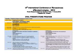
[email protected] BACKGROUND: The Wnt family of proteins plays an important role in morphogenesis and cellular differentiation in many tissues [1]. It is well-known that Wnt/ß-catenin signaling plays multiple roles in various stages of tooth morphogenesis [2]. However, little is known about the involvement of Wnt/ß-catenin signaling in odontoblast differentiation. The tooth root is important part of tooth together with surrounding periodontium to maintain tooth in alveolar socket [3]. While the molecular and cellular mechanisms of early tooth development and crown morphogenesis have been extensively studied, little is known about the molecular mechanisms controlling tooth root formation. METHODOLOGY: We generated and analyzed mice with tissue-specific inactivation of ßcatenin, an obligatory transducer of canonical Wnt signaling, in developing odontoblasts to directly investigate the role of Wnt/ß-catenin signaling in root formation. PRINCIPAL FINDINGS: Here, we show that ßcatenin is strongly expressed in odontoblast lineage cells, and it is required for root formation (Fig. A-B). Tissue-specific inactivation of ßcatenin in developing odontoblasts produced molars lacking roots and aberrantly thin incisors (Fig. C-D). At the beginning of root formation in the mutant molars, the cervical loop epithelium extended apically to form Hertwig’s epithelial root sheath (HERS), but root odontoblast differentiation was disrupted and followed by the loss of some HERS inner layer cells. However, outer layer of HERS extended without the root (Fig. EF), and the mutant molars finally erupted. The periodontal tissues invaded extensively into the dental pulp (Fig. G-H).
Legend: Expression of ß-catenin in the odontoblast lineage cells and tooth phenotypes in mice with tissuespecific inactivation of ß-catenin in developing odontoblasts. (A) Localization of ß-catenin in the odontoblast lineages during root formation in mouse molar (B) OC-Cre activities in the differentiating odontoblasts during root formation. (C-D) Molars lacking roots and thin incisors in OC-Cre:Ctnnb1CO/CO mice (P28). (E-F) Disrupted differentiation of root odontoblasts in OC-Cre:Ctnnb1CO/CO mice at the beginning of root formation (P8). (G-H) Erupted molars without root formation (P28).
DISCUSSION and CONCLUSIONS: In this study, we investigated the functional significance of Wnt/ß-catenin signaling in the dental mesenchyme during tooth root formation. OCCre;Ctnnb1CO/CO mice exhibited remarkable tooth phenotypes characterized by erupted molars lacking roots and thin incisors. This abnormality was closely associated with odontoblast differentiation. The results of the present study further imply that Wnt/ß-catenin signaling in the dental mesenchyme is required to differentiate odontoblasts for root formation. REFERENCES: [1] Logan and Nusse. Annu Rev Cell Dev Biol (2004). [2] Liu and Millar. J Dent Res (2010). [3] Thomas HF. Int J Dev Biol (1995). Funding: This work was supported by National Research Foundation of Korea (NRF) grants, funded by the Korean government (MEST) (Nos. 2009-0085733 and 2012R1A1A2000919).
______________________________________________________________________________ Monday May 27: 13.30 → 14.30 Poster presentation PA1
TMD 2013 Tooth Development ______________________________________________________________________________
The pattern of cementum formation on the developing roots in mice during stages P12–P36 Valentina Woth*, Isabel Nowak, Herbert Renz, Ralf. J. Radlanski Charité -Campus Benjamin Franklin at Freie Universität Berlin, Center for Dental and Craniofacial Sciences, Dept. of Craniofacial Developmental Biology, Berlin, Germany * presenting author: valentina.woth@charité.de BACKGROUND: Cementum formation is essential for the formation of the tooth-bone interface, because of the insertion of the periodontal fibers. Different types of cementum, primarily as acellular cementum on the cervical root and cellular cementum covering the apical root have been studied by many authors [2,3,4]. However, it has not been described in detail, how the temporospacial formation and morphogenesis of dental cementum takes place on the root. The purpose of this study is to elucidate the pattern of dental cementum formation on the surfaces of developing roots in mice. METHODOLOGY: 13 Mice, ranging from postnatal day 12-36, were prepared as serial histological sections (thickness 10 μm) and stained with H.E., Alcianblue and Trichrom. 3-D- reconstructions were made using the software AnalySIS (Olympus, Berlin). Regions of dental cementum on the developing roots of mandibular M1, M2, M3 were identified in the histological sections, marked and reconstructed in 3-D together with dentin and pulp. PRINCIPAL FINDINGS: Alcianblue turned out to be a specific staining for identifying acellular cementum. A thin layer of acellular cementum could be found on the root surface as soon as it was formed itself (stage P12). However, formation of cellular cementum lagged behind the formation of the root, starting at stage P 20. Therefore, large areas of root surface remain uncovered from cellular cementum.
Legend: M1-M3 at P36. Cellular cementum is shown in blue-grey.
plays an important role for further development of fiber insertion leading to tooth-bone- osseointegration. Insights like these are crucial for a possible, future implantation of extracorporal cultured tooth germs into an adult jaw. REFEFENCES: [1] Foster Int. J. of Oral Sci. (2012); [2] Nanci et al. Periodontology (2006); [3] Radlanski Entwicklungsbiologie, Quintessenz Verlag (2011); [4] Schroeder The Periodontium, Springer Verlag (1986); [5] Shukla J. Forensic Odontostomatology (2012). FUNDING: Supported by grants Ra 428/1-9. Deutsche Forschungsgemeinschaft. We thank Mrs. B. Danielowski, Mrs. I. Schwarz for their most valuable and skillfull assistance.
DISCUSSION and CONCLUSIONS: Our study showed that cementum formation seems not to be a uniformly layered process – instead, we revealed an insular formation pattern for specific regions of cementum. This specific pattern and location of dental cementum formation of the root
______________________________________________________________________________ Monday May 27: 13.30 → 14.30 Poster presentation PA2
TMD 2013 Tooth Development ______________________________________________________________________________
Precise chronology of differentiation of developing human primary dentition Xuefeng Hu1*, Shan Xu1, Bingmei Wang1, YiPing Chen1,2, Yanding Zhang1 1
Fujian Key Laboratory of Developmental and Neuro Biology, College of Life Science, Fujian Normal University, Fuzhou, China; 2Department of Cell and Molecular Biology, Tulane University, New Orleans, LA, USA * presenting author:
[email protected]
BACKGROUND: While correlation of developmental stage with embryonic age of the human primary dentition has been well documented, the available information regarding the differentiation timing of the primary teeth varies significantly. In this study, we aimed to document precise differentiation timing of the developing human primary dentition. METHODOLOGY: Dental tissues were collected from chemically-terminated human embryos with well defined ages. Tissue sections were subjected to Hematoxylin/Eosin staining for standard histological analysis, and to dichromic staining for detection of mineralized tissues, and for immunohistochemical staining for expression of molecular markers. PRINCIPAL FINDINGS: We systematically examined the expression of odontogenic differentiation markers along with the formation of mineralized tissue in each developing maxillary and mandibular tooth from human embryos with welldefined embryonic age. We show that, despite that all primary teeth initiate development at the same time, odontogenic differentiation begins in the maxillary incisors at the 15th week and in the mandibular incisors at the 16th week of gestation, followed by the canine, the first primary premolar, and the second primary premolar at a week interval sequentially. Despite that the mandibular primary incisors erupt earlier than the maxillary incisors, this distal to proximal sequential differentiation of the human primary dentition coincides in general with the sequence of tooth eruption.
ing human embryogenesis should be rather consistent. However, current information regarding the differentiation timing of the human primary dentition varies dramatically. Our results presented here provide an accurate chronology of odontogenic differentiation in the developing human primary dentition, which could serve as a standard base for future studies of human tooth development and could also be used in textbook as a standard reference. REFERENCES: [1] Welbury et al. Pediatric Dentistry. 4th Ed. (2012) Oxford Press; [2] Schoenwolf et al. Larsen’s Human Embryology. 4th Ed. (2009). Churchill Livingston, Elsevier, Philadelphia, PA. Funding: The 973 Project of China (2010CB944800); National Natural Science Foundation of China (81100730, 81271102); The Natural Science Foundation of Fujian Province (2012J01119).
DISCUSSION and CONCLUSIONS: Although timing of eruption of each primary tooth differs significantly between each individual, the gestation ages of tooth development at each stage dur-
______________________________________________________________________________ Monday May 27: 13.30 → 14.30 Poster presentation PA3
TMD 2013 Tooth Development ______________________________________________________________________________
The role of Fibrillin during tooth root and periodontal ligament tissue development Kyoko Oka1*, Michiko Kira1, Eichi Tsuruga2, Hidemitsu Harada3, Naoki Fujiwara3, Yoshihiko Sawa2, Masao Ozaki1 1
Pediatric Dentistry, Department of Oral Growth and Development, 2Functional structure, Department of Morphological Biology, Fukuoka Dental College, Fukuoka Japan; 3Developmental Biology and Regenerative Medicine, Department of Anatomy, Iwate Medical University, Yahaba, Japan * presenting author:
[email protected]
BACKGROUND: Fibrillin microfibrils are unique architectural elements of the extracellular matrix (ECM) that endow connective tissues with specific physical properties, either as obligatory constituents of elastic fiber or as elastin-free assemblies. Mutation of fibrillin genes result in the severe heritable connective tissue diseases such as Marfan syndrome. The oxytalan fibers are defined as fibrillin microfibril assemblies without elastin deposition and are specially located in periodontal ligament tissues (PDL) [1]. The biological function of oxytalan fibers in periodontal ligaments of an erupted tooth has been reported in several articles [2]. However, the role of fibrillin during tooth development has not been fully understood. Furthermore, fibrillin can bind TGF-βs through latent transforming growth factors βbinding proteins (LTBPs). The aim of this study is to examine and compare the localization and distribution of fibrillin-1 and -2 and TGF-βs during tooth development. METHODOLOGY: Samples in vivo. Pregnant mice were prepared for collect embryos from embryonic stage 14.5 (E14.5) to post neonatal day 10 (P10). The dissected mouse head or mandibular bones without chemical fixation and decalcification were embedded in super cryoembedding medium, and rapid-frozen by the hexane-dry ice method. The samples were cut with a cryostat into 8-µm-thick sections. Post neonatal samples were prepared by using the Film transfer method. In vitro: Murine HERS cells (HERS01a) were established by the limiting dilution-culture technique. They were cultured in DMEM/HAMF12 medium with supplement in a humidified 5% CO2 atmosphere at 37°C [3]. Immunofluorescence: Anti-Fibrillin-1, -2, TGF-β3, and Cytokeratin were used as the primary antibodies.
Legend: The expression of Fibrillin-1 and -2 at P5 in mouse.
The immunoreaction was visualized on sections with anti-IgG antibody conjugated with Alexa Fluor. Immuno-stained sections and cells were then counterstained with DAPI. PRINCIPAL FINDINGS: In vivo analysis, Fibrillin-1 expression started in periodontal ligament tissue around P10. This fiber-like expression was attached with root surface (cementum) but not with alveolar bone. Interestingly, Fibrillin-2 was expressed in dental epithelium since E14.5. This expression was observed in the cervical loop and Hertwig’s epithelial sheath during root development. TGF-β3 was also expressed in Hertwig’s epithelial sheath. In vitro analysis, HERS01a cells clearly expressed Fibrillin2. DISCUSSION and CONCLUSIONS: These findings suggest that Fibrillin-2 protein not only plays a part in forming oxytalan fibers in PDL, but also potentially serves as a regulator for root formation via regulation of TGF-β signaling during tooth development. REFERENCES: [1] Tsuruga et al. J Periodontal Res. (2002); [2] Kondo et al. Acta Histchemica. (2011); [3] Akimoto et al. Biochem Biophys Res Commun. (2011). Funding: Grant-in-Aid for Scientific Research from the Japanese Ministry of Education, Culture, Sports, Science and Technology [KAKENHI (24593116) to KO].
______________________________________________________________________________ Monday May 27: 13.30 → 14.30 Poster presentation PA4
TMD 2013 Tooth Development ______________________________________________________________________________
Spatio-temporal expression and possible function of tuftelin during mouse embryonic craniofacial development Dekel Shilo1, Yoav Leiser1, Boaz Shay1, Asher Ornoy2, Anat Blumenfeld1, Dan Deutsch1* 1
Dental Research Laboratory, Faculty of Dental Medicine, 2Laboratory of Teratology, Department of Anatomy and Cell Biology, The Hebrew University of Jerusalem - Hadassah, Jerusalem, Israel * presenting author:
[email protected]
BACKGROUND: Tuftelin is expressed in the epithelial ameloblasts at an early stage of amelogenesis. Tuftelin cDNA sequences were detected in morula and embryonic stem cells as well as in many different soft tissues, normal and cancerous. The tuftelin protein was also detected in different soft tissues such as eye, brain, kidney, lung and testis. Little was known about tuftelin expression in the developing embryo, where tuftelin was proposed to be involved in epithelial mesenchymal reciprocal interactions. In addition, tuftelin-soaked agarose beads induced recruitment and proliferation of embryonic mesenchymal cells. METHODOLOGY: Embryos from pregnant CD-1 mice aged E10.5-E18.5 and newborns aged P3 were used in this study. All experiments were approved by the Animal Care Ethical Committee of The Hebrew University of Jerusalem. Heads were removed and mRNA and protein were extracted. Real-time quantitative PCR was performed using TaqMan® AOD. Indirect immunohisto- chemistry and In-situ hybridization were performed on craniofacial sections and Western blot was performed on the extracted protein. PRINCIPAL FINDINGS: (1). The expression of tuftelin mRNA in the cranio-facial complex at E10.5 up to E16.5, was analyzed by RT-PCR, followed by cDNA sequencing and compared to endogenous control Actin. Two known tuftelin isoforms were detected along the different stages of mouse embryonic cranio-facial development; one containing all 13 exons of tuftelin and one lacking exon 2. (2). Tuftelin mRNA expression level increased during mouse embryonic craniofacial development from E10.5 up to E16.5. Tuftelin protein was also detected. (3). Tuftelin is expressed in various tissues of the developing
mouse embryonic craniofacial complex such as brain, eye, ganglion, cartilage and bone, and is already expressed at E10.5 in the brain and eye, long before the initiation of tooth formation. Tuftelin protein expression was detected in the tooth germ (dental lamina) already at E12.5, much earlier than previously reported (E17). DISCUSSION and CONCLUSIONS: In the developing tooth, tuftelin protein is already expressed from the initiation of tooth germ formation at E12.5 and is continuously expressed throughout tooth development, at least up to P3 (the latest age tested). The expression pattern of tuftelin protein and mRNA exhibits dynamic spatio-temporal changes in the various tissues. Tuftelin protein expression was detected in the craniofacial complex already at E10.5 (brain, eye), long before the initiation of tooth formation, suggesting possible additional role(s) for tuftelin other than those suggested in the past in epithelialmesenchymal reciprocal interactions during tooth germ development. At younger embryonic age tuftelin protein expression was detected mainly in the cytoplasm while at later embryonic ages and post-nataly its expression concentrates in the perinuclear/nuclear region. Tuftelin expression seems to shift to the nucleus with developmental progression. The present results, combined with our previous results regarding tuftelin involvement in HIF1α and NGF pathways, may suggest that tuftelin has a role in cell signalling also during tooth development. REFERENCES: [1] Zeichner-David et al. Int J Dev Biol (1997); [2] MacDougall et al. J Dent Res (1998); [3] Deutsch et al. Connect Tissue Res (2002); [4] Shay et al. Protein Expr Purif (2009); [5] Leiser et al. J Cell Physiol (2011). Funding: Israel Science Foundation (ISF) 75/08
______________________________________________________________________________ Monday May 27: 13.30 → 14.30 Poster presentation PA5
TMD 2013 Tooth Development ______________________________________________________________________________
Overactivation of Wnt/β-catenin signalling pathway induces Fgf4 overexpression in tooth development Maitane Aurrekoetxea, Jon López, Igor Irastorza, Patricia García-Gallastegui, Gaskon Ibarretxe, Fernando Unda* Department of Cell Biology and Histology, Faculty of Medicine and Dentistry, University of the Basque Country, Leioa 48940, Bizkaia. Spain * presenting author:
[email protected] BACKGROUND: Previous results about overactivation of the Wnt/β-catenin signalling pathway in mesenchymal cells are contradictory and depend on the tissue being studied. Some authors have found a direct relationship between increased cell proliferation or tumour formation and Wnt/β-catenin overactivation [1, 2]. METHODOLOGY: In this work we have provoked the overactivation of the Wnt pathway at key moments of dental development through inhibition of glycogen synthase kinase 3 (GSK-3) by means of 6-bromoindirubin-3'-oxime (BIO).
DISCUSSION and CONCLUSIONS: This work shows that epithelial-mesenchymal interactions, and in particular Wnt-dependent dental epithelial FGF4 production, are essential for dental mesenchymal cell proliferation, tooth morphogenesis and correct dental development. REFERENCES: [1] Miyake et al. Pathol Int. (2006); [2] Siriwardena et al. Oral Oncol. (2009); [3] Aurrekoetxea et al. Biol Cel. (2012). FUNDING: UFI11/44, GUI09/70, Basque Government (SAIOTEK), Jesús Gangoiti Barrera Foundation.
PRINCIPLE FINDINGS: In tooth morphogenesis and odontoblast differentiation stage, H3P immunohistochemistry demonstrated that Wnt/βcatenin overactivity promoted proliferation of dental mesenchymal cells. This increase in the proliferation reflects on an enlargement of the dental piece, but ameloblast and odontoblast cell differentiation is delayed with respect to control [3]. However, when overactivation of the route is induced in MDPC-23, a dental mesenchymal cell line, cell proliferation decreased significantly after BIO treatment. These data suggest that increased proliferation in mesenchymal cells could depend on the activation of the Wnt/β-catenin in dental epithelium. In this context, in molar teeth exposed to BIO and overactivating the Wnt/βcatenin pathway, Fgf4 expression increased dramatically and extended along the inner dental epithelium. The increase of expression of Fgf4 in epithelium seems to be related with the previously observed enhancement of proliferation in the dental mesenchyme.
______________________________________________________________________________ Monday May 27: 13.30 → 14.30 Poster presentation PA6
TMD 2013 Tooth Development ______________________________________________________________________________
Msx2 is required to regulate cell differentiation in stratum intermedium Mitsushiro Nakatomi*, Hiroko Ida-Yonemochi, Hayato Ohshima Division of Anatomy and Cell Biology of the Hard Tissue, Niigata University Graduate School of Medical and Dental Sciences, Niigata, Japan * presenting author:
[email protected] BACKGROUND: Msx2 encodes a homeodomain type transcription factor and mutations of the MSX2 gene are known to be responsible for amelogenesis imperfecta in humans. In accordance with this phenotype, Msx2 null homozygous mice exhibit disrupted ameloblast differentiation and hypoplastic enamel formation [1]. Msx2 is expressed in the enamel organ including ameloblasts and stratum intermedium (SI) cells during amelogenesis as well as in the enamel knot during the early phase of tooth morphogenesis. Though several target genes of Msx2 related to tooth development have been reported to date such as Amelogenin, Bmp4 and Laminin 5 alpha 3 [2,3,4,5], Msx2 function for amelogenesis have not yet been fully understood and some unsolved questions remain to be elucidated. Among those is whether loss of Msx2 affects ameloblast differentiation cell-autonomously or secondarily due to abnormal SI function. To gain further insight into the specific role of Msx2 for amelogenesis, we investigated the detailed dental phenotypes of Msx2 null mice. METHODOLOGY: Post-natal 3d, 5d, 7d, 9d, 10w, 20w and 25w Msx2+/+, Msx2+/- and Msx2-/mice were dissected and analyzed by H&E staining, section and whole-mount in situ hybridization, immunohistochemistry, micro-CT, TEM, semi-thin section, electron probe microanalysis and conventional RT-PCR. PRINCIPAL FINDINGS: In the early process of ameloblast differentiation, some marker genes including Sox2, Shh, Patched1, Dspp, ALP, Mmp20 and Klk4 were almost normally expressed in mutants, implying that Msx2 is not essential for initial ameloblast differentiation. However, notably, SI cells became multi-layered and ectopically expressed Heat-shock protein (Hsp) 25. The signal intensity of Hsp25 in mutant SI cells was
identical to what is commonly seen in the skin and oral mucosa in wild-type.
Legend: Sagittal sections of the labial side of the upper incisor of P10w mice. The incisal side is to the right. While anti-Hsp25 immunoreactivity is detectable in ameloblasts and almost negative in SI cells in WT, Hsp25 is remarkably expressed ectopically in mutant SI cells (arrows).
DISCUSSION and CONCLUSIONS: Our data suggest that Msx2 deficiency may alter the cell character of SI to what resembles keratinized tissues and raise a possibility that aberrant SI formation might affect the maintenance of differentiated state of ameloblasts. REFERENCES: [1] Satokata et al. Nat Genet (2000); [2] Zhou et al. J Biol Chem (2000); [3] Bei et al. Dev Dyn (2004); [4] Aïoub et al. Bone (2007); [5] Molla et al. Am J Pathol (2010). Funding: Grant-in-Aid for Scientific Research (B) (no. 22390341 to H.O.) from JSPS, Japan.
______________________________________________________________________________ Monday May 27: 13.30 → 14.30 Poster presentation PA7
TMD 2013 Tooth Development ______________________________________________________________________________
Absence of the transcription factor osterix in zebrafish disturbs dentinogenesis but does not affect tooth replacement Ann Huysseune1*, Erika Kague2, Mieke Soenens1, P. Eckhard Witten1, Shannon Fisher2 1
Evolutionary Developmental Biology, Ghent University, Belgium; 2University of Pennsylvania, Philadelphia, USA * presenting author:
[email protected]
BACKGROUND: The transcription factor osterix (osx) is an important regulator of bone development [1]. Using an osterix-deficient zebrafish (Danio rerio) model, we tested the hypothesis that osx is required for tooth development and replacement and that osx deficiency differentially affects the teeth and the bone. METHODOLOGY: We prepared serial semithin (1 µm) plastic sections of 5 week and 6 month old zebrafish homozygous for osx deficiency. PRINCIPAL FINDINGS: Patterning of teeth is unaffected in osx -/- mutant zebrafish. Like in the WT, eleven tooth families are present on each side, each family consisting of two tooth germs. Within each tooth family, the larger tooth germ is considered to be the “functional tooth”, its tip occasionally protruding into a crypt of the pharyngeal epithelial lining. The other tooth germ, considered to be its successor, is observed to be in a ventral position and connected to the base of the crypt. Remarkably, unlike in the WT, the pulp cavity of predecessor and successor are connected. Moreover, several teeth display an anomalous orientation, with the tip pointing laterally rather than medially. Unlike patterning, tooth development is severely disturbed in the absence of osterix. An enamel organ forms properly, and becomes organized into an inner and an outer dental epithelium. The inner dental epithelium differentiates into polarized ameloblasts, which develop even into ruffled-bordered ameloblasts. A cap of enameloid is laid down. Subsequently a small amount of dentine is deposited, and odontoblast processes can be found in the thin layer of dentine. Still, odontoblasts never acquire a high cylindrical shape, and in none of the teeth a layer of dentine thicker than 6 to 8 µm could be observed. Thus, development appears to be arrested
in early dentinogenesis. No attachment bone is ever formed and none of the teeth is attached to the underlying ceratobranchial bone. DISCUSSION and CONCLUSIONS: The data revealed by this mutant demonstrate that neither attachment nor eruption is required as a stimulus for initiation of the new tooth, unlike what was formerly suggested based on observations in WT zebrafish [2]. This suggests that downstream targets of osterix are either not involved in repeated downgrowth of the epithelium, or are activated via an alternative pathway. Moreover, not even defective differentiation of the tooth prevents a successional tooth to be formed. These data do not necessarily challenge the assumption of local control over tooth replacement; rather they reveal a hitherto unknown mechanism for triggering replacement tooth formation. The data furthermore show that teeth and bones have a distinct developmental program, in support of data published before on other mutants [3]. REFERENCES: [1] Fong. Nature Rev Molec Cell Biol (2012); [2] Huysseune. Int J Dev Biol (2006); [3] Schilling et al. Development (1996).
______________________________________________________________________________ Monday May 27: 13.30 → 14.30 Poster presentation PA8
TMD 2013 Tooth Development ______________________________________________________________________________
The role of Nogo-A in orofacial development and regeneration Pierfrancesco Pagella1,2*, Maria Alexiou1, Martin E. Schwab3, Thimios Mitsiadis1 1
Institute of Oral Biology, Center of Dental Medicine, University of Zurich, Switzerland; 2Molecular Life Sciences PhD Program, University of Zurich and ETH Zurich, Switzerland; 3Brain Research Institute, University of Zurich and ETH Zurich, Switzerland. * presenting author:
[email protected]
BACKGROUND: The role of innervation in tooth and orofacial development is still poorly understood. Accumulating evidence supports an active role of innervation in the development of orofacial organs [1-2]. Nogo-A was identified as a strong regulator of neurite outgrowth and maturation, and it was mainly studied for its role in CNS repair and regeneration [3]. Nogo-A may play an important role in orofacial innervation and development. Moreover, Nogo-A role outside the nervous system has never been investigated. METHODOLOGY: Description of Nogo-A expression in the orofacial complex. Nogo-A knockout characterization. Organ cultures and cocultures (cranial ganglia/neurons and target organs), Nogo-A inhibition and overexpression. Nogo-A in tooth injury in vivo.
Legend: Working hypothesis. Nogo-A may regulate tooth and other organs development through its effect on innervation or independently of innervation. REFERENCES: [1] Oakley and Witt. J.Neurocyt. (2004); [2] Knox et al. Science (2010); [3] Schwab. Nat. Rev. Neurosc. (2010). Funding: University of Zurich
PRINCIPAL FINDINGS: a) Development of co-culture methods for trigeminal ganglia and target organs. b) Description of Nogo-A expression in orofacial organs, particularly teeth, at different developmental stages. Nogo-A is expressed in ameloblasts and odontoblasts postnatally. c) Characterization of Nogo-A knockout mice – focus on the orofacial area and teeth in particular; expression of markers is altered in Nogo-A knockout teeth. DISCUSSION and CONCLUSIONS: Nogo-A is expressed both in nerve fibers and in teeth. Analysis of Nogo-A expression and knockout mice suggests that Nogo-A may play innervationindependent roles in tooth development. Nogo-A knockout mice are currently being analysed. Moreover, Nogo-A function on tooth development is being assessed in vitro, using tooth organ cultures and co-cultures of trigeminal ganglia and different orofacial target organs.
______________________________________________________________________________ Monday May 27: 13.30 → 14.30 Poster presentation PA9
TMD 2013 Tooth Development ______________________________________________________________________________
Role of Barx1 during molar tooth development Srishti Datta*, Julie de Keyser, Adam Kara, Isabelle Miletich, Paul T. Sharpe Craniofacial Development and Stem Cell Biology, Guy’s Hospital, Guy’s Tower Floor 27, London, United Kingdom * presenting author:
[email protected] BACKGROUND: The Barx1 gene, which encodes a homeobox transcription factor, is expressed at all stages of molar tooth development but is not expressed at any stage of incisor development. Prior to tooth development (E9-E10), Barx1 is expressed in ectomesenchyme cells of the developing mandibular and maxillary primordia in a small region that corresponds to the position where molar tooth buds will form. Ectopic expression of Barx1 at E10 in the region where incisors develop, results in incisor primordia developing into teeth with molar crown characteristics [1-2]. By the bud stage of tooth development Barx1 expression localizes to the condensing mesenchyme of molar buds. Barx1 expression patterns, together with the results of the ectopic expression experiments, strongly implicate Barx1 in molar morphogenesis. METHODOLOGY: In situ hybridization was carried out with riboprobes labeled with digoxygenin on paraffin sections of paraformaldehydefixed tissue. Anti-phospho-Smad 1/5/8 antibodies (Cell Signaling Technology) and secondary antibodies conjugated with biotin (Vector) were used. Fluorescent signal was amplified with TSA Fluorescein system (PerkinElmer). PRINCIPAL FINDINGS: In the absence of Barx1, molar teeth exhibit a 24 hour delay of development at the bud stage [3]. Molar tooth development subsequently restarts and accelerates, catching up with the rest of the embryo. Developmental stalling is associated with a sharp drop in Bmp4 transcription and Bmp signaling in the molar dental mesenchyme. It has been previously shown that a multiprotein complex involving Msx1 and Pax9 is critical for the bud-tocap transition as well as maintenance of Bmp4 expression levels within the dental mesenchyme and we demonstrated that Barx1 genetically and
physically interacts with Msx1. We have also identified new downstream targets of Barx1.
Legend: Molar tooth development in a wild-type (A) and Barx1 homozygous mutant (B).
DISCUSSION and CONCLUSIONS: Barx1 positively regulates BMP signalling activity in molar tooth bud mesenchyme, which has been reported to play a role in the régulation of cusp formation [4]. Further proof that Barx1 has a specific role in cuspal morphogenesis comes from observations of expression of Barx1 in the development of premolar but not canine (unicuspid) teeth, in shrews that are mammals with intermediate tooth crown shapes. Evidence in shrew premolar development indicates a level of Barx1 expression that is lower than that observed in molar development [1], which may be indicative of the mechanism of reduced cusp number in premolars. REFERENCES: [1] Tucker et al., Science (1998); [2] Miletich, I et al., J Anat (2005); [3] Miletich et al. PNAS (2011); [4]. Plikus et al., Evol Dev (2005). FUNDING: The Wellcome Trust and Research Councils UK supported this research.
______________________________________________________________________________ Monday May 27: 13.30 → 14.30 Poster presentation PA10
TMD 2013 Tooth Development ______________________________________________________________________________
Biomineralisation in pig molars 1,2*
Susanna Sova
, Aki Kallonen3, Keijo Hämäläinen3, Jukka Jernvall1, Pasi Heikkilä2
1
Institute of Biotechnology, University of Helsinki, Helsinki, Finland; 2Department of Geoscience and Geography, University of Helsinki, Helsinki, Finland; 3Department of Physics, University of Helsinki, Helsinki, Finland * presenting author:
[email protected] BACKGROUND: Enamel is the hardest part of mammalian body and the only one that is fully mineralized. Even if tooth development is better known than that of many other organs, the maturation of enamel is still not completely understood. Enamel formation is biologically a slow process. For humans, the mineralization of enamel for the first permanent molars starts at the time of birth, and the mineralization still continues after the eruption of the tooth in the age of 6. Any discontinuities during the enamel formation or later on will become permanent, as enamel does not reform. The aim of this work is to document the enamel maturation of the molars of 6 months old pigs. METHODOLOGY: Domestic pig (Sus Scrofa domesticus) molars were scanned using custombuilt MicroCT and 3D electron density models were constructed using ImageJ software. Polished thin sections of the teeth and Vickers hardness tested epoxy mounts were compared to the electron density values of the MicroCT models. The crystallographic orientation of the mineralized prisms and the degree of mineralisation were studied using X-ray diffraction method.
Legend: Compiled plane polarized thin section image of a non-erupted m2. The most mineralized part is visible in brown. REFERENCES: [1] Tonge & MacCane; J. of Anat. (1973). Funding: Academy of Finland and University of Helsinki
RESULTS: The MicroCT models of the nonerupted molars show that the electron density of the enamel starts to increase the enamel-dentine junction of the cusp tips. In erupted teeth the highest electron density can be found from the surface of the tooth cusp. DISCUSSION and CONCLUSIONS: For modern domestic pigs of the study, the mineralisation of m3 started earlier than anticipated [1]. MicroCT proved out to be a valuable starting point for the traditional destructive methods used in the study of biomineralisation.
______________________________________________________________________________ Monday May 27: 13.30 → 14.30 Poster presentation PA11
TMD 2013 Dental Clinics ______________________________________________________________________________
Alveolar bone maintenance after teeth extractions Jean Raphäel Nefussi1,2*, Raquel Benfredj, Marie Karrelle Riviere3, Ariane Berdal1,2 1
Laboratoire de Physiologie Orale Moléculaire, INSERM, UMRS 872, Equipe 5, Centre de Recherche des Cordeliers Université Paris-Diderot 15 rue de l'Ecole de Médecine 75270 PARIS cedex 06, France; 2 APHP Hopital Rotchschild, Service d’Odontologie, Paris, France; 3 INSERM, U872, Equipe 22, Centre de Recherche des Cordeliers, Université Paris 5, Université Paris 6, Paris, France. * presenting author:
[email protected]
BACKGROUND: Clinical observations of patients presenting missing teeth years after teeth extractions showed alveolar ridges with various bone resorption without relationship of mobile prosthetic restoration(1). If it has been established that alveolar bone formation is developed during tooth formation (2), bone resorption after teeth extraction appeared to be variable producing conflicting results (3). Therefore, this work limited to the mandible bone has been undertaken in order to evaluate if teeth extraction is always correlated to mandible bone height resorption. METHODOLOGY: The present study was performed in Rothschild Hospital in Paris, France. Measures of alveolar (MC) and basal bone (MB) height at the mandible foramen and at the first molar were recorded using a Kodak 8000 panoramic X-ray. Distance (MP) between apical first molar when present and alveolar canal was also measured. Three groups have been constituted: group 1 with 30 patients aging from 18 to 70 years old (16 women and 14 men) presenting complete dentures; group 2 with 78 patients aging from 26 to 80 years old (44 women and 34 men) presenting unilateral edentulous sector and finally group 3 with 14 patients aging from 45 to 75 years old (8 women and 6 men) presenting complete edentulous mandible arch.
PRINCIPAL FINDINGS: Investigation for gold standard proportions measurements between mandible alveolar bone and basal bone height have been researched in group 1. We found regardless of age 70 % and 60 % of alveolar bone height respectively at the first molar level and at the mandible foramen. Alveolar bone height resorption was then recorded in group 2 and 3 and compared to this gold standard reference. In both groups, we noted various alveolar bone height
proportions with an alveolar bone resorption ranging from less than 5% to over 65% with three picks distribution. Strong correlations were found between MP and MC alveolar bone height (R = 0,83, p < 0,0001) (group 1) and between MP and MC alveolar bone ridge height (R = 0,70, p < 0,01) (group 2). DISCUSSION and CONCLUSIONS: This pioneer study shows that (1) gold standard proportions exist between mandible alveolar bone and basal bone height (2) alveolar bone height resorption can be evaluated on one panoramic X-Ray without originally passed X-Rays (3) teeth extraction are not always followed by bone resorption showing an indirect link between presence of teeth and maintenance of mandible alveolar bone (4) predicable alveolar bone resorption after teeth extraction can be determinate before surgery using MP measurement. REFERENCES: [1] Tallgren J Prosthet Dent (2003); [2] Thesleff Am J Med Genet A. (2006); [2] Bodic et al. Joint bone spine (2005).
______________________________________________________________________________ Monday May 27: 13.30 → 14.30 Poster presentation PA12
TMD 2013 Dental Evolution ______________________________________________________________________________
Masticatory and dental functions in early mammals Julia A. Schultz* Vertebrate Palaeontology, Steinmann-Institut für Geologie, Mineralogie und Paläontologie, Rheinische Friedrich-Wilhelms-Universität Bonn, Bonn, Germany * presenting author:
[email protected] BACKGROUND: During mammalian evolution the primitive orthal jaw movement of tetrapods has been modified into different chewing movements. In combination with the modification of the postcanine teeth complex mastication patterns for food reduction evolved. The best known example for taking advantage of the transversal jaw movement is the tribosphenic molar [1]. Modern placentals and marsupials developed various versatile masticatory functions from this molar type. Before this significant molar type evolved, different efficient chewing patterns for food comminution already existed in the Mesozoic Era. In this study various pre-tribosphenic molar types were investigated in order to reconstruct chewing cycles and to compare the ability of food reduction. METHODOLOGY: Striation analysis and virtual simulation of the relative movements of the molars using the newly developed software “Occlusal Fingerprint Analyser” (OFA) [2] demonstrate functional differences. 3D surface models of different molar types, generated from µ-CT scans, are the basis for the analysis of masticatory movements with the OFA. Quantification of shearing planes and collision areas allow estimating the efficiency of various molar types. PRINCIPAL FINDINGS: The hypoflexid groove is of great importance for the food reduction in dryolestids, while it is less involved in occlusal contacts in tribosphenids. Two directions of striations on dryolestid molars indicate a chewing cycle consisting of two phases: an initial piercing-cutting phase followed by a shearing phase, ending in centric occlusion [3]. In tribosphenids, centric occlusion is followed by a grinding phase in the talonid basin. The chewing patterns of tritylodontids and multituberculates are fundamentally different. The postcanine morphology in both groups is triggered by the palinal (mesial to distal) chewing movement [4]. Despite the similarity of the occlusal surface adapted to
Legend: Collision areas plotted against timesteps in a) a multituberculate, and b) a tritylodontid. In multituberculates multiple cusp rows are active simultaneously, while two cusp rows are active in tritylodontids. c) Three steps of the OFA simulated chewing movement of multituberculate molars with coloured collision areas (upper transparent, lower non-transparent).
herbivory both groups achieved different strategies of mastication. Food particles are cut along a series of blade-like edges in tritylodontids, while in multituberculates food is mainly sheared between large shearing areas. DISCUSSION AND CONCLUSIONS: The OFA allows the study of functional evolution of chewing abilities within pre-tribosphenic dryolestid mammals and other mammaliaforms. For the first time masticatory movements and the original wear pattern of fossil teeth can be integrated in a quantitative 3D-surface analysis. It shows that the ability of precise occlusion is mandatory to evolve highly efficient dentitions long before the tribosphenic molar appeared in the mammalian fossil record. REFERENCES: [1] Crompton, Zool. J. Linn. Soc. (1971); [2] Kullmer et al., J. Hum. Evol. (2013); [3] Schultz & Martin, Paläontol. Z.. (2011); [4] Lazzari et al., Paläontol. Z. (2010). Funding: Deutsche Forschungsgemeinschaft (DFG); DFG research unit 771 ‘Function and performance enhancement in the mammalian dentition’ (project D1; MA 1643/16-1).
______________________________________________________________________________ Monday May 27: 13.30 → 14.30 Poster presentation PA13
TMD 2013 Dental Evolution ______________________________________________________________________________
New evidence for a lamprey-like motion of euconodonts’ feeding elements Nicolas Goudemand1*, Guido Roghi2, Manuel Rigo2,3, Morgane Brosse1, Maximiliano Meier1, Paul Tafforeau4, Séverine Urdy5 1
Paleontological Institute and Museum, Zurich, Switzerland; 2IGG, Padova, Italy; 3Depart. of Geosciences, Padova, Italy; 4ESRF, Grenoble, France; 5CWI, Amsterdam, Netherlands. * presenting author:
[email protected]
BACKGROUND: Conodonts were marine chordates with an eel-like shape. The general architecture of their oral skeleton is a bilaterally symmetrical array of usually 15 phosphatic elements. Exceptional fused clusters of elements of Novispathodus, an Early Triassic conodont, led Goudemand et al. [1] to revise the positional homologies of the feeding elements within the conodont’s head (Figure). Goudemand et al. [2] re-interpretated some uncommon bedding-plane natural assemblages as potentially recording rare but alternative living configurations of the elements, and proposed an animated reconstruction of the feeding apparatus at work. The reconstructed movements are best explained by the presence of a lingual cartilage about which the elements were rotated by pairs of antagonistic muscles, i.e. a pulley-like mechanism also present in extant cyclostomes (hagfishes and lampreys). One of the predictions of this feeding model is that the unpaired S0 element and the pair of anterior and obliquely pointed M elements performed a synchronized pinching movement (Figure). This unfrequent configuration is apparently recorded by some bedding-plane natural assemblages. Yet, their preservation renders them somewhat equivocal. In threedimensional fused clusters either one of the Ms or the S0 element is usually missing. PRINCIPAL FINDINGS: We have recently imaged some newly discovered and exceptionally preserved fused clusters of elements of the Norian Mockina (Cypridodella) using propagation phasecontrast X-ray synchrotron microtomography. The analysis of these almost complete clusters lends further support to the revised positional homologies [1] and to the proposed model [2]. In particular, the recorded positions of the M elements relative to the S elements add new
constraints that are compatible with the model and one cluster even shows direct evidence of the predicted pinching configuration.
Legend: Oblique anterior views of the reconstructed feeding apparatus of Novispathodus in opened (left) and pinching (right) configurations.
CONCLUSIONS: Contrary to the previous model [3] this conodont feeding model was derived independently of any assumption about the affinity of conodonts. Hence it can be put forward to suggest that the proposed pulley-like mechanism, which is also shared by cyclostomes and possibly some ‘ostracoderms’ was probably the plesiomorphic condition of vertebrates before the apparition of jaws. REFERENCES: [1] Goudemand et al. Palaeontology (2012); [2] Goudemand et al. PNAS (2011); [3] Purnell, Donoghue. Phil. T. Roy. Soc. B (1997). Funding: N.G., M.B. and M.M are funded by the Swiss SNF project 135446 (to H. Bucher). We acknowledge the ESRF for provision of synchrotron radiation facilities and for granting access to beamline ID19 (proposal ec1024).
______________________________________________________________________________ Monday May 27: 13.30 → 14.30 Poster presentation PA14
TMD 2013 Dental Evo-Devo ______________________________________________________________________________
The African bichir (Polypterus senegalus): an attractive model for comparative studies on tooth replacement Sam Vandenplas1*, Adelbert De Clercq1, Ann Huysseune1 1
Evolutionary Developmental Biology, Ghent University, Ghent, Belgium * presenting author:
[email protected]
BACKGROUND: Most actinopterygians replace their teeth continuously throughout life. Major differences are nevertheless observed in the spatial and temporal relationship between a developing replacement tooth and its predecessor. For example, species differ in the presence or absence of a distinct epithelial downgrowth, termed successional dental lamina. If present, this lamina develops from the functional tooth and gives rise to the new replacement tooth [1,2]. To address the question of where and how replacement teeth form in actinopterygians, it is advisable to investigate well-chosen representatives within the lineage. The African bichir, Polypterus senegalus, occupies a basal position within the actinopterygians. Its well characterized dentition [3], together with its phylogenetic position, make this species an attractive model to answer the following questions: (1) when and where does the replacement tooth form and how is it connected with the dental organ of the predecessor, and (2) is there any evidence for the presence of epithelial stem cells, hypothesized to play a role in replacement [4]? METHODOLOGY: We investigated stages of tooth development and replacement in juvenile bichirs of approx. 12 cm, using serial semithin (2 µm) Technovit sections. A BrdU pulse-chase experiment was conducted on 20 juvenile Polypterus senegalus. BrdU was injected 5 times in 72 h and chased for 1, 2, 4 and 8 weeks. Dentaries were serially sectioned. Confocal laser scanning microscopy was used to visualize labeled cells.
genesis stage. Replacement teeth in cytodifferentiation stage showed proliferation in their cervical loop and the postero-lingual side of the outer dental epithelium. This is the site of the initiation of a new replacement tooth. No clear successional lamina was observed to connect the functional tooth and its successor. No label retaining cells were observed in the dental organ after 4 and 8weeks chase time. A weak and mostly scattered BrdU signal was nevertheless observed in the basal layer of the oral epithelium. DISCUSSION AND CONCLUSIONS: Similar to the euteleost Atlantic salmon (Salmo salar), another species that we include in our survey, but different from many other actinopterygians, a distinct successional lamina is absent. Like in salmon, label retaining cells are absent from the dental organ of the replacement tooth. We furthermore compare our results in Polypterus senegalus with data previously obtained on Salmo salar in terms of proliferation patterns, tooth growth and organization of tooth families. REFERENCES: [1] Huysseune et al. Evol Dev (2008); [2] Smith et al. J Anat (2009); [3] Wacker et al. Ann. anat. (2001); [4] Huysseune et al. BioEssays (2004). Acknowledgements: We thank Mieke Soenens and Dennis Vlaeminck for their superior paraffin and Technovit sections, and Prof. dr. P. Eckhard Witten, together with the other EDB lab members, for many fruitful discussions.
PRINCIPAL FINDINGS: Dentary teeth are organized in one row. Each tooth family was observed to house three members: one functional tooth showing resorption near the tooth base, its successor in late cytodifferentiation, close to attachment, and a secondary successor in morpho-
______________________________________________________________________________ Monday May 27: 13.30 → 14.30 Poster presentation PA15
TMD 2013 Dental Evo-Devo ______________________________________________________________________________
The inhibitory cascade in marsupials Alistair Evans1*, Karlena Proctor1, Jesse Vitacca1, Md Roysul Islam1 1
School of Biological Sciences, Monash University, Melbourne, Australia * presenting author:
[email protected]
BACKGROUND: The inhibitory cascade (IC) is a developmental and macroevolutionary model for the evolution of relative molar sizes in mammals [1]. It predicts that the relative sizes of molars follow a simple formula, with the middle of three molars being 1/3 of the total row area. We present the first comprehensive test of the model in the second-largest radiation of extant mammals, the marsupials. Marsupials have four molars rather than the general maximum of three in eutherians, in which the model was developed.
the inhibitory cascade is therefore a major controlling factor in molar size patterning in mammals but not the only developmental factor.
METHODOLOGY: We digitised tooth outlines for over 300 individuals in over 100 species of extant and extinct marsupials. We considered molars 1-3 as the first series of three teeth and molars 2-4 as the second series. PRINCIPAL FINDINGS: We found substantial deviation from the predicted IC pattern in some species. In a few species, e.g. koala Phascolarctos cinereus, all four teeth were close to the same size and so both series fell in the centre of the morphospace. In others, both series were close to the IC line, with the first further to the top right than the second. Several species showed one series on the IC line and the other series below it (e.g. sugar glider Petaurus breviceps), and in others, both series sat an equal distance below the line (such as Antechinus). The special case of the nabarlek, the pygmy rock-wallaby with continually replacing teeth, showed the first two series fell to the top-right of the morphospace, and the remainder close to the centre. The South American marsupials were more conservative in their variation around the IC. Molar ratios for fossil species examined largely followed their closest extant relatives.
Legend: The IC morphospace for lower molars of extant marsupials. Two series for each tooth row (series one small dot, series two large dot) joined by a line. Mx1 = 1st tooth in series, Mx2 = 2nd, etc. REFERENCES: [1] Kavanagh et al. Nature (2007). Funding: Australian Research Council and Monash University.
DISCUSSION AND CONCLUSIONS: The inhibitory cascade line appears to form an upper bound in the morphospace in marsupials, but some deviation below the line for one or both series is common. For species such as P. breviceps only the anterior half of the first molar deviates from the IC pattern. We conclude that
______________________________________________________________________________ Monday May 27: 13.30 → 14.30 Poster presentation PA16
TMD 2013 Dental Evo-Devo ______________________________________________________________________________
Comparative dynamics of sequential molar row and molar cusps formation in mouse and hamster Anne Lambert1, Manon Peltier1, Vincent Laudet1, Marie Sémon1§, Sophie Pantalacci1§* 1
Molecular Zoology, IGFL, Lyon, France; § co-direction * presenting author:
[email protected]
BACKGROUND: Activation-inhibition mechanisms have been involved in the sequential formation of molar row1 and molar cusps2. Kavanagh proposed the "cascade model" where the balance between activators and inhibitors control the timing of initiation of the next tooth, and ultimately the proportions of the three molars found in adult. Together with the team of R. Peterkova, we proposed that the vestigial buds initiate this cascade of sequential formation of the tooth row. Salazar-Ciudad developed an in silico model of molar morphogenesis3, where reactiondiffusion mechanisms coupled with tissue morphogenesis control the sequential formation and final size of cusps. Mouse (Mus musculus) and hamster (Mesocricetus auratus) differ in terms of molar proportions (Fig 1A) and number of main cusps (mouse displaying an additional row of lingual cusps at the upper molar). In order to gain insights into the evolution of the abovementioned developmental mechanisms, we compared the dynamics of molar row and molar cusps formation in these two rodents. METHODOLOGY: In situ hybridization (ISH) against markers of primary and secondary enamel knots (Shh, fgf4, Edar) were performed on series of carefully staged mouse and hamster embryos. PRINCIPAL FINDINGS: Vestigial buds are found in the hamster molar row development as well, although with a different spatio-temporal dynamics. The latter may explain the difference in the timing of formation of the anterior cusps within (lower/upper) and between species. On top of this finding, we found interesting similarities and differences in the timing of cusp development in the two species. Interestingly, in both mouse and hamster, the first cusp to form is a labial one (Protoconid in the lower molar, Paracone in the upper molar), the second is more lingual (Meta-
Legend: A. molar rows of hamster and mouse; B. dissociated epithelium of the developing first molars at the 3 cusps stage (ISH against fgf4).
conid, Protoconid), but the third cusp to form is labial in the lower molar and lingual in the upper molar (Fig 1B). Overall, the timing of cusp formation is more similar for the lower than for the upper first molar, in agreement with the cusp number difference between mouse and hamster upper molars. Indeed, although the two additional cusps of the upper mouse molar are the last to be added, the relative timing of formation of other cusps is also different. Finally, we found that the formation of the second molar is anticipated in mouse as compared with hamster. DISCUSSION and CONCLUSIONS: Our results on timing of cusp formation may highlight an intrinsic difference in the development of upper versus lower molars, which could date back to the tribosphenic molars of early mammals. The mouse upper molar morphology with two additional cusps probably results not from a simple terminal addition but from a more global change in cusp formation dynamics. Finally, we discuss our results in frame with the "cascade" model. REFERENCES: [1] Kavanagh et al. Nature 2007; [2] Prochazka et al. PNAS 2010; [3] Salazar-Ciudad & Jernvall, PNAS 2002. Funding: Agence Nationale de la Recherche (France)
______________________________________________________________________________ Monday May 27: 13.30 → 14.30 Poster presentation PA17
TMD 2013 Dental Stem Cells ______________________________________________________________________________
Human epithelial cell source for tooth bioengineering Ana Angelova-Volponi1,4, Maiko Kawasaki1,2, Paul Sharpe1,3,4* 1
Department of Craniofacial Development and Stem Cell Biology, Dental Institute 2Division of BioProsthodontics, Niigata University Graduate School of Medical and Dental Sciences, Niigata-city, Niigata, Japan 3MRC Centre for Transplantation 4NIHR GSTT/KCL Comprehensive Biomedical Research Centre, Kings College London * presenting author:
[email protected] BACKGROUND: The first demonstration of the success of adult non-dental cells being used to form a biotooth came from recombinations between embryonic tooth epithelium and adult bone marrow stromal cells [1]. Subsequent studies have focussed on the use of embryonic cells and although it is clear that embryonic tooth primordia cells can readily form teeth following dissociation into single cell populations and subsequent recombination, such cell sources have little relevance for the development of a clinical therapy [2]. What is required is the identification of adult sources of human epithelial and mesenchymal cells that can be obtained in sufficient numbers to make biotooth formation a viable alternative to dental implants. METHODOLOGY: Gingival epithelium was cut into pieces and plated in 6 well dishes. The cells were grown in feeder-free and serum-free conditions using progenitor cell targeted media CNT24 (CellNTec media) at 37°C. After 2 days in culture outgrow of typical cobblestone epithelial cells around the explant was evident and there were no mesenchymal cells present. At 90% of confluence, the cells were trypsinised and centrifuged at 400g for 5 minutes. The cell pellet was then resuspended in CNT24 (CellNTec™ media) and plated 6x10³ per sm2 on Hydrocell low binding plates (Nunc). After 3 days in culture, cell clusters were collected using pipettes and gently pipetting used in order to break the clusters into single cells, the cells were centrifuged at 400g for 5 minutes and the pellet was used in reassociation experiment with mouse tooth germ mesenchymal cells.
chyme, as long as such mesenchyme has stem cell-like properties. Similarly, tooth bud mesenchyme is able to induce tooth formation in a nondental epithelium as long as the epithelial cells are undifferentiated. The first experiments using adult bone marrow stroma containing mesenchymal stem cells as a source showed that the embryonic inductive tooth epithelium could induce tooth formation in an adult non-dental mesenchymal cells population [1]. We now show here that the reciprocal inductive interaction between human adult epithelial cells isolated from gingival tissue and embryonic tooth-inducing mesenchyme will also produce teeth with roots following growth in renal capsules, where all tooth epithelial cells are derived from the adult epithelial cells. DISCUSSION and CONCLUSIONS: We describe here the isolation and culture of a population of human adult epithelial cells from oral mucosa that when combined with mouse embryonic tooth inducing mesenchyme cells, form teeth. The epithelial cell contribution to these teeth includes ameloblast-like cells and Rests of Mallasez. Thus in addition to being able to respond in an embryonic-like manner to the mesenchymal inducing signals, the adult epithelial cells are also able to differentiate into appropriate specialised epithelial cell derivatives and fully contribute to tooth structure. REFERENCES: [1] Ohazama et al. J. Dent. Res. (2004); [2] Hu et al. Arch. Oral Biol. (2005). Funding: NIHR GSTT/KCL Comprehensive Biomedical Research Centre.
PRINCIPAL FINDINGS: In heterotypic tissue recombinations, early dental epithelium is able to induce tooth formation in a non-dental mesen-
______________________________________________________________________________ Monday May 27: 13.30 → 14.30 Poster presentation PA18
TMD 2013 Dental Stem Cells ______________________________________________________________________________
The use of antimicrobials accelerates the dental pulp regeneration following intentionally-delayed tooth replantation in mice Angela Quispe-Salcedo*, Hiroko Ida-Yonemochi, Hayato Ohshima Division of Anatomy and Cell Biology of the Hard Tissue, Department of Tissue Regeneration and Reconstruction, Niigata University Graduate School of Medical and Dental Sciences, Niigata, Japan * presenting author:
[email protected] BACKGROUND: The mixture of ciprofloxacin, metronidazole, and minocycline has been reported to be effective against oral bacteria from carious and endodontic lesions in vitro and in vivo1. Recently, the use of these antibiotics in the regenerative endodontic therapy for necrotic immature teeth has been demonstrated to reduce the size of periapical lesion and to improve root development2. However, its potential use in tooth replantation procedures remains to be clarified. METHODOLOGY: To elucidate the effect of the antibiotic mixture on the pulpal regeneration process after intentionally-delayed tooth replantation3, the upper first molars of 3-weeks-old ICR mice were extracted and immersed in an experimental solution containing ciprofloxacin (0.1 mg), metronidazole (0.2 mg) and minocycline (0.1 mg) for 30-60 minutes, in addition to PBS alone (control). Immunohistochemistry for nestin and Ki-67 and TUNEL assay were performed to assess the progression of the dental pulpal healing from 0-14 days after operation. Furthermore, the gene expressions were analyzed by RT-PCR using Dspp, ALP, Cyclin D1, Caspase3, and the stem cell markers Oct 3/4A and B primers. PRINCIPAL FINDINGS: In the experimental group, a considerable amount of tertiary dentin was induced in the dental pulp at Week 2. In contrast, the regenerative process was still on-going in the dental pulp in the control at the same time point, when matrix-like structures were seen in some areas, particularly in the root pulp. The number of Ki-67- and TUNEL-positive cells in the experimental group consistently decreased from Day 7 onward compared to the control. The expression of the stem cells marker Oct3/4 B, related to cell differentiation, was first detected in the experimental group at Day 3, while in the
Legend: Nestin-positive newly-differentiated odontoblast-like cells (Ob) are observed beneath tertiary dentin (TD) in the regenerated dental pulp (DP) at Week 2. (D): preexisting dentin.
control the intensive signal of Oct 3/4 B and ALP were observed at Day 5. Furthermore, at Day 7, Dspp signal was intensely expressed in the experimental group. DISCUSSION and CONCLUSIONS: The use of antimicrobials seems to be helpful for the dental pulp regeneration. Thus, the results suggest that the combination of ciprofloxacin, metronidazole, and minocycline triggers stem/progenitor cell-mediated cell differentiation that leads to the pulpal regeneration and accelerates the rate of dentinogenesis following intentionally-delayed tooth replantation. REFERENCES: [1] Hoshino E et al. Int Endod J (1996); [2] Cvek M et al. Endod Dent Traumatol (1990); [3] Hasegawa T et al. Cell Tissue Res (2007). Funding: This work was supported by Grant-in-Aid for Scientific Research (B) (no.22390341 to H.O.) from JSPS and Grant for Promotion of Niigata University Research Projects (no.24H086).
______________________________________________________________________________ Monday May 27: 13.30 → 14.30 Poster presentation PA19
TMD 2013 Dental Stem Cells ______________________________________________________________________________
Dental pulp-derived mesenchymal cells in supernumerary teeth Momoko Sato, Yuko Akiyama, Taku Toriumi, Keitaro Isokawa, Masaki Honda* 1
Department of Anatomy, Nihon University School of Dentistry, Tokyo, Japan * presenting author:
[email protected]
BACKGROUND: A population of post-natal stem cells in the human dental pulp of both permanent and exfoliated deciduous teeth has been proposed as a promising source for dental regenerative medicine. These cells were characterized by self-renewal, colony forming capacity, and multipotent differentiation potential in vitro. Recently, a new cell source for MSCs was identified in dental pulp from supernumerary tooth. Since supernumerary teeth produce occlusal and dental problems, they may be easily accessible and noninvasive source of MSCs. The objective of this study was the comparative analysis of the growth potential, gene expression pattern, immunophenotypic, in vitro osteo/odontogenic and adipogenic differentiation characteristics of MSCs derived from dental pulp of permanent, deciduous, and supernumerary teeth. METHODOLOGY: Human dental pulp (hDP) were isolated from mesiodens supernumerary teeth at the Hospital of Nihon University School of Dentistry (n=10). When the cells that grew out from the explants had reached pre-confluence, they were harvested and seeded on the culture dish. All cells were used between the second and fourth passage in culture. The phenotype of the hDP-derived cells obtained was identified using standard FACS techniques to assay typical cell surface markers. The hDP-derived cells were also examined the gene expressions of embryonic stem cell markers, Oct4, Nanog, SOX2. In addition, the colony-forming efficiency was calculated as the number of colonies. Cell cycle analysis was performed using Click-iTTM EdU Flow Cytometry Assay Kits For the assessment of odontogenic/osteogenic and adipogenic differentiation potential, cells were cultured by exposure to selective culture media.
pulp mesenchymal cell population contains the cells that display MSC properties, however, the variation between patient samples were observed. In particular, the average of CD105-positive cells showed inter-individual variability in the dental pulp-derived cells from supernumerary teeth. In addition, there were scarcely any cells with positive Oil-Red-O staining in the culture from supernumerary teeth. DISCUSSION and CONCLUSIONS: Totally, all property of dental pulp-derived mesenchymal cells from supernumerary tooth was similar to that of other mesenchymal cells from either permanent or deciduous tooth-derived dental pulp in our culture condition. REFERENCES: [1] Huang AH et al.J Oral Pathol Med (2008). Funding: This work was supported in part by Grantin-Aid for Scientific Research (B) (21390528 & 24390447 to MH), and Nihon University Research Grant for 2011 and 2012 (MH).
PRINCIPAL FINDINGS: All cultures from the mesiodens supernumerary tooth-derived dental
______________________________________________________________________________ Monday May 27: 13.30 → 14.30 Poster presentation PA20
TMD 2013 Dental Stem Cells ______________________________________________________________________________
Estradiol Promotes Proliferation and Odontogenic Differentiation of Human Dental Pulp Cells Ji-Yeon Jung*, Su-Mi Woo, Min-Seok Kim, Jin-Hyoung Cho, Won-Jae Kim Department of Oral Physiology, Dental Science Research Institute, Medical Reserch Center for Biomineralization Disorders, Chonnam National University School of Dentistry, Gwangju, Republic of Korea * presenting author:
[email protected] BACKGROUND: Human dental pulp cells (HDPCs) are isolated from dental pulp tissues, which are known as stem cells with a high capacity for proliferation and multi-potency, and these cells have the ability to differentiate into odontoblast-like cells and form dentin-like structures. The differentiated and undifferentiated cells within dental pulp may contribute to the dentinal regeneration process. Estrogens (female strogenic hormones such as 17β-estradiol) are candidates for extrinsic regulators that influence on proliferarion and differentiation by regulating differentiated or undifferentiated pulp cells. The aim of this study is to investigate the effects of estradiol on the proliferation and odontoblastic differentiation of HDPCs and their signal transduction pathways. METHODOLOGY: For differentiation condition, cells were cultured in differentiation inductive medium including 100μM/L ascorbic acid and 10 mM/L β-glycerophosphate. Cell viability, proliferation, levels of messenger RNA for differentiation-related genes, alkaline phosphate (ALP) activity, and mineralization were assessed in HDPCs treated with various concentrations of estradiol. PRINCIPAL FINDINGS: Estradiol stimulated cell viability in a dose-dependent manner. BrdU positive cells and expression of proliferating cell nuclear antigen (PCNA) were increased in estradiol-treated HDPCs, compared with control. However, estradiol increased the ALP activity, and enhanced formation of mineralized nodule. Moreover, estradiol upregulated odontoblastic markers including ALP, bone sialoprotein (BSP), dentin matrix protein-1 (DMP-1) and dentin sialophosphoprotein (DSPP). In addition, estradiol increased phosphorylation of Erk, p38 and JNK in differentiation condition.
A
B
Legend: (A) Estradiol increased cell viability in HDPCs. The cells were incubated with different concentration of estradiol for indicated days (1, 3, 5 or 7 days). The cell viability was determined using a MTS assay. (B) Alizarin red S stain in estradiol-treated HDPCs. The cells were cultured in differentiation inductive medium supplementally with 0.1, 1 and 10 μM estradiol for 14 days and then stained using alizarin red S.
DISCUSSION and CONCLUSIONS: Taken together, estradiol enhanced proliferation and promoted odontoblastic differentiation of HDPCs, as evidenced by up-regulation of odonto/osteoblastic markers, enhanced ALP activity and accelerated mineralized nudules formation. Thus, estradiol is an important factor in HDPCs, with clinical implications for pulp injuries or the regeneration of human dental tissues for tissue engineering. REFERENCES: [1] Agata et al. Differentiation (2008); [2] Gronthos et al. Cell Biology (2000); [3] Okada et al. Int J Mol Sci (2010). Funding: Basic Science Research Program through the National Research Foundation of Korea (NRF) funded by the Ministry of Education, Science and Technology (2009-0075373 and 2010-0006901) and National Research Foundation of Korea (NRF) grants funded by the Korea government (MEST) (20110030761).
______________________________________________________________________________ Monday May 27: 13.30 → 14.30 Poster presentation PA21
TMD 2013 Dental Stem Cells ______________________________________________________________________________
HtrA1 may enhance the osteogenesis of human periodontal ligament cells Qi Zhang12*, Ran Li2, Xianyu Li2, Nana Han2, Mi Zhou2 1
School of Stomatology, Tongji University, Shanghai, PR China; 2School & Hospital of Stomatology, Wuhan University, Wuhan, PR China * presenting author:
[email protected]
BACKGROUND: HtrA1 (high-temperature requirement protein A1) is a key regulator of physiological and pathological matrix mineralization. Periodontal ligament (PDL) cells possess osteoblastic differentiation ability to regenerate alveolar bone and cementum. In our previous study, we have found that HtrA1 is expressed in human periodontal ligament cells (hPDLCs) and may regulate hPDLCs osteogenic differentiation. However, the exact role of HtrA1 in hPDLCs differentiation has not been defined yet. This study aimed to investigate the effect of HtrA1 on hPDLCs osteogenic differentiation by overexpression and reduction of HtrA1. METHODOLOGY: hPDLCs were obtained from extracted healthy premolar teeth and cultured in mineralizing medium containing dexamethasone, ascorbic acid and β-glycerophosphate. Lentivirus-mediated overexpression and reduction of HtrA1 level was used to explore effects of the protease on hPDLCs osteogenic differentiation in vitro. Alkaline phosphatase (ALPase) activity was measured with p-nitrophenol phosphate assay and mineralized nodules formation was detected by Alizarin Red staining. The growth of hPDLCs was characterized using CCK-8 assay. PRINCIPAL FINDINGS: HtrA1 overexpression inhibited the proliferation of hPDLCs (Fig. 1A), while the proliferation rate of hPDLCs was increased after down-regulation of HtrA1 (Fig. 1B). During 14 days of osteogenic differentiation, ALPase activity showed a time-dependent increase. Interestingly, the ALPase activity in HtrA1 overexpression group (HtrA1) was higher compared with that in negative control group (NC) at day 7 and day 14 (Fig. 2A), while downregulation of HtrA1 led to lower ALPase activity at day 14 (Fig. 2B). By Alizarin Red staining, we found that HtrA1 enhanced the formation of mi
Fig. 1 (A) HtrA1 overexpression inhibited the proliferation of hPDLCs. (B) Down-regulation of HtrA1 increased the proliferation of hPDLCs. NC, negative control group ; HtrA1, HtrA1 overexpression group; rHtrA1, reduction of HtrA1 group. * pT / c.117+1G>T Heterozygous Mother c.117+1 G>T / N Heterozygous Father c.117+1 G>T / N
Legend: DNA sequencing chromatogram
zygous mutation c.117+1G>T in the second intron for the affected girl. Each parent was heterozygous for this mutation (G/T). Sequencing of the RT-PCR amplicons revealed the absence of exon 2 for the patient, while it was present in the control. This mutation causes the destruction of exon 2 splice donor site, and exon 3 is assembled in phase. DISCUSSION and CONCLUSIONS: The finding of this new mutation confirms the diagnosis of KTS for this patient and could provide further information towards the understanding of ROGDI role in enamel formation, in epilepsy and in development. REFERENCES: [1] Schossig et al. Eur J Med Genet (2012); [2] Mory et al. Am J Hum Genet (2012); [3] Tucci et al. Hum Mutat (2012); [4] http://www. ensembl.org (2012/10); [5] http://frodo.wi.mit.edu /primer3 (2012/10). Funding: Offensive Sciences, a Science Initiative in the Trinational Metropolitan Region of the Upper Rhine, FEDER (European fund for regional development), Programme INTERREG IV Upper Rhine Project A27 "Oro-dental manifestations of rare diseases".
PRINCIPAL FINDINGS: Bidirectional sequencing analysis of ROGDI identified a homo
______________________________________________________________________________ Tuesday May 28: 13.30 → 14.30 Poster presentation PB26
TMD 2013 Dental Stem Cells ______________________________________________________________________________
Human dental pulp stem cells (DPSC) can contribute to bone-like tissue in cell and organotypic culture Igor Irastorza, Maitane Aurrekoetxea, Jon Luzuriaga, Jon López, Verónica Uribe-Etxebarria, Gaskon Ibarretxe*, Fernando Unda Department of Cell Biology and Histology, Faculty of Medicine and Dentistry, University of the Basque Country (UPV/EHU), Leioa 48940, Bizkaia. Spain. *Presenting author:
[email protected] BACKGROUND: Stem cells can be found in different sources of the human body, including the pulp of adult (Gronthos et al. 2000) and deciduous teeth (Miura et al., 2003). Teeth are reported to harbor a very active stem cell population with a particularly high differrentiation potential, and a neural crest stem cell phenotype (Ibarretxe et al., 2012). Dental pulp stem cells (DPSC) are a very attractive option for its use in tissue engineering. The aim of our research is to explore the osteogenic potential of these cells to enhance bone regeneration. METHODOLOGY: DPSC culture and marker expression. We isolated and cultured DPSC from third molars of healthy adult human donors. Marker expression in cultures was performed by RT-PCR and immunocytochemistry. Cells were exposed in culture to dexamethasone, ascorbate and β-glycerolphosphate. Formation of mineralized tissue was quantified by Alizarin Red staining. Cells marked with fluorescent DiI/DiO lipophilic tracers were transplanted to bell-stage mouse molar teeth, and mouse mandibular bone explants. Organotypic cultures were kept for 2 weeks in vitro and examined by H/E histology, fluorescence microscopy and scanning electron microscopy. PRINCIPAL FINDINGS: Freshly-plated DPSCs in DMEM + 10%FBS formed adherent colonies, which proliferated to eventually create a uniform collagen I-containing monolayer. We examined the expression of pluripotency and osteogenic/dentinogenic markers in these conditions. DPSC were positive for bone markers collagen I, osteocalcin and osteonectin. Interestingly, the same cells also expressed a wide range of pluripotent stem cell markers, such as Oct-4, Sox-2 and Nanog. We next
differentiated DPSC to osteogenic lineages based on reported protocols (Laino et al., 2006). Cells in the presence of osteogenic differentiation factors during 5 days significantly increased the production of mineralized tissue, quantified by Alizarin Red staining up to more than 3-fold, compared to controls. Finally, we transplanted DiI/DiO-labeled DPSC, either undifferentiated or treated with osteogenic differentiation factors, to bone and tooth organotypic cultures. Cells survived well after transplantation, and contributed to the generation of new integrated tissue, in both cases. In bone explants, the generation of bone-like tissue containing transplanted cells was observed. In tooth explants, DPSC integrated within the host dental pulp and periodontal tissues. DISCUSSION and CONCLUSIONS: Our results corroborate recent reports that DPSC express pluripotency markers in culture, and demonstrate that these cells can be readily induced to generate bone-like mineralized tissue. Owing to their high accessibility and easy manipulation, the clinical use of these cells constitutes an interesting approach to boost bone formation. REFERENCES: [1] Gronthos et al. PNAS (2000); [2] Miura et al. PNAS (2003); [3] Ibarretxe et al. Stem Cells Int (2012); [4] Laino et al. J Craniofac Surg. (2006). Funding: UFI11/44, GUI09/70, Basque Government (SAIOTEK), Jesús Gangoiti Barrera Foundation.
______________________________________________________________________________ Tuesday May 28: 13.30 → 14.30 Poster presentation PB27
TMD 2013 Dental Stem Cells ______________________________________________________________________________
Comparative measurements of osseointegration of implants into rat tail vertebrae with non-invasive and invasive methods Sándor Farkasdi1*; Katalin Perczel-Kovách1; Beáta Kerémi1; Márta Fülöp Papp1; Bence Szabó2; Róbert Rácz1; Csaba Dobó-Nagy2; Gábor Varga1; József Blazsek1 1
2
Department of Oral Biology, Department of Oral Diagnostic, Semmelweis University, Budapest, Hungary
* presenting author:
[email protected] BACKGROUND: Osseointegration was introduced by Branemark PI as a rigid fixation of a dental implant within the alveolar bone. Noninvasive and invasive methods are available for measuring the strength of integration of implants [1]. An experimental model was developed in our department for evaluation of osseointegration and new-bone formation, named OSSI [2]. Until recent time during evaluation of osseointegration we could only measure a tangential fixation force. In the past few months we succeeded in the adaptation of OSSI model to a non-invasive method resonance frequency analysis (RFA) [3]. METHODOLOGY: During our experiments female Wistar rats (Crl(Wi)Br, Charles River; 250–370 g) were used (ethical permission for animal experiments No: 1799/003/2004). The surgical procedure was performed according to the OSSI model [2]. The tail was disinfected and ligatured in order to control bleeding during surgery. At the level of caudal C4–C5 joint vertebrae a circular incision was made on the skin. After the skin retraction vertebrae were dissected under sterile conditions. In the exposed joint surface of C4, axial cavity for screw type titanium implant placement was formed (Full-Tech Ltd, Hungary). Evaluation in previous experiments took place at 8, 12 and 16 weeks after the surgery. Animals were sacrificed at different time points. The axial removal force of the implant from the vertebra was evaluated with a force measurement system (extraction force with Tenzi, pull-out test). The extraction force is expressed in Newtons (N). New bone formation was also examined using a micro-CT instrument. Now, after a successful adaptation of non-invasive measurements, before using invasive evaluation method (pull-out test), we can measure implant stability with resonance
frequency analysis (RFA, Osstell ISQ). For that we fixed the vertebrae into plaster. PRINCIPAL FINDINGS: The strength of osseointegration increased gradually with time as measured by the force needed to extract the implant. It reached a plateau at 36 weeks after implantation (extraction force on week 6, 18 and 36 were 30.0±5.9N, 107.9±22.7N and 148.9±29.2N, respectively). Micro-CT investigations confirmed these functional observations showing a gradual increase of bone hard tissue structure during the investigated period. We found that there is a correlation between extraction force and RFA values. DISCUSSION and CONCLUSIONS: Our OSSI model allows us to quantitatively study osseointegration and new bone formation around implants. In addition, we successfully adapted the RFA non-invasive technique to our experimental model, which is comparable to the pull-out test. We can study correlation between the results of two different methods for evaluating osseointegration. REFERENCES: [1] Chang et al. Clin Oral Implants Res (2010); [2] Blazsek et al. Pathol Oncol Res (2009); [3] Gupta et al. Indian J Dent Res (2011). Funding: OTKA–NKTH-CK80928; TAMOP-4.2.1/B09/1/KMR-2010-0001; TAMOP-4.2.2/B-10/1-20100013.
______________________________________________________________________________ Tuesday May 28: 13.30 → 14.30 Poster presentation PB28
TMD 2013 Dental Development ______________________________________________________________________________
N-cadherin is required for cytodifferentiation during zebrafish odontogenesis Barbara Verstraeten1, Jolanda van Hengel2,3, Ellen Sanders1,2,3, Frans Van Roy2,3, Ann Huysseune1* 1
Evolutionary Developmental Biology, Ghent University, Belgium; 2Molecular Cell Biology Unit, Department for Molecular Biomedical Research, VIB Ghent, Belgium; 3Department of Biomedical Molecular Biology, Ghent University, Ghent, Belgium * presenting author:
[email protected]
BACKGROUND: N-cadherin is a well-studied classic cadherin involved in multiple developmental processes [1]. It is also known to have a signaling function. Using the zebrafish (Danio rerio) as a model, we tested the hypothesis that tooth morphogenesis is accompanied by dynamic changes in distribution of N-cadherin, encoded by the gene cdh2, and that absence of N-cadherin disturbs tooth development. We also examined whether E-cadherin, normally expressed throughout odontogenesis in the epithelial-derived part of the tooth [2], is affected by the absence of Ncadherin in mutant zebrafish. METHODOLOGY: We used immunohistochemistry combined with thin section histology to examine the detailed cellular distribution of Ncadherin during tooth development in larval stages, as well as during replacement in adults. We also examined tooth development in the zebrafish parachute (pac) mutant [3,4], deficient for Ncadherin. PRINCIPAL FINDINGS: N-cadherin is absent during initiation and morphogenesis stages of both primary (first-generation) and replacement teeth. However, N-cadherin is upregulated at the onset of differentiation of cells of the inner dental epithelium and of the dental papilla, i.e., the ameloblasts and odontoblasts, respectively. In the inner dental epithelium, N-cadherin is coexpressed with E-cadherin. In pac mutants, the first tooth, 4V1, forms but its development is arrested at the early cytodifferentiation stage. Ncadherin deficiency also completely inhibits the development of the other first-generation teeth. No replacement teeth are observed, despite the fact that mutants survive up to 5dpf.
DISCUSSION and CONCLUSIONS: Both in zebrafish and in humans, N-cadherin expression is associated with differentiation of odontogenic cells. Yet, unlike in humans, where E- and Ncadherin have mutually exclusive patterns of expression [5], E- and N-cadherin are co-expressed in the zebrafish inner dental epithelium, excluding a possible cadherin switching. While early lethality of N-cadherin knockout mice prevents any functional study of N-cadherin in murine odontogenesis, zebrafish N-cadherin deficient mutants survive beyond the age when primary teeth normally start to form. Tooth development is arrested at the onset of cytodifferentiation, suggesting a vital requirement for N-cadherin in odontoblast and ameloblast differentiation. We furthermore speculate that N-cadherin deficiency in tooth 4V1 leads to failure in FGFR-mediated signaling and failure of the formation of adjacent teeth. REFERENCES: [1] Hulpiau, Van Roy. Int J Biochem Cell Biol (2009); [2] Verstraeten et al. BMC Dev Biol (2010); [3] Jiang et al. Development (1996); [4] Lele et al. Development (2002); [5] Heymann et al. Am J Pathol (2002). Funding: We acknowledge a GOA research grant (BOF08/ GOA/019) to JvH, ES, FVR and AH, grants from VIB (Flanders Institute for Biotechnology) to FVR, and from the Agency for Innovation by Science and Technology (IWT) to BV.
______________________________________________________________________________ Tuesday May 28: 13.30 → 14.30 Poster presentation PB29
TMD 2013 Dental Development ______________________________________________________________________________
Comparative analysis of different genes in crown and root dentin formation Hyun-Jung Oh*, Hye-Kyung Lee, Joo-Cheol Park Department of Oral Histology-Developmental Biology, School of Dentistry, Seoul National University * presenting author:
[email protected] BACKGROUND: Tooth organ development involves crown and root formation through sequential interaction between dental epithelial and mesenchymal cells. There are many fundamental differences between crown dentin and root dentin, including the biochemical components, and morphology in crown and root odontoblasts. Also, crown dentin is covered by enamel from ameloblasts, but root dentin is covered by cementum. Moreover, crown formation completes after birth in mice, but root formation begins following crown formation to 3 weeks. The dental pulp stem cells (DPSCs) are capable to differentiate into odontoblast lineages. The Wnt/beta-catenin signalling is required in tooth morphogenesis and dentin formation. Also, the nuclear factor I (NFIC) is known to be an important transcription factor for odontoblast differentiation and tooth root formation. In this study, we investigated whether NFI-C is involved in the root dentin formation and compared with the potential of crown or root dentin formation in DPSCs. METHODOLOGY: Mesenchymal cells from human third molar were isolated and expanded in vitro. They were compared in respect to proliferation (MTT assay), morphology and STRO-1 expression. Expression of beta-catenin and NFI-C protein was evaluated by Immunohistochemistry analysis in postnatal day of mice and by RT-PCR and Westernblot analysis in odontoblast cells. PRINCIPAL FINDINGS: The DPSCs isolated from dental pulp, and they were separated two parts of DPSCs, including crown area and root area. These cells were positive for the early mesenchymal stem/progenitor cell marker STRO-1. We checked various genes expression in crown pulp and root pulp by real time PCR and Western blot assay. These genes showed different expression patterns in crown pulp and root pulp cells.
Legend: Expression of NFI-C and b-catenin.
Among these genes, the beta-catenin and NFI-C expression were greater in each part of crown and root pulp. Moreover, the beta-catenin was expressed in the differentiated odontoblast and ameloblasts in future cusp area at postnatal day 1 (P1) and NFI-C was expressed in the differentiated odontoblasts and preodontoblasts, when molar root formation is in progress (P7–P16) by Immunohistochemistry. During odontoblast differentiation (day 0-14) in MDPC-23 cells, the betacatenin was expressed in early time (day 0–4) and NFI-C was unregulated after 7 day. DISCUSSION and CONCLUSIONS: Therefore, our results suggest that beta-catenin and NFI-C were involved in crown and root dentin formation during dentinogenesis. REFERENCES: [1] Huang et al. JDR (2009); [2] Bakopoulou et al. AOB (2011); [3] Huang et al. JBMR (2009); [4] Lee et al. JBC (2009).
______________________________________________________________________________ Thursday May 30: 13.30 → 14.30 Poster presentation PC1
TMD 2013 Dental Development ______________________________________________________________________________
Expression of SHH signalling molecules in the developing human tooth germ Yide Huang1*, Xuefeng Hu1, Guimiao Chen1, Chensheng Lin1, Zhen Huang1, YiPing Chen1,2, Yanding Zhang1 1
Fujian Key Laboratory of Developmental and Neuro Biology, College of Life Science, Fujian Normal University, Fuzhou, China; 2Department of Cell and Molecular Biology, Tulane University, New Orleans, U.S.A. * presenting author:
[email protected] BACKGROUND: Our current knowledge on tooth development derives primarily from studies in mice. Very little is known about gene expression and function during human odontogenesis. Sonic Hedgehog (SHH) signalling has been demonstrated to play crucial roles in the development of multiple organs in mice, including the tooth. However, if SHH signalling molecules are expressed and function in the developing human tooth remain unknown.
REFERENCES: [1] Hardcastle et al. Development (1998); [2] Lin et al. Dev Dyn (2007). Funding: ‘973’ Project of China (2010CB944800); National Natural Science Foundation of China (81100730, 81271102); The Natural Science Foundation of Fujian Province (2012J01119).
METHODOLOGY: Microarray assay was conducted to reveal the expression profile of SHH signaling pathway molecules. In situ hybridization was used to validate and reveal spatial and temporal expression patterns of a number of selected molecules, including SHH, PTC1, SMO, GLI1, GLI2, and GLI3, in the developing human tooth germs, and compared them with that in mice. PRINCIPAL FINDINGS: SHH signalling molecules are expressed in the developing human tooth germs at a higher level compared to the adjacent tissue. Several key molecules of SHH signalling including SHH, PTC1, SMO, GLI1-3 exhibit similar expression patterns in the developing human and mouse tooth germs. GLI and PTC1 genes show slightly distinct expression in the developing human tooth germ as compared to that in mice. DISCUSSION and CONCLUSIONS: Our results demonstrate an active SHH signalling that operates in the developing human tooth and suggest a conserved function of SHH signalling pathway during human odontogenesis.
______________________________________________________________________________ Thursday May 30: 13.30 → 14.30 Poster presentation PC2
TMD 2013 Dental Development ______________________________________________________________________________
The functional significance of glycogen accumulation in cell differentiation during amelogenesis Hiroko Ida-Yonemochi1*, Hayato Ohshima1, Hidemitsu Harada2 1
Division of Anatomy and Cell Biology of the Hard Tissue, Department of Tissue Regeneration and Reconstruction, Niigata University Graduate School of Medical and Dental Sciences, Niigata, Japan; 2 Division of Developmental Biology and Regenerative Medicine, Department of Anatomy, Iwate Medical University, Iwate, Japan * presenting author:
[email protected] BACKGROUND: Cells play important roles in the production, degradation, secretion and transport of substances in organogenesis. It is considered that the energy for the functional activity is supported by glucose via bloodstream. Recently, we found that some cells accumulate transiently glycogen in the cytoplasm according to the advance of their action. Here, we focused on amelogenesis and provided novel findings regarding glucose-glycogen metabolism in the process of cell differentiation. METHODOLOGY: Distribution of glycogen and its related molecules in the ameloblast lineage cells of murine tooth germ was studied by immunohistochemistry. To understand the significance of glycogen accumulation, we examined the effect of activator/inhibitor molecules related glycogen metabolism and growth factors on ameloblast differentiation under tooth germ organ and cell culture. PRINCIPAL FINDINGS: Glycogen synthase changed from inactive to active types in the transition between preameloblasts and secretory ameloblasts, and subsequently glycogen accumulation occurred in concomitant with the advance of active enamel matrix secretion. Glucose-free condition inhibited the growth of tooth germ, but we could observe only the differentiation from preameloblasts to ameloblasts under the culture of 1st molar germ of E16. The inhibition of glycogen synthesis/degradation disturbed ameloblast differentiation and enamel matrix secretion. While the activation of Akt signaling by IGF-1 promoted glycogen accumulation and ameloblast differentiation.
Legend: Double immunohistochemical staining with glycogen (red) and p-glycogen synthase (inactive form; green) in the maxillary incisor. am: ameloblast, e: enamel, d: dentin, ob: odontoblast.
DISCUSSION and CONCLUSIONS: The timing of glycogen synthesis, accumulation and degradation is tightly associated with the process of ameloblast differentiation. Furthermore, transitory glycogen accumulation is regulated by the signaling pathway of IGF and some other growth factors. In conclusion, the similar glycogen accumulation in the process of osteoblast differentiation and salivary gland development suggests that the accumulation of a potent energy source made from glycogen is an important system for cell differentiation. REFERENCES: [1] Ohshima et al. Cell Tissue Res (1999); [2] Ida-Yonemochi et al. Dev Biol (2012). Funding: This work was supported in part by a Grantin-Aid for Exploratory Research (no. 24659810 to H.I.-Y.).
______________________________________________________________________________ Thursday May 30: 13.30 → 14.30 Poster presentation PC3
TMD 2013 Dental Development ______________________________________________________________________________
Compositional and Structural Heterogeniety of Peritubular Dentin Alix Black1, Jason R. Dorvee1, Stuart R. Stock2, Lauren Gerkowicz1, Arthur Veis1* 1
Department of Cell and Molecular Biology, 2Department of Molecular Pharmacology and Biochemistry, Feinberg School of Medicine, Northwestern University, Chicago, IL, USA, * presenting author:
[email protected]
BACKGROUND: Peritubular dentin (PTD) is deposited in the space surrounding the dentinal processes (OP) within the dentinal tubules [1,2], separating the cellular process from the bulk intertubular dentin (ITD) collagen-rich phase. The ITD and PTD differ in contents of carbonated hydroxylapatite (cAP), collagen, non-collagenous proteins and proteolipids [3,4,5]. The intimate entanglement of collagen fibril matrix and tubules has made it been difficult to analyze the PTD and ITD without destructive extraction procedures. In this work we have used procedures to retain the intact structures for imaging and analysis, referenced to the macroscopic position of the PTD within the tooth. METHODOLOGY: Intact fresh mandibular teeth from 18 mo. old Aberdeen Angus cattle were sliced serially at 2-3 mm intervals ⊥ to the tooth long axis. Blocks 2x2 mm were cut and polished such that one surface had its exposed tubule cross sections ⊥ to the tubule long axis. Each block was indexed in position along its slice. SEM was used to compare PTD distributions in deciduous and adult teeth, crystal texture, size and intratubular morphology. EDX and TOFSIMS compared elemental co-localizations. PRINCIPAL FINDINGS: The PTD, with a rough surface texture, was easily distinguished from the surrounding ITD. The a-b plane of the PTD platy cAP crystals was equiaxid at ~25x25 nm. The PTD is not distributed as a uniform collar but appears preferentially located along one edge of the tubule. The PTD is highly fenestrated with pore diameters up to ~75 μm. Lateral branches from the OP penetrate some of the PTD pores. The SEM structure and EDX and TOFSIMS elemental distributions on the same section, Fig., 1, showed agreement of the Ca distributions, reflected the porosity, the coarse structure of the PTD and its eccentric placement within in the tubule. The reduced C content confirmed the low collagen content of the PTD, while the O distribu
Legend: Correlation of SEM and EDX analyses of a single tubule containing PTD and its surrounding ITD. The left panels show the texture of the mineral phase, the right panels, are the SEM and EDX as noted. The red line is the extent of the ITD-PTD boundary. The yellow dashed line is the boundary of the empty tubule lumen and PTD.
tion, correlating with the cAP phosphate, emphasized the PTD porosity. The average tubule diameter was 3.02±0.40 μm. DISCUSSION and CONCLUSIONS: Tubules originate at the DEJ are initially smaller in diameter than in the deeper portion of the dentin, and have little PTD. PTD accumulates early in development as the tubule diameter increases at a depth of 150 μm from DEJ. In the minimally treated teeth, the rough textured PTD is easily distinguishable from the smoother ITD. Pores traverse the PTD but do not continue into the ITD. The pores do house lateral branches of the OP, and may thus anchor the process to some extent, preventing OP retraction. The porosity of the high mineral-low protein PTD allows the intrinsically brittle material to become a high energy absorber, enhancing its material properties. The porosity of the PTD allows for flux of materials between the living dentin matrix of the ITD and the odontoblasts. The compositional and structural heterogeneity of the PTD appears to be relevant to biological functionality. REFERENCES: [1] Frank, Arch. Oral Biol. (1959); [2] Gotliv et al. J. Struct Biol (2006); [3] Gotliv & Veis. Calcif. Tiss. Int. (2007); [4] Takuma.J. Dent. Res. (1960); [5] Takuma & Edo. J Dent Res. (1966). Funding: NIH-NIDCR R01-DE001374
______________________________________________________________________________ Thursday May 30: 13.30 → 14.30 Poster presentation PC4
TMD 2013 Dental Development ______________________________________________________________________________
Caspase-7 in odontogenesis 1,2*
Eva Svandova
, Abigail S. Tucker3, Paul T. Sharpe3, Hervé Lesot4, Tom Vanden Berghe5, Eva Matalova1,2
1
Department of Physiology, University of Veterinary and Pharmaceutical Sciences, Brno, Czech Republic, Laboratory of Animal Embryology, Institute of Animal Physiology and Genetics CAS, v.v.i., Brno, Czech Republic, 3Department of Craniofacial Development and Stem Cell Biology, King´s College London, UK, 4 INSERM U977,University of Strasbourg, Strasbourg, France, 5Department of Molecular Biology, Ghent University, Ghent, Belgium 2
* presenting author:
[email protected] BACKGROUND: Apoptosis in odontogenesis requires caspase-9 and caspase-3. The role of other caspases, such as caspase-7, has not been investigated yet. Caspase-7 belongs to the proapoptotic and proinflammatory caspases and is a member of executioner trio (caspase-3, 6, 7). In addition to their apoptotic roles, non-apoptotic functions of caspases have been reported in others system but has not bee examined in the tooth. Therefore, this research focuses on apoptotic and non-apoptotic effects of caspase-7 in prenatal and postnatal odontogenesis. METHODOLOGY: Serial histological sections of mouse heads (prenatal) and mouse mandibles (postnatal) were used to evaluate apoptosis based on TUNEL assay, and to colocalize apoptotic cells with caspase-7 activation based on immunohistochemistry. Moreover, comparative analysis of wild type vs. caspase-7 deficient mice was performed. Along with morphological evaluations, microCT scans were applied to reveal any changes in the dental hard tissues. PRINCIPAL FINDINGS: Activated caspase-7 was associated with apoptotic cells within the enamel knot but did not appear to be essential for programmed death of these cells. Activated caspase-7 was also found in differentiated odontoblasts and ameloblasts, where expression did not correlate with apoptosis. Analysis of caspase-7deficient mice revealed a thinner layer of hard tissue in the adult teeth. Indicating that caspase-7 might be involved in functional cell differentiation and in the regulation of mineralization.
Legend: Immunohistochemistry of cleaved caspase-7 in odontoblasts (brown) and negative dental pulp (blue).
chanism of how these apoptotic molecules can influence cell-fate decision and regulate differentiation, survival or proliferation remains unclear. One hypothesis includes a role in cleavage of proteins typical associated with undifferentiated cells, such as OCT4 and NANOG, or participation in the activation of NF-κB. Subcellular localization of caspases also appears to influence their apoptotic vs. non-apoptotic effect. Analyses based on PCR Arrays may help reveal this molecular network. FUNDING: The research was funded by the Grant Agency of the Czech Republic (P502/12/1285). International cooperation was supported by the Academy of Sciences of the Czech Republic (project M200451201). The IAPG lab runs under RVO: 67985904.
DISCUSSION and CONCLUSIONS: It has been shown that some caspases are required for differentiation of a variety of cell types. The me-
______________________________________________________________________________ Thursday May 30: 13.30 → 14.30 Poster presentation PC5
TMD 2013 Dental Development ______________________________________________________________________________
The roles of RhoA and Rac1 in palatal shelf movement Qinghuang Tang, Han-Sung Jung* Division in Anatomy and Developmental Biology, College of Dentistry, Yonsei University, Seoul * presenting author:
[email protected] BACKGROUND: During secondary palate development in mammals, the elevation of palatal shelves from a vertical to a horizontal plane in the oral-nasal cavity is a complex process that involves the coordination of a series of cellular arrangement. Since members of the GTPase family are involved in mechanical force, cell migration and cell arrangement [1], we decided to study their putative roles in the process of palatal shelves. METHODOLOGY: We firstly examined the expression pattern of the members of Rho GTPases family in developing palate. We then treated palate in with lentivirus overexpression vector in vitro to evaluat the effects of RhoA and Rac1 on palate elevation and the cell arrangement involved. PRINCIPAL FINDINGS: Our results revealed that RhoA, Rac1, pMLC2, Fibronectin and βcatenin are enriched in the regional palate shelves which faced the tongue at early embryonic day 13 (E13). At E13.5, the RhoA, Rac1 and β-catenin intensive cells were found in the dorsal part of the palatal shelves base, where palatal shelves connect to the maxillary process. Palate failed to elevate after RhoA was inhibited.
Legend: pMLC2, RhoA and Rac1 localization in palate before elevation were analyzed by immunofluoresence microscopy. (A) pMLC2 enrich in palate epithelium facing the tongue (yellow arrow) and weaken in the palate epithelium facing the mandible (white arrow) at E13. (B) RhoA is intensive in mesenchymal cells in dorsal part of the palate base (yellow arrow), from which palate originated from the maxillay process. RhoA exhibit faint in the mesenchyme on the tip of the palate shelf (white arrow). (C) Rac1 shows a similar expression pattern with RhoA. REFERENCES: [1] Thomas et al. Developmental biology (2010); [2] Loebel et al. Development (2012). Funding: This work was supported by the grant for the Bio & Medical Technology Development Program (2012M3A9B4028738) funded by the National Research Foundation of Korea (NRF) of the Ministry of Education, Science and Technology (MEST), Republic of Korea.
DISCUSSION and CONCLUSIONS: The present study provided evidence that the balance of RhoA and Rac1 pathway plays a pivotal role in linking actin cytoskeleton to ECM protein, Fibronectin and Paxillin, which provided a mechanical palatal elevation.
______________________________________________________________________________ Thursday May 30: 13.30 → 14.30 Poster presentation PC6
TMD 2013 Dental Development ______________________________________________________________________________
Differences in the early development of the mouse upper and lower incisors Lucie Smrckova1,2*, Katerina Lochovska1,4, Herve Lesot3, Renata Peterkova1, Maria Hovorakova1 1
Department of Teratology, Institute of Experimental Medicine, Academy of Sciences of the Czech Republic, Prague, Czech Republic, 2Department of Cell Biology, Faculty of Natural Science, Charles University, Prague, Czech Republic,, 3INSERM UMR 1109 and Dental School, Université de Strasbourg, France, 4 Department of Anthropology and Human Genetics, Faculty of Natural Science, Charles University, Prague, Czech Republic * presenting author:
[email protected] BACKGROUND: The mouse incisor development has been documented on histological sections and 3D reconstructions in the lower [1] and upper jaws [2]. A detailed description of initial stages of the incisor development using combination of histology, whole mount in situ hybridization, 3D reconstructions and fluorescent microscopy have been recently made for the lower jaw [3]. Using Shh as a marker of odontogenesis, we compared the early stages of the upper and lower incisor development in wild type mice. The aim was to search for differences in the temporospatial expression of Shh as well as in the morphology of the oral and dental epithelia.
lopmental relationships of the structures in the incisor region and on the differences between the upper and lower jaw in normal mice, are essential for the understanding of abnormal incisor development in various genetic disorders. REFERENCES: [1] Kieffer et al. Int. J. Dev. Biol. (1999); [2] Peterkova et al. Int. J. Dev. Biol. (1993); [3] Hovorakova et al. J. Exp. Zool. (Mol. Dev. Evol.) (2011). Funding: This work was supported by the Grant Agency of the Czech Republic (grant P305/12/1766).
METHODOLOGY: Whole mount in situ hybridizations of the upper and lower jaws from finely staged wild-type mouse embryos were combined with computer-aided 3Dreconstructions of the dental epithelium in the incisor region and with fluorescent microscopy to visualize Shh expression. PRINCIPAL FINDINGS: There were similarities in the antero-posterior pattern of the Shh signalling domains between the upper and lower incisor area. However, the fate of cells of the first signalling domains (corresponding to the rudimentary tooth primordia) showed differences when comparing the upper and lower jaws. In the lower jaw, the descendants of cells originating in the anterior (firstly appearing) signalling center contributed to the formation of the vestibular lamina. In the upper jaw (where such a vestibular lamina is not present), the descendants of corresponding cells took part in adjacent structures formation. DISCUSSION and CONCLUSIONS: A detailed knowledge on the development and deve-
______________________________________________________________________________ Thursday May 30: 13.30 → 14.30 Poster presentation PC7
TMD 2013 Dental Development ______________________________________________________________________________
Characterization of DSPP-Cerulean/DMP1-Cherry reporter mice Mina Mina1*, Sean Ghassem-Zadeh1, Barbara Rodgers1, Karen Sagomonyants1, Yu Fu2, Xi Jiang2, Peter Maye2 Departments of Craniofacial Sciences1 and Reconstructive Sciences2 School of Dental Medicine, University of Connecticut, Farmington, CT, * presenting author:
[email protected] BACKGROUND: Terminal differentiation of odontoblasts from dental papilla involves several intermediate steps and changes in the transcriptional profile and expression of proteins secreted by cells in the odontoblast lineage. During this process dental papilla differentiates into preodontoblasts then functional odontoblasts and finally terminally differentiated odontoblasts. In mice the steps between the formation of preodontoblasts and highly differentiated odontoblasts are completed within 6–10h. Transgenic mouse lines in which Green Fluorescence Protein (GFP) expression is under the control of tissueand stage specific promoters have provided powerful experimental tools for identification and isolation of cells at specific stages of differentiation along many lineages including odontoblast lineage. Our previous studies showed utilization of pOBCol3.6GFP and pOBCol2.3GFP animals for isolation and identification of odontoblasts at early and late stages of polarization respectively [1]. Our further studies on DMP1-GFP transgenic animal showed that DMP1-GFP transgene can be used for identification and isolation of functional odontoblasts [2]. These studies also showed that the 2.3-GFP, 3.6-GFP and DMP1-GFP transgenes similar to expression of the endogenous proteins, are expressed by both odontoblasts and osteoblasts [1,2] and therefore make it difficult to distinguish between the two cell types. To overcome this difficulty, we have generated a DSPPCerulean/DMP1-Cherry transgenic mice. OBJECTIVE: To generate a new fluorescent protein reporter transgenic mice that allows identification and isolation of odontoblasts at later stages of differentiation and to distinguish between odontoblasts and osteoblasts. MATERIALS and METHODS: DSPPCerulean /DMP1-Cherry transgenic mice were generated using a bacterial recombination strate-
gy with the mouse BAC clone RP24-258g7. The expression of both fluorescent reporters in the mandibular molars and incisor was examined in frozen sections at various stages of tooth development. Primary pulp cultures from these animals were used to examine the stage-specific activation of both fluorescent reporters during in vitro mineralization. Reporter gene expression was compared to the expression of endogenous Dspp and Dmp1. RESULTS and PRINCIPLE FINDINGS: DSPP-Cerulean and DMP1-Cherry were not expressed in pre-odonblasts, ameloblasts or dental pulp. The expression of both reporter genes was first detected in functional odontoblasts associated with pre-dentin and increased in newly differentiated odontoblasts. Fully differentiated odontoblasts exhibited high levels of DSPPCerulean and reduced levels of DMP1-Cherry expression. Unlike DSPP-Cerulean, DMP1Cherry was also expressed at high levels in the osteoblasts and osteocytes within the alveolar bone. In primary pulp cultures DSPP-Cerulean expressing cells appeared within the mineralized regions. There was close correlation between the expression of DSPP-Cerulean and endogenous Dspp. CONCLUSIONS: In DSPP-Cerulean/DMP1Cherry transgenic mice DSPP-Cerulean transgene can be used for identification and isolation of secretory/functional odontoblasts and DMP1Cherry for isolation of functional odontoblasts and osteocytes. This transgenic animal provides a new experimental model to study the molecular mechanisms that regulate the progression of progenitors into odontoblasts vs. osteoblasts /osteocytes. REFERENCES: [1] Balic et al. Bone. (2010); [2] Balic and Mina; Bone (2011). FUNDING: Supported by a Grant R01-DE016689.
______________________________________________________________________________ Thursday May 30: 13.30 → 14.30 Poster presentation PC8
TMD 2013 Dental Development ______________________________________________________________________________
Modification of molar tooth development in K14-Spry4 mice Pauline Marangoni1*, Cyril Charles1, Youngwook Ahn2, Andrew Jheon3, Kerstin Seidel3, Robb Krumlauf2, Laurent Viriot1, Ophir Klein3 1
Evo-Devo of Vertebrate Dentition, IGFL, Lyon, France; 2The Krumlauf Lab, Stowers Institute of Medical Research, Kansas City, USA; 3The Klein Lab, CMB-UCSF, San Francisco, USA * presenting author:
[email protected]
BACKGROUND: The FGF signaling pathway is a central component of the precise molecular networks that drive odontogenesis [1]. Three genes in this superfamily are known to be expressed in the dental epithelium: Fgf3, Fgf4 and Fgf8 and Fgf3 [2]. The Sprouty family of FGF antagonists modulates FGF signaling and thus ensures correct patterning and development of the oral cavity and the teeth [3]. We have investigated the precise role of the FGF pathway in tooth morphogenesis using the newly generated mouse line K14-Spry4. METHODOLOGY: K14-Spry4 transgenic mice were produced at the Stowers Institute for Medical Research, Kansas City (USA). The mouse Spry4 cDNA was inserted downstream of a human K14 promoter, leading to the overexpression of this gene in the epithelium. Histological analyses and in situ hybridization were performed according to standard procedures. Adult dental phenotype was analyzed using micro-computed tomography with a voxel size of 3µm. PRINCIPAL FINDINGS: Down-regulation of epithelium-expressed genes as a result of antagonism of FGF signaling led to modifications of molar tooth development, which resulted in modifications of tooth crown morphology. Molars displayed a reduction or absence of the distalmost cusps (posteroconid) in about 50% of the transgenic animals. Tooth proportions and outlines were significantly different from the wildtype mice. From a molecular point of view, Fgf gene inhibition caused a delayed formation of the enamel knot and modified the signaling networks arising from it. Specifically, we found downregulation of Shh gene expression in the mandibular germs. In addition to cusp defects during embryonic development, we also found a defect
in some embryos in keeping the upper and lower jaws separated.
Legend: Adult lower molar row in transgenic and WT 6-week-old adults. Adult morphology of lower molars in the K14-Spry4 line (right) and in the WT mice (left). Dotted circles show the reduction of the distalmost cusps respectively in m1 (up) and m3 (down). (scale bar: 0.7mm).
DISCUSSION and CONCLUSIONS: This study further delineates the role of the FGF signaling pathway in development of the oral and dental epithelium. Phenotypes in the K14-Spry4 transgenic line were predominant in the mandibular dental germ, which explains the adult phenotypes. The craniofacial defect of sealing of upper and lower jaws is a very interesting finding because of its possible insights into a rare defect in humans. REFERENCES: [1] Tucker and Sharpe. Nat Rev Genet (2004); [2] Jernvall and Thesleff. Mech Dev (2000); [3] Klein et al. Dev Cell (2006). Funding: These studies were supported by the National Institutes of Health and by the French ANR program.
______________________________________________________________________________ Thursday May 30: 13.30 → 14.30 Poster presentation PC9
Dental Development ______________________________________________________________________________
Deletion of Bmp2 or Bmp7 from dental epithelium cause enamel defects Zeba Malik*, Anastasiia Kozlova, Maria Alexiou, Thimios Mitsiadis, Daniel Graf Section of Orofacial Development and Regeneration, Institute of Oral Biology, ZZM, Faculty of Medicine, University of Zurich, Zurich, Switzerland * presenting author:
[email protected]
METHODOLOGY: Conditional alleles for Bmp2 [2] and Bmp7 [3] were crossed to a Keratin14-Cre driver to ablate either gene in tooth epithelium. A histological and morphometric microCT analysis was performed at various stages of tooth development and in adult teeth. PRINCIPAL FINDINGS: Neither ablation of Bmp2 nor Bmp7 from the tooth epithelium interrupted the early stages of tooth development. Adult mice showed a normal dentition. Morphometric analysis of the lower incisor in adult mice revealed non-redundant functions during enamel formation. Deletion of Bmp2 resulted in a delay in mineralization and a shorter dental pulp chamber. Histological analysis revealed an altered morphology of cells in the stratum intermedium.
M2
M1
Bmp2 fl/wt
M3
d
c
Bmp2 fl/ko ; K-14 Cre
BACKGROUND: Tooth development is successfully used as a genetic model to study formation of organs that derive through reciprocal epithelial-mesenchymal interactions. These interactions involve reciprocal signaling loops of only a few conserved signaling networks, including the Bone Morphogenetic Protein (BMP) family. BMPs are secreted proteins belonging to the TGFß superfamily. Several BMPs have been implicated at various stages of tooth formation [1] though their precise roles remain elusive. Both BMP2 and BMP7 are expressed in the dental lamina at initiation of tooth development and remain expressed in tooth epithelium up to the cap stage of developing molars. Both molecules are also found in epithelial structures of the continuously growing incisor indicating a function of these molecules also for enamel formation. Here we present evidence that BMP2 and BMP7 are redundant for early stages of tooth development but have non-redundant roles in the regulation of enamel formation and mineralization.
Delayed enamel mineralization in the absence of Bmp2. Serial sections through a microCT recording of the lower incisor reveal a delay in mineralization when Bmp2 is deleted from the dental epithelium.
DISCUSSION AND CONCLUSIONS: Individual loss of Bmp2 or Bmp7 did not compromise tooth development indicating a functional redundancy. This is in line with the overlapping expression pattern of these molecules during early stages of tooth development and suggests that BMP2/7 heterodimers might be operational. A non-redundant function for BMP2 was revealed during enamel formation, where enamel mineralization was delayed. The histological disruption of cells of the stratum intermedium indicates a role for BMP2 in organizing this supporting cell layer, which in turn controls enamel mineralization. REFERENCES: [1] Aberg et al. Dev Dyn (1997); [2] Graf & Economides, Birkhauser Verlag (2008); [3] Zouvelou et al. Int J Dev Biol (2009); [4] Miyoshi et al. J Med Invest (2008). Funding: Swiss National Science Foundation (SNSF), Swiss Dental Association (SSO), University of Zurich.
______________________________________________________________________________ Thursday May 30: 13.30 → 14.30 Poster presentation PC10
TMD 2013 Dental Development ______________________________________________________________________________
Quantitative X-ray microprobe analysis of fluorotic amelogenin-null enamel Donacian M. Lyaruu1*, Jing Guo1, Carolyn W. Gibson2, Yoshiro Takano3, Pamela DenBesten4, Antonius LJJ Bronckers1 1
Oral Cell Biology, ACTA, VU, Amsterdam, the Netherlands; 2Anatomy and Cell Biology, UPSDM, Philadelphia, USA; 3Tokyo Medical and Dental University Graduate School, Tokyo, Japan,4Oral Sciences, UCSF, San Francisco, USA * presenting author:
[email protected]
BACKGROUND: Fluorotic enamel is characterized by the presence of a hypermineralized surface and a porous hypomineralized subsurface. We hypothesized that enamel fluorosis develops by enhanced acid formation when mineralization is stimulated by F. Amelogenins can bind protons and may be involved in buffering the enamel fluid to sustain mineral growth. We tested whether formation of enamel crystals was changed in absence of amelogenins and what the effect of F would be on enamel formation in amelogenn null mice. METHODOLOGY: Four groups of mice (n=4-5 per group) were examined: groups 1 and 2; wild type controls and groups 3 and 4; amelogenin knockout (Ako) mice. Groups 2 and 4 were exposed to 100m ppm F in drinking water for 6 weeks. The lower jaws were freeze-dried, anhydrously embedded in MMA and then examined by back-scattered electron detector (BSD) and quantitative X-ray micro probe analysis. PRINCIPAL FINDINGS: The enamel layer of incisor of Ako mice was very thin. In Ako mice at late maturation stage the mineral content in enamel was lower and sulphur content (a measure for organic matrix) was higher than in wild type controls. Exposure of Ako mice to F enhanced the fluorine content 3-fold but in contrast to fluorotic wild type controls this slightly enhanced mineral content. In wild type littermates exposure to F reduced mineral content more than the absence of amelogenins did in Ako mice. Backscattered electron detector (BSD) images furthermore confirmed that the subsurface layers of fluorotic wild type enamel were indeed severely hypomineralized below an hypermineralized surface. In fluorotic Ako mice however, hypomineralization did not occur in spite of high F levels
Legend: Effect of absence of amelogenins and exposure to F on composition of enamel
and mineral content on the other hand increased to that found in WT mice and reached values similar to dentin and bone. In WT mice, F reduced mineral density in maturation stage enamel more than 50% in but in AKO mice it did not change the mineral density. DISCUSSION and CONCLUSIONS: The data show that the hypomineralisation found in fluorotic enamel depends on the presence of amelogenins in the enamel space. Fluorotic effects in enamel (increased mineral -and F content) are enhanced in the absence of amelogenins suggesting amelogenins control the rate of crystal formation and may be involved in buffering of enamel fluid. Funding: This study was supported by NIH grant DE13508.
______________________________________________________________________________ Thursday May 30: 13.30 → 14.30 Poster presentation PC11
TMD 2013 Dental Evolution ______________________________________________________________________________
New non-model fish for the study dentition diversity and evolution Johan Malègue1*, Philippe Janvier2, Laurent Viriot1 1
2
Evo-Devo of Vertebrate Dentition, IGFL, Lyon, France Paleobiodiversity: history and dynamics, MNHN, Paris, France. * presenting author:
[email protected]
BACKGROUND: Fish display an amazing diversity in tooth implantation sites within the oropharyngeal cavity, along with a tremendous variety of tooth shape as well as various patterns of continuous dental replacement. Otophysi is the most important group of freshwater fish, encompassing 9,741 valid species (i.e. about 72% of the extant known freshwater species in the world). Among them, the monophyletic Characiphysi brings together Gymnotiforms (electric fish, 194 species), Siluriforms (cat fish, 3529 species) and Characiforms (characin fish, 1995 species). Characiphysi appears to be an excellent candidate in order to (1) find new fish models for the study of dental diversity and evolution, (2) analyze dental patterns in the otophysian phylogenetic frame, and (3) confront tooth shape and arrangement with diet. METHODOLOGY: These questions require the analysis of the dental phenotype at the genus level for the majority of characiphysian genera and then the study of some groups of interest, at the genus or species level. We used conventional Xray microtomography in order to extract maximal information of the specimens without damaging them, with a voxel size ranging from 2 to 10 µm2. PRINCIPAL FINDINGS: The family Loricariidae includes good candidates to study tooth shape as well as continuous dental replacement (CDR). In particular, Peckoltia viattata has a treadmill CDR located inside the jaw bone. As the number of specimens scanned increase, coherent dental patterns emerge on the phylogeny, especially on the Gymnotiforms and Siluriforms which can be divided into to 2 main groups with different dental patterns.
Legend: 3D reconstruction of the silurid genus Pangasius showing oral, palatal, and pharyngeal teeth.
shape, in terms of implantation sites and in terms of patterns of continuous dental replacement. To keep screening the whole group at the genus level will enable to link dental pattern with evolutionary history of the different families as well as find new models for further studies in tooth evolution. REFERENCES: [1] Nakatani et al. BMC Evolutionary Biology (2011); [2] Wei-Jen Chen et al, Evolution (2013); [3] Briggs. Journal of Biogeography (2005); [4] Geerinckx et al. Journal of morphology (2007). Funding: PhD funded by the ministry of research and higher education.
DISCUSSION and CONCLUSIONS: Characiphysi show a much more important dental diversity than expected, diversity in terms of tooth
______________________________________________________________________________ Thursday May 30: 13.30 → 14.30 Poster presentation PC12
TMD 2013 Dental Evolution ______________________________________________________________________________
Immunohistochemical observations on ganoine in the scales of Lepisosteus oculatus Ichiro Sasagawa1*, Mikio Ishiyama2, Hiroyuki Yokosuka2, Masato Mikami3, Takashi Uchida4 1
Advanced Research Center, 2Department of Histology, 3Depaertment of Microbiology, Nippon Dental University, Niigata, Japan; 4Department of Oral Biology, Graduate School of Biomedical Sciences, Hiroshima University, Hiroshima, Japan. * presenting author:
[email protected] BACKGROUND: Although skin denticles (scales) have been suggested as the homologous organ of teeth, the origin of teeth remains controversial. It is important to compare teeth with scales when we consider the origin and evolution of teeth in lower vertebrates. The ganoine is a well-mineralized layer existing on the surface of ganoid scales, and corresponds to tooth enamel [1]. In Polypterus, the preganoine (ganoine matrix), as well as collar enamel exhibited immunoreactivity for anti-mammalian amelogenin antibodies [2, 3]. However, there have been no histochemical findings on the preganoine in Lepisosteus. In this study, the preganoine in Lepisosteus oculatus was immunohistochemically examined using several anti-mammalian amelogenin antibodies and antisera. METHODOLOGY: Four Lepisosteus oculatus, spotted gar, (TL. 16-23 cm), an actinopterygian, were used in this study. The tooth-bearing jaws were removed after anesthesia and decapitation, and were placed in 4% paraformaldyhyde- 0.2% glutaraldyhyde fixative for 3 h at 4°C. Selected samples were demineralized with EDTA-2Na. Specimens were dehydrated and embedded in LR-White resin. Semithin sections were cut and mounted on glass slides for light microscopy. Serial ultrathin sections were cut from the same resin block and mounted on nickel grids for transmission electron microscopy. The protein Agold method was employed for immunohistochemistry. The antibody against bovine amelogenin, antiserum against porcine 25 kDa amelogenin, and region-specific antibodies or antiserum against the C-terminus, central region and Nterminus of porcine amelogenin were used in this study. PRINCIPAL FINDINGS: The preganoine showed intense immunoreactivity to the antibody
Legend: Light micrograph showing immunoreactivity against the porcine 25kDa amelogenin antiserum (a). Panel b shows a part of the longitudinal section of a scale stained with toluidine blue during ganoine formation. B: bony plate, IGE: inner ganoine epithelial cells, PG: preganoine. Bar=20µm.
against bovine amelogenin, the antiserum against porcine amelogenin, and central and C-terminus region-specific antibodies, but not to the Nterminus region-specific antibody. DISCUSSION and CONCLUSIONS: The findings of the present study suggest that the preganoine in Lepisosteus possesses amelogenin-like proteins containing a domain that closely resembles the C-terminal and central region of porcine amelogenin, and that the proteins in the preganoine are similar to the enamel proteins of Lepisosteus [4]; however, the amelogenin-like proteins in Lepisosteus are different from mammalian amelogenin. These results support the ganoine being an enamel-like tissue. REFERENCES: [1] Sire, Anat Rec (1994); [2] Zylberberg et al. Anat Rec (1997); [3] Sasagawa et al. Cell Tissue Res (2012); [4] Ishiyama et al. Arch Histol Cytol (1999). Funding: Grant-in-Aid for Scientific Research, 16591844, from the Ministry of Education, Science, Sports and Culture, Japan, and by a Research Promotion Grant (NDUF-12-12, 10-13, 09-18, 08-19, 07-12) from Nippon Dental University.
______________________________________________________________________________ Tuesday May 28: 13.30 → 14.30 Poster presentation PC13
TMD 2013 Dental Evo-Devo ______________________________________________________________________________
Evolution of teeth and tooth-like structures in vertebrates Mélanie Debiais-Thibaud1*, Silvan Oulion1, Franck Bourrat2, Patrick Laurenti3, Didier Casane3, Véronique Borday-Birraux3 1
ISEM, Montpellier, France; 2INAF, Gif-sur-Yvette, France; 3LEGS, Gif-sur-Yvette, France * presenting author:
[email protected]
BACKGROUND: Teeth and tooth-like structures, together named odontodes, are repeated organs thought to share a common evolutionary origin. These structures can be found in gnathostomes at different locations along the body: oral teeth in the jaws, teeth and denticles in the oral-pharyngeal cavity, and dermal denticles on elasmobranch skin. METHODOLOGY: We present a detailed comparison of the first developing teeth and dermal denticles (caudal primary scales) of the lesser spotted catshark (Scyliorhinus canicula). The precise Dlx gene expression patterns are compared during tooth and dermal denticle development.
Legend: Adult jaw in the lesser spotted catshark
PRINCIPAL FINDINGS: We show that both odontodes develop through identical stages that correspond to the common stages of oral and pharyngeal odontogenesis. We identified six Dlx paralogs in the catshark and examined their expression patterns in oral teeth and dermal denticles in order to test the hypothesis of serial homology between these odontodes. Our histological and expression data strongly suggest that teeth and dermal denticles develop from the same developmental module and under the control of the same set of Dlx genes. DISCUSSION AND CONCLUSIONS: Teeth and dermal denticles should therefore be considered as serial homologs developing through the initiation of a common gene regulatory network (GRN) at several body locations. This mechanism of heterotopy supports the 'inside and out' model [1] that has been recently proposed for odontode evolution. REFERENCES: [1] Fraser et al. Bioessays (2010)
______________________________________________________________________________ Thursday May 30: 13.30 → 14.30 Poster presentation PC14
TMD 2013 Dental Evo-Devo ______________________________________________________________________________
Transgenic PPII rich bos amelogenin enhances enamel matrix sub-unit compaction in a mouse model David A. Reed1, Thomas G.H. Diekwisch1* Oral Biology, The University of Illinois at Chicago, USA * presenting author:
[email protected] BACKGROUND: The evolution of the feeding system from basal vertebrates to mammals corresponds with specializations for mastication as the principal mechanism of food reduction. Ruminants represent an ideal model for studying these specializations as they average daily feeding times between 6-10 hrs. On a materials level, cyclic loading of the tooth occlusal surface for extended periods of time necessitates unique design criteria to resist fracture and abrasion. To understand the changes in enamel growth facilitating changes in the mechanical surface properties, we have examined the patterning of the organic enamel matrix as the primary crystal nucleation site. Previous studies have demonstrated that the number of polyproline tri-peptide repeats in the principal enamel matrix protein, amelogenin substantially increase when comparing mammals to non-mammals and is especially pronounced in ruminants [1]. We have thus asked the question whether a polyproline-rich amelogenin enamel matrix affects matrix subunit dimensions when compared to mouse and non-mammalian counterparts. Such a change in matrix organization could explain a gradual change in enamel crystal dimension and organization during vertebrate evolution. METHODOLOGY: A transgenic mouse overexpressing the PPII rich bos (bovine) amelogenin was created. Enamel protein supramolcular assembly is assessed with transmission electron microscopy. Stippled enamel matrix subunits are imaged in two transgenic and control mice. Matrix subunits are measured from three locations, with 10-20 subunits measured from each image. PRINCIPAL FINDINGS: The three day postnatal transgenic mouse produced significantly smaller subunits within the electron dense stippled material of the enamel matrix when compa-
Legend: Enamel matrix ultrastructure in three day postnatal mice. (A) is from a wild-type control and (B) is from a transgenic mouse expressing bos amelogenin. Scale bar equals 100 nm. Numbers in the blow up region reflect averages taken from multiple mice and sections. Note that the subunits in the bovine transgenic mouse are more compact when compared to the control.
red to a WT control (16.65 nm, 22.27 nm respectively; p